Clinicoradiological Session
Case 5/2007 – A Five-Year-Old Child with Atrial Septal Defect
Edmar Atik
Instituto do Coração do Hospital das Clínicas – FMUSP – São Paulo, SP - Brazil
Mailing address: Edmar Atik •
InCor – Av. Dr. Enéas de Carvalho Aguiar, 44 05403-000 – São Paulo, SP – Brazil E-mail: [email protected]
Clinical data
A five-year-old white female child had been diagnosed with a heart murmur in a routine examination when she was 18 months old, remaining asymptomatic and quite active. However, since birth she had shown difficulty in gaining weight. On physical examination she was eupneic, with normal pulses and a good skin color. She weighed 14.2 kg and was 110 cm tall. Her blood pressure was 90/60 mm Hg and her heart rate, 96 bpm. The aorta was not palpable in the suprasternal notch. Chest examination showed mild systolic impulses along the left sternal border and a valve and muscular apical impulse located at the 4th left intercostal space limited
by one finger breadth. Heart sounds were very loud, and the second sound was split, with both components of equal intensity. There was an early systolic click ++ at the left sternal border. A harsh, early, systolic murmur + was heard at the 2nd
and 1st left intercostal spaces. The liver was not palpable.
The ECG showed sinus rhythm and signs of right ventricular overload with polyphasic QRS complexes in V1 and V2 and wide S waves from V4 to V6. QRS duration was 0.08 seconds. P-axis:+ 50o , QRS-axis:+ 150o, T-axis:+ 20o.
Radiographic examination
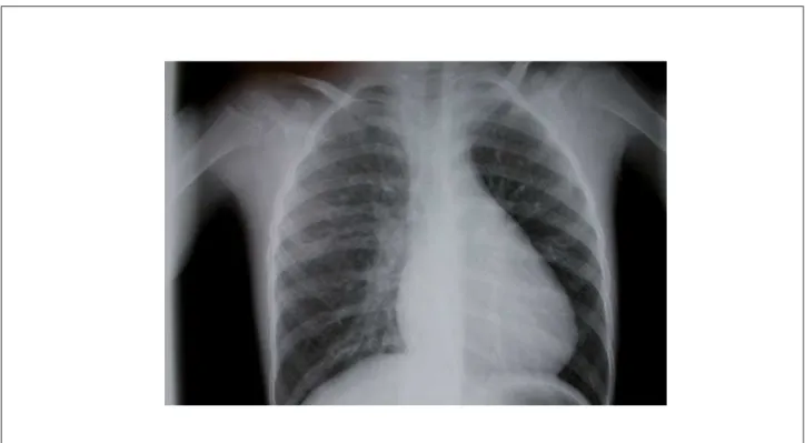
Radiographic findings included an enlarged cardiac silhouette secondary to right atrial and right ventricular dilation, a bulged mid-arch, and increased pulmonary vascularity (Figure 1).
Diagnostic impression
This image suggests the diagnosis of atrial septal defect.
Differential diagnosis
Other abnormalities may share similar features, such as partial atrioventricular septal defect with ostium primum ASD, associated with a cleft in the mitral valve causing mild mitral regurgitation; partial anomalous pulmonary venous drainage into the right atrium; and coronary artery-to-cardiac chamber arteriovenous fistulas, also into the right atrium.
Diagnostic confirmation
Clinical findings are consistent with the diagnosis of atrial septal defect, considering the split second heart sound, the soft murmur in the pulmonary area, the right ventricular systolic impulses at the left sternal border, and the right ventricular volume overload on electrocardiogram, in addition to the distinctive radiographic image. The echocardiogram (Figure 2) showed a large ostium secundum-type ASD measuring 28 mm and right atrial and ventricular enlargement. Other measurements were as follows: right ventricle 21 mm, left ventricle 29 mm, left atrium 20 mm, aorta 16 mm, and septum 5 mm. Myocardial fractional shortening was 38%.
Management
The 30-mm interatrial communication was closed via a trans-sternal minithoracotomy using a pericardial patch, and the patient evolved satisfactorily.
Key words
Child; heart defects, congenital; heart murmurs; heart septal defects, atrial.
Clinico Radiological Session
Edmar AtikArq Bras Cardiol 2007; 89(4) : 248-249
Fig. 1 - Chest x-ray showing the classic radiographic features of atrial septal defect, namely, enlarged right atrium and ventricle, increased pulmonary mid-arch and increased flow through the pulmonary vascular bed.
Fig. 2 - Echocardiogram showing the large ostium secundum atrial septal defect (ASD) in a subcostal view (A), and a left-to-right shunt on color flow mapping in the apical four-chamber view (B). RA: right atrium, LA: Left atrium, RV: right ventricle, LV: left ventricle.