Inhibition of L-type Calcium Current by Tramadol and Enantiomers in
Cardiac Myocytes from Rats
Emiliano Medei
2, Juliana M. Raimundo
1, José Hamilton M. Nascimento
2, Margarete M. Trachez
3, Roberto T.
Sudo
1, Gisele Zapata-Sudo
1Programa de Desenvolvimento de Fármacos, Instituto de Ciências Biomédicas, Universidade Federal do Rio de Janeiro1; Instituto de Biofísica Carlos Chagas Filho, Universidade Federal do Rio de Janeiro2, Rio de Janeiro, RJ; Serviço de Anestesiologia, Universidade Federal Fluminense3, Niterói, RJ, Brazil
Abstract
Background: Tramadol is a centrally acting analgesic, whose mechanism of action involves opioid-receptor activation. Previously, we have shown that tramadol and its enantiomers had a negative inotropic effect on the papillary muscle in which the (+)-enantiomer is more potent than (-)- and (±)-tramadol.
Objective: In this study, we investigated the effects of tramadol and its enantiomers on L-type calcium current (ICa-L).
Methods: The experiments were carried out in isolated Wistar rat ventricular myocytes by using the whole cell patch clamp technique.
Results: Tramadol (200 µM) reduced the peak amplitude of ICa-L at potentials from 0 to +50 mV. At 0 mV, ICa-L was reduced by 33.7 ± 7.2%. (+)- and (-)-tramadol (200 µM) produced a similar inhibition of ICa-L, in which the peak amplitude was reduced by 64.4 ± 2.8% and 68.9 ± 5.8%, respectively at 0 mV (p > 0.05). Tramadol, (+)- and (-)-tramadol shifted the steady-state inactivation of ICa-L to more negative membrane potentials. Also, tramadol and (+)-tramadol markedly shifted the time-dependent recovery curve of ICa-L to the right and slowed down the recovery of ICa-L from inactivation. The time
constant was increased from 175.6 ± 18.6 to 305.0 ± 32.9 ms (p < 0.01) for tramadol and from 248.1 ± 28.1 ms to 359.0 ± 23.8 ms (p < 0.05) for (+)-tramadol. The agonist of µ-opioid receptor DAMGO had no effect on the ICa-L.
Conclusions: The inhibition of ICa-L induced by tramadol and its enantiomers was unrelated to the activation of opioid receptors and could explain, at least in part, their negative cardiac inotropic effect. (Arq Bras Cardiol XXXX;XX(X):000-000)
Keywords: Tramadol; calcium channels, l-Type; myocytes, cardiac; rats.
Mailing Address: Gisele Zapata-Sudo •
Programa de Desenvolvimento de Fármacos, Universidade Federal do Rio de Janeiro, Centro de Ciências da Saúde, Instituto de Ciências Biomédicas, Bloco J, Sala 14 - 21941-590 - Rio de Janeiro, Brazil
E-mail: [email protected]
Manuscript received November 04, 2010, revised manuscript received November 04, 2010; accepted April 08, 2011.
low affinity with µ-opioid receptors and inhibits norepinephrine uptake3. The complementary and synergistic actions of both enantiomers improve the analgesic profile of the racemate4. The main metabolite of tramadol, O-desmethyltramadol, seems to contribute to the analgesic effect because it has approximately a 300-fold greater affinity with µ-opioid receptors than tramadol5. Tramadol has been shown to affect 5-HT6, muscarinic7,8, nicotinic9, NMDA and GABAA
10 receptors. Also, voltage-dependent K+ and
Na+ channels are involved in the antinociceptive and anesthetic effect of tramadol, respectively11,12. Limited information about the effects of tramadol on systems other than the central nervous system is available and few studies have compared the effects of tramadol and its enantiomers. In a previous study, we showed that tramadol and its enantiomers induced relaxation of precontracted rat aorta, which was stereoselective to (+)-tramadol13. Also, we have shown that tramadol reduced the contractility of rat cardiac muscle13. The possible mechanism involved in this phenomenon could be the inhibition of L-type calcium current (ICa-L), related to the activation of a receptor that modulates ICa-L and/or to a direct effect on the channel.
In order to evaluate the role of L-type Ca2+ channels on the negative cardiac inotropic action of tramadol and its enantiomers,
Introduction
we investigated their effects on the L-type Ca2+ currents (I Ca-L) of rat ventricular myocytes. Comparisons among these compounds were done using the concentration (200 µM) that had induced negative inotropic effect on rat cardiac muscle13.
Methods
The Animal Care and Use Committee at Universidade Federal doRio de Janeiro approved the protocols used.
Isolation of Cardiomyocytes
Hearts from male Wistar rats (250-350 g) were rapidly removed and mounted on a modified Langendorff system. They were retrogressively perfused through the aorta for 5 min at 10 ml.min-1 with oxygenated Tyrode solution (in mM: 132.0 NaCl, 1.0 CaCl2, 1.2 MgCl2, 4.0 KCl, 10.0 HEPES, and 5.0 glucose; pH 7.3) at 33-35ºC. Ventricular myocytes were enzymatically isolated after 10 min perfusion with nominally Ca2+-free solution containing collagenase type II (Worthington Biochemical, Lakewood, NJ; 150 U/ml). The enzyme was washed out by perfusion with Ca2+-free Tyrode solution. Isolated myocytes were maintained in Kb solution (in mM: 1.0 MgCl2, 30.0 KCl, 10.0 KH2PO4, 10.0 HEPES, 10.0 glucose, 70.0 glutamic acid, 0.3 EGTA, 20.0 taurine; pH 7.3) at room temperature until use. They were placed in the recording chamber mounted on the stage of an inverted microscope (Axiovert 40 CFL, Zeiss) and perfused at 5 ml.min-1 with Tyrode solution at 35ºC.
Electrophysiological studies
Recordings of Ca2+ currents were obtained by using the whole-cell configuration of the patch-clamp technique14 through an Axopatch 200B amplifier (Axon Instruments, Foster City, CA). Voltage pulses were generated by pClamp software and a digital interface (Digidata 1200, Axon Instruments, Foster City, CA). Micropipettes were prepared with borosilicate glass
capillaries and had a resistance of 4-7 MΩ when filled with
pipette solution (in mM: 110.0 CsCl, 5.0 ATP-Mg, 0.1 GTP, 10.0 EGTA, 10.0 HEPES, and 30.0 TEA-Cl; pH 7.1). Currents were low-pass filtered at 1 KHz and digitized at 2 KHz. Current-voltage relationships were determined through 500ms Current-voltage step from a holding potential of -40 mV to test potentials ranging from -50 to +60 mV, in 10 mV increments during 500 ms before and after 5 min of perfusion with 200 µM tramadol. Peak ICa-L value was expressed as relative to cell capacitance (pA/pF) and presented as mean ± SEM. Steady-state activation (d) and steady-state inactivation (f) curves were fitted with a Boltzmann function: d (or f) = 1/[1 + exp (Vm–V0.5)/k], where Vm was the membrane potential, V1/2 was the potential of half maximum activation/inactivation and k the slope factor.
Substances
Racemic tramadol and its enantiomers were generously donated by Cristália Produtos Químicos e Farmacêuticos (Itapira, São Paulo, Brazil) and were dissolved in distilled water at a stock concentration of 50 mM. Collagenase type II, taurine, tetraethylammonium chloride (TEA-Cl), HEPES,
glutamic acid, EGTA, CsCl, MgATP, GTP and tetrodotoxin (TTX) were purchased from Sigma.
Statistical analysis
Data were presented as mean ± SEM. Intergroup comparisons were performed using ONE-WAY ANOVA with Bonferroni’s multiple comparison test (selected pairs of column). While, intragroup comparisons were performed using repeated measures ONE-WAY ANOVA with Bonferroni’s multiple comparison test (compare selected pairs of column). Statistical differences were considered significant when p < 0.05.
Results
Effect of tramadol and enantiomers on current-voltage relationship of ICa-L
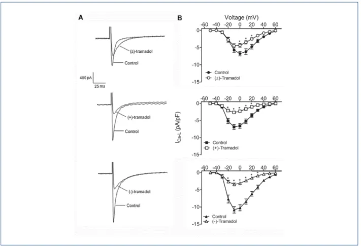
Figure 1A shows a representativetracing of the inward Ca2+ currents recorded from rat ventricular myocytes at 0 mV in the absence and presence of 200 µM (±)-tramadol (Figure-1 top) and its enantiomers (Figure-1 middle and bottom). Mean values of ICa-L at different potentials (-50 to 60 mV) were plotted in I-V curves before and after treatment with (±)- (Figure-1B top), (+)- (Figure-1B middle) and (-)-tramadol (Figure-1B bottom). Upon perfusion ofeach enantiomer, the peak amplitude of ICa-L was significantly (p < 0.05)reduced at potentials more depolarized than -20 mV, while with (±)-tramadol the reduction was significant at potentials more positive t4han 0 mV (p < 0.05). (±)-Tramadol significantly inhibited the peak amplitude of ICaL which was reduced by 33.7 ± 7.2% at 0 mV. There was no difference in the peak current inhibition induced by the enantiomers, (+)-tramadol (64.4 ± 2.8%) and (-)-tramadol (68.9 ± 5.8%). However, the inhibition of ICa-L induced by both enantiomers was significantly greater than that induced by (±)-tramadol (p < 0.01 vs (+)-tramadol; p < 0.001 (-)-tramadol). The reduction was partially reversed after a 10 min washout.
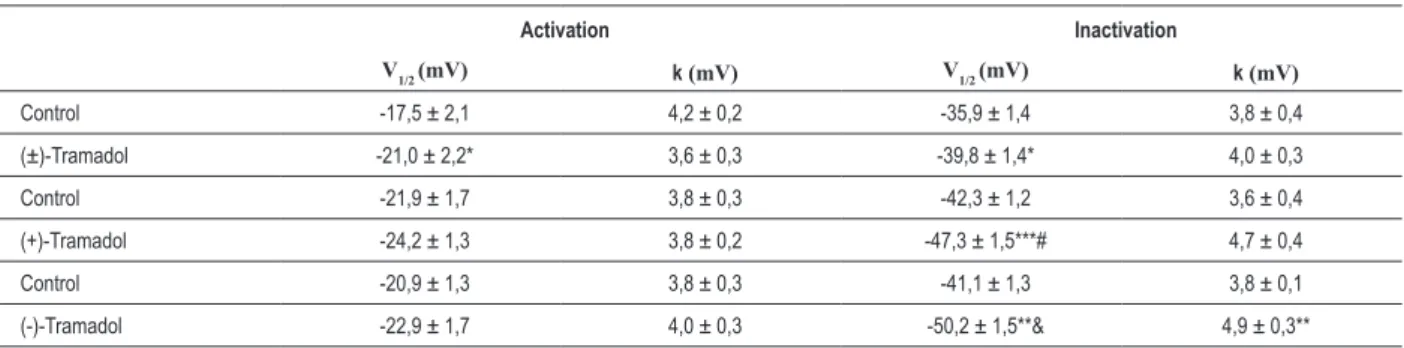
Effect of tramadol and enantiomers on activation of ICa-L Activation curves were constructed from the current-voltage relationship by dividing the amplitude of ICa-L at each potential by the driving force (Figure 2). (±)-Tramadol decreased the V1/2 of the steady-state activation curve of ICa-L, with no changes in k value. In the control condition, V1/2 was -17.5 ± 2.1 mV and k was 4.2 ± 0.2 mV and in the presence of (±)-tramadol, V1/2 was -21.0 ± 2.2 mV (p < 0.05) and k was 3.6 ± 0.3 mV (p> 0.05). On the other hand, the enantiomers did not change the steady-state activation curve of ICa-L (Table 1).
steady-state inactivation curves to more negative potentials (Figure 3) and changed the V1/2 of inactivation (Table 1).
In fact, the V1/2 of inactivation of enantiomers was significant different when compared to (±)-Tramadol ((+)-tramadol p < 0.01 and (-)-tramadol p < 0.001 compared with (±)-tramadol). However, only (-)-tramadol altered the k value (control, 3.8 ± 0.1 mV; (-)-Tramadol, 4.9 ± 0.3 mV; p < 0.01). No differences were observed in the mean of the k value among groups.
Effect of tramadol and enantiomers on recovery of ICa-L from inactivation
The effects of (±)-tramadol and its enantiomers on the kinetics of recovery of ICa-L from inactivation were studied using a double-pulse protocol consisting of a 500 ms prepulse to 0 mV followed by a 500 ms test pulse to 0 mV, after a variable inter-pulse interval (0 to 2500 ms) from a holding potential of -40 mV, every 10 s. (±)- and (+)-tramadol shifted the time-dependent recovery curve to the right and slowed down the recovery from inactivation (Figure 4). The recovery from inactivation could be fitted by a single exponential, where the time constant (t) was increasedfrom 175.6 ± 18.6 ms to 305.0 ± 32.9 ms (p < 0.01)for(±)-tramadol and from 248.1
± 28.1 ms to 359.0 ±23.8 ms (p < 0.05) for (+)-tramadol. (-)-Tramadol did not modify the time course for recovery of ICa-L. No differences were observed in the mean of the time constant (t) value among groups.
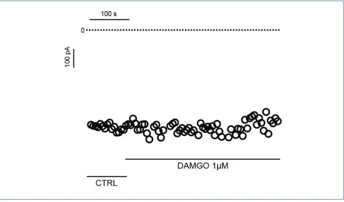
Effect of the agonist of µ-opioid receptor (DAMGO) on the ICa-L of rat cardiomyocites
In this study, our results have shown that tramadol and its enantiomers inhibit the ICa-L. The possible mechanism
involved in this effect could be due to the activation of µ-opioid receptors. To verify this hypothesis, we tested whether the agonist of the µ-opioid receptor (DAMGO) modulates the ICa-L in rat cardiomyocytes. Thus, figure 5 shows that DAMGO has no effect on the calcium currents activated by depolarizing pulses to 0 mV (Control: -449.7 ± 4.4 pA vs DAMGO: -416.9 ± 2.7 pA; n=3; p> 0.05).
Discussion
In this study, our data have shown different effectiveness of tramadol enantiomers to inhibit ICa-L (~60%) when compared to racemic mixture (~30 %). In contrast, in our previous study, the negative inotropic effect on rat papillary muscles
Figure 1 – Effects of tramadol and enantiomers on L-type Ca2+ current (ICa-L) in isolated rat ventricular myocytes. In A, representative current traces following a
depolarizing pulse from -40 to 0 mV, in the absence and presence of (±)- (top), (+)- (middle) and (-)-tramadol (bottom). In B, current-voltage relationships of ICa-L obtained
observed at 200 µM presented the following order of potency (IC50): (+)-tramadol > (-)-tramadol > (±)-tramadol13. This difference between cell and tissue responses could be due
to a possible inhibition of monoamine reuptake by tramadol and their enantiomers on the electrically-stimulated papillary muscle. It has been demonstrated that the electrically evoked
R
esu
lt
ad
o
s
lCal-L
Figure 2 – Effects of tramadol and enantiomers on the steady-state activation kinectics. Voltage-activation curves of ICa-L were obtained from the current-voltage
relationship, in the absence and presence of 200 µM (±)-, (+)- and (-)-tramadol (A, B and C, respectively). Only (±)-tramadol shifted the voltage of half-activation curve. Intragroup comparisons were performed using repeated measures ONE-WAY ANOVA with Bonferroni’s multiple comparison test (selected pairs of column). *p <0.05, **p < 0.005, ***p < 0.0005 compared with control in the same group (ONE-WAY ANOVA repeated measures with Bonferroni’s compared selected pairs of columns). #p<0.01, &p<0.001 compared with (±)-Tramadol, intergroup comparison (ONE-WAY ANOVA with Bonferroni’s multiple comparison test compared all pairs of columns). V1/2 - potential of half maximum activation/inactivation; κ - slope factor.
Table 1 – Effects of tramadol and its enantiomers on the kinetics of activation and inactivation of ICa-L
Activation Inactivation
V1/2 (mV) k (mV) V1/2 (mV) k (mV)
Control -17,5 ± 2,1 4,2 ± 0,2 -35,9 ± 1,4 3,8 ± 0,4
(±)-Tramadol -21,0 ± 2,2* 3,6 ± 0,3 -39,8 ± 1,4* 4,0 ± 0,3
Control -21,9 ± 1,7 3,8 ± 0,3 -42,3 ± 1,2 3,6 ± 0,4
(+)-Tramadol -24,2 ± 1,3 3,8 ± 0,2 -47,3 ± 1,5***# 4,7 ± 0,4
Control -20,9 ± 1,3 3,8 ± 0,3 -41,1 ± 1,3 3,8 ± 0,1
(-)-Tramadol -22,9 ± 1,7 4,0 ± 0,3 -50,2 ± 1,5**& 4,9 ± 0,3**
Figure 3 – Effects of tramadol and enantiomers on steady-state inactivation kinectics. Voltage inactivation curves of ICa-L were obtained using a double pulse protocol,
which consisted of conditioning voltage pulses to membrane potentials from –60 to + 60 mV followed by a test pulse to 0 mV. Test pulse currents were normalized to the maximum value. In A, B and C, steady-state inactivation curves of ICa-L were obtained in the absence and presence of (±)-, (+)- and (-)-tramadol (200 µM), respectively.
release of noradrenaline by sympathetic nerve endings in isolated cardiac muscle and the amplitude of contraction are decreased by local anesthetics15. Different activities of tramadol have shown to be stereoselective, including opioid receptor activation3,16, inhibition of monoamine reuptake17,18, analgesic effect3 and vascular relaxation13,19. However, this study demonstrated that tramadol enantiomers (200 µM) decreased ICa-L (~60%), indicating a non-enantiomer-specific blockade of L-type Ca2+ channels. The racemic tramadol produced a minor inhibition of ICa-L (~30%) when compared to its enantiomers which could be correlated to the small potency of racemic to reduce cardiac
contractility13. Tramadol and enantiomers differently altered the kinectics of ICa-L. (±)- and (+)-tramadol shifted the steady-state inactivation curve for ICa-L to more negative membrane potentials and markedly slowed the recovery of ICa-L from inactivation. (-)-Tramadol significantly altered only the inactivation of ICa-L. The effects of tramadol on ICa-L may be related to the activation of a receptor that modulates ICa-L and/or to a direct effect on the channel. However, the effect of tramadol on ICa-L seems not to be related to the activation of opioid receptors. Tramadol binds preferentially to m-receptors, which have been demonstrated not to be present in the mammalian heart20-23. Indeed, our results
Figure 4 – Effects of tramadol and enantiomers on ICa-L recovery from inactivation. The recovery of ICa-L from inactivation was determined using a double pulse protocol consisting of two pulses to 0 mV with a variable inter-pulse interval (0 – 2500 ms), in the absence and presence of 200 µM (±)-, (+)- and (-)-tramadol (A, B and C, respectively). The time course for recovery of ICa-L was signiicantly altered by (±)-, (+)-tramadol. Intragroup comparisons were performed using repeated measures ONE-WAY ANOVA with Bonferroni’s multiple comparison test (selected pairs of column).
Figure 5 – The agonist of µ-opioid receptor (DAMGO) had no effect on the ICa-L. Figure 5 shows a representative experiment where the ICa-L time-course was recorded
showed no effect of the m-receptor agonist DAMGO on the cardiac ICa-L. Moreover, it has been shown that the cardiac effects of opioids could be or not be dependent on opioid receptors. The effects of morphine on ionic currents in cardiac myocytes have shown not to depend on opioid receptor24 or to be mediated via d- and k-receptors25. However, its cardioprotective effect is known to be due to the activation of d-receptors26,27. Studies have shown that opioids like dextropropoxiphene, pethidine and leucine enkephalin reduce ICa-L but only the inhibition induced by d-receptor agonist (leucine enkephalin) was blocked by naloxone28,29. The effects of tramadol on other ionic currents also suggest a direct effect on L-type Ca2+ channels. Haeseler et al12 showed that tramadol, sufentanil and fentanil but not morphine blocked sodium currents of heterologously expressed NaV1.2 neuronal Na+ channels. It is important to note that the potency to block sodium current was irrespective of the relevant opioid receptor potency of the compound. Tramadol has also been reported to block delayed rectifier K+ current (I
K(DR) in NG108-15 neuronal cells in a concentration-dependent manner30. As observed in our study for ICa-L, tramadol shifted the steady-state inactivation curve of IK(DR to more negative potentials30.
Conclusion
The effectiveness of tramadol enantiomers to inhibit ICa-L was twice the one observed with (±)-tramadol and such effect seems unrelated to the activation of opioid receptors.
Acknowledgements
The authors thank Cristália Produtos Químicos e Farmacêuticos Ltda. (BR), Conselho Nacional de Desenvolvimento Cientifico e Tecnológico (CNPq, BR), Coordenação de Aperfeiçoamento de Pessoal de Nível Superior (CAPES, BR), Fundação Universitária Jose Bonifácio (FUJB, BR), Fundação Carlos Chagas Filho de Amparo à Pesquisa do Estado do Rio de Janeiro (FAPERJ, BR) for finantial support and fellowships from CAPES (to JMR) and CNPq (to GZS, RTS). None of the authors have any conflict of interest or financial interest to disclose.
Potential Conflict of Interest
No potential conflict of interest relevant to this article was reported.
Sources of Funding
This study was partially funded by CNPq, CAPES and FAPERJ.
Study Association
This article is part of the thesis of Doctoral submitted by Juliana M. Raimundo, from Universidade Federal do Rio de Janeiro.
References
1. Raffa RB, Friderichs E, Reimann W, ShanK RP, Codd EE, Vaught JL. Opioid and non-opioid components independently contribute to the mechanism of action of tramadol, an atypical opioid analgesic. J Pharmacol Exp Ther. 1992;260(1):275-85.
2. Garrido MJ, Valle M, Campanero MA, Calvo R, Trocóniz IF. Modeling of the in vivo antinociceptive interaction between an opioid agonist, (+)-O-desmethyltramadol, and a monoamine reuptake inhibitor, (-)-O-desmethyltramadol, in rats. J Pharmacol Exp Ther. 2000;295(1):352-9.
3. Raffa RB, Friderichs E, Reimann W, Shank RP, Codd EE, Vaught JL, et al. Complementary and synergistic antinociceptive interaction between the enantiomers of tramadol. J Pharmacol Exp Ther. 1993;267(1):331-40. 4. Grond S, Sablotzki A. Clinical pharmacology of tramadol. Clin
Pharmacokinet. 2004;43(13):879-923.
5. Gillen C, Haurand M, Kobelt DJ, Wnendt S. Afinitty, potency and efficacy of tramadol and its metabolites at the cloned human m-opioid receptor. Naunyn-Schmiedeberg’s Arch Pharmacol. 2000;362(2):116-21.
6. Ogata J, Minami K, Uezono Y, Okamoto T, Shiraishi M, Shigematsu A, et al. The inhibitory effects of tramadol on 5-hydroxytryptamine type 2c receptors expressed in Xenopus oocytes. Anesth Analg. 2004;98(5):1401-6.
7. Shiraishi M, Minami K, Uezono Y, Yanagihara N, Shigematsu A. Inhibition by tramadol of muscarinic receptor-induced responses in cultured adrenal medullary cells and in Xenopus laevis oocytes expressing cloned M1 receptors. J Exp Pharmacol Ther. 2001;299(1):255-60.
8. Shiga Y, Minami K, Shiraishi M, Uezono Y, Murasaki O, Kaibara M, et al. The inhibitory effects of tramadol on muscarinic receptor-induced responses in Xenopus oocytes expressing cloned M3 receptors. Anesth Analg. 2002;95(5):1269-73.
9. Shiraishi M, Minami K, Uezono Y, Yanagihara N, Shigematsu A, Shibuya I. Inhibitory effects of tramadol on nicotinic acetylcholine receptors in adrenal chromaffin cells and in Xenopus oocytes expressing a7 receptors. Br J Pharmacol. 2002;136(2):207-16.
10. Hara K, Minami K, Sata T. The effects of tramadol and its metabolite on glycine, g-aminobutyric acidA, and N-methyl-D-aspartate receptors
expressed in Xenopus oocytes. Anesth Analg. 2005;100(5):1400-5. 11. Yalcin I, Aksu F. Involvement of potassium channels and nitric oxide in
tramadol antinociception. Pharmacol Biochem Behav. 2005;80(1):69-75. 12. Haeseler G, Foadi N, Ahrens J, Dengler R, Hecker H, Leuwer M.
Tramadol, fentanyl and sufentanil but not morphine block voltage-operated sodium channels. Pain. 2006;126(1-3):234-44.
13. Raimundo JM, Sudo RT, Pontes LB, Antunes F, Trachez MM, Zapata-Sudo G. In vitro and in vivo vasodilator activity of racemic tramadol and its enantiomers in Wistar rats. Eur J Pharmacol. 2006;530(1-2):117-23. 14. Hamill OP, Marty A, Neher E, Sakmann B, Sigworth J. Improved
patch-clamp techniques for high-resolution current recording from cells and cell-free membrane patches. Pflugers Arch. 1981;391(2):85-100. 15. Joseph A, Montiague R, Effendi AR, Urbanska RA, Vogel S, Winnie AP, et al.
Effect of bupivacaine and levobupivacaine on exocytotic norepinephrine release from rat atria. Anesthesiology. 2005;102(5):977-84.
16. Lai J, Ma SW, Porreca F, Raffa RB. Tramadol, M1 metabolite and enantiomer affinities for cloned human opioid receptors expressed in transfected HN9.10 neuroblastoma cells. Eur J Pharmacol. 1996;316(2-3):369-72.
18. Halfpenny DM, Callado LF, Hopwood SE, Bamigbade TA, Langford RM, Stamford JA. Effects of tramadol stereoisomers on norepinephrine efflux and uptake in the rat locus coerulus measured by real time voltammetry. Br J Anaesth. 1999;83(6):909-15.
19. Shin IW, Sohn JT, Park KE, Chang KC, Choi JY, Lee HK, et al. A supraclinical dose of tramadol stereoselectively attenuates endothelium-dependent relaxation in isolated rat aorta. Anesth Analg. 2006;103(2):366-71.
20. Krumins SA, Faden AI, Feuerstein G. Opiate binding in rat hearts: modulation of binding after hemorrhagic shock. Biochem Biophys Res Commun. 1985;127(1):120-8.
21. Ventura C, Bastagli L, Bernardi P, Caldarera CM, Guarnieri C. Opioid receptors in rat cardiac sarcolemma: effect of phenylephrine and isoproterenol. Biochim Biophys Acta. 1989;987(1):69-74.
22. Zhang WM, Jin WQ, Wong TM. Multiplicity of kappa opioid receptor binding in the rat cardiac sarcolemma. J Mol Cell Cardiol. 1996;28(7):1547-54. 23. Wittert G, Hope P, Pyle D. Tissue distribution of opioid receptor gene
expression in the rat. Biochem Biophys Res Commun. 1996;218(3):877-81.
24. Hung CF, Tsai CH, Su MJ. Opioid receptor independent effects of morphine on membrane currents in single cardiac myocytes. Br J Anaesth. 1998;81(6):925-31.
25. Xiao GS, Zhou JJ, Wang GY, Cao CM, Li GR, Wong TM. In vitro electrophysiologic effects of morphine in rabbit ventricular myocytes. Anesthesiology. 2005;103(2):280-6.
26. Ela C, Barg J, Vogel Z, Hasin Y, Eilam Y. Distinct components of morphine effects on cardiac myocytes are mediated by the k and d opioid receptors. J Mol Cell Cardiol. 1997;29(2):711-20.
27. McPherson BC, Yao Z. Signal transduction of opioid-induced cardioprotection in ischemia-reperfusion. Anesthesiology. 2001;94(6):1082-8.
28. Xiao RP, Spurgeon HA, Capogrossi MC, Lakatta EG. Stimulation of opioid receptors on cardiac ventricular myocytes reduces L type Ca2+ channel current.
J Mol Cell Cardiol. 1993;25(6):661-6.
29. Wu C, Fry CH, Henry J. The mode of action of several opioids on cardiac muscle. Exp Physiol. 1997;82(2):261-72.