Review
Global Diversity and Review of Siphonophorae (Cnidaria:
Hydrozoa)
Gillian M. Mapstone*
Department of Life Sciences, The Natural History Museum, London, United Kingdom
Abstract: In this review the history of discovery of siphonophores, from the first formal description by Carl Linnaeus in 1785 to the present, is summarized, and species richness together with a summary of world-wide distribution of this pelagic group within the clade Hydrozoa discussed. Siphonophores exhibit three basic body plans which are briefly explained and figured, whilst other atypical body plans are also noted. Currently, 175 valid siphonophore species are recognized in the latest WoRMS world list, including 16 families and 65 genera. Much new information since the last review in 1987 is revealed from the first molecular analysis of the group, enabling identification of some new morphological characters diagnostic for physonect siphonophores. Ten types of nematocysts (stinging cells) are identified in siphonophores, more than in any other cnidarian; these are incorporated into batteries in the side branches of the tentacles in most species (here termed tentilla), and tentilla are reviewed in the last section of this paper. Their discharge mechanisms are explained and also how the tentilla of several physonect siphonophores are modified into lures. Of particular interest is the recent discovery of a previously unknown red fluorescent lure in the tentilla of the deep sea physonect Erenna, the first described example of emission of red light by an invertebrate to attract prey.
Introduction
Siphonophores are a small group of complex fragile polymor-phic and mostly elongate colonial hydrozoans currently compris-ing 175 valid species [1] (the present author is the main editor of the Siphonophora section of the WoRMS world list). Most siphonophore species are pelagic and restricted to oceanic waters, and generally live well below the surface to avoid turbulence. Small active species inhabit the epipelagic zone (0-c. 300 m), where they lie in wait for copepods and other zooplankton, and rapidly spread their tentacles to entrap prey. Larger, though mostly more fragile, species live in the deeper and tranquil mesopelagic zone (300–1000 m), where they passively extend an enormous feeding net of tentacles to ensnare prey [2,3]. A few genera are neritic with most of their species restricted to coastal waters (Muggiaea, Sphaeronectes). One family, the Rhodaliidae, is epibenthic with a short corm-like stem and tentacles that extend out in all directions for anchorage to the substrate [4]. Siphonophores make a significant contribution to complex trophic links in the deep sea ‘jelly web’, of which gelatinous zooplankton can contribute up to 25% of the total pelagic biomass [3]. The geographical distribution of most siphonophores is cosmopolitan with species inhabiting all oceans [5]. However, some are restricted to particular latitudinal ranges or oceanic areas, and a few are so far known only from a single location. Siphonophores are extremely difficult to capture, with the best specimens collected
and observed from submersibles or with blue-water SCUBA equipment.
Siphonophores are holoplanktonic, except for rhodaliids which can transiently attach their tentacles to the substrate, and thus lack the true benthic stage that is characteristic of the life cycle of many hydromedusae and other colonial cnidarians. Larvae are some-times collected, and a few species have been successfully reared in the laboratory [40], but larvae of most species are still unknown. The yolky planula soon develops a stem and in most species begins budding zooids from two growth zones [6]. As the stem extends, more zooids form, the colony matures and various morphological axes can be identified [6,7]. Upon maturity, this asexual life stage may release egg or sperm masses directly into the water [8], or release gametes either from sexual gonophores that remain attached to the stem, or form eudoxids (sexual life stages) from the end of the stem which are later released. These life stages are explained in a recently published glossary of siphonophore terminology [6].
For many decades, siphonophore systematics was based primarily on the classification of Totton [9], including the last review of the group [2]. Totton divided siphonophores into three suborders: Cystonectae, Physonectae and Calycophorae. More recently, however, the first molecular analysis of siphonophores [10] revealed a major new phylogeny in which cystonects, without nectosomal swimming bells, are sister to all other siphonophores with bells. This latter clade is known as the bell-bearers, or Codonophora, and these taxa differ from the Cystonecta in one important respect: the feeding and sexual zooids (gastrozooids and gonodendra, or gonophores) of each iterative group (cormidium) on the stem form from a single probud, except for a few secondary zooids which arise from primary zooids in some species [11]. In cystonects gastrozooids and gonodendra develop separately and directly on the stem, not from a probud [8]. This important difference is reflected in the new phylogeny, as summarized by Mapstone (p. 75 [6]).
Previously unknown red fluorescent lures have also been discovered in a new deep sea physonect species of the genus Erenna [12]. In addition, new families and sub-families of siphonophores have been identified and others reviewed
Citation:Mapstone GM (2014) Global Diversity and Review of Siphonophorae (Cnidaria: Hydrozoa). PLoS ONE 9(2): e87737. doi:10.1371/journal.pone.0087737
Editor:Andreas Hejnol, Sars International Centre for Marine Molecular Biology, Norway
PublishedFebruary 6, 2014
Copyright:ß2014 Gillian M. Mapstone. This is an open-access article distributed under the terms of the Creative Commons Attribution License, which permits unrestricted use, distribution, and reproduction in any medium, provided the original author and source are credited.
Funding:No current funding sources for this study.
Competing Interests:The author has declared that no competing interests exist.
[13,14,15,16,17,18,19], several new species have been described [1], and body axes standardized for all siphonophores [6,7]. All these new findings are discussed below, together with a summary of the history of discovery of siphonophores and, for the first time, an assessment of siphonophore nematocysts, tentilla and lures.
Results and Discussion
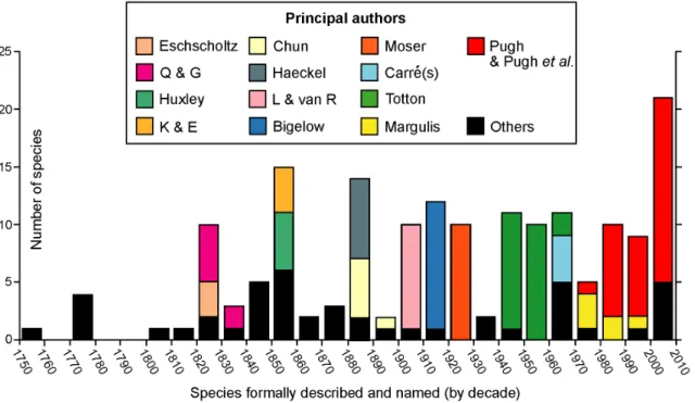
History of Discovery
Most non-specialist biologists know only one species of siphonophore, the Portuguese Man O’War (Physalia physalis), because it has a large and colourful float on the sea surface propelled by the wind. Not surprisingly, this was the first siphonophore to be formally described and introduced, as long ago as 1758, by Carl Linnaeus. Only four additional valid species were described during the rest of that century. In the nineteenth century, however, 56 new species were introduced (Figure 1).
The first half of the 19thcentury saw a flowering of voyages of discovery. Collection of fauna and flora provided ships with a free passport to otherwise hostile anchorages controlled by various European maritime powers; distant lands were discovered and charts made of their coastal waters. Marine fauna collected often included the almost exclusively holoplanktonic group Siphono-phora. Specimens were found in surface waters in these early days, some of which arrived there via upwelling events. Eschscholtz (Figure 1) circumnavigated the world twice in the Russian brigs Rurik (1816–1818) and Enterprise (1823–1826) and brought back the first specimens ofAgalma okeniandChelophyes appendiculatafrom the tropical north Pacific Ocean. He included formal descriptions of these species, and another 12 valid species he had introduced earlier in his 1829 volume ‘System der Acalephen’ [20]. All were placed in a new order Siphonophora, which at that time also included the ‘‘chondrophores’’ (Porpita and Velella, see below). Eschscholtz’s 1829 work was published just after the first observations on siphonophores by Quoy and Gaimard in 1827.
The latter authors sailed to the Pacific Ocean in the Astrolabe (1826–1829); they found five new species in the Strait of Gibraltar, shortly after the ship left Toulon [21], whilst the full zoological report of the ‘zoophytes’ discovered during the voyage (cnidarians and echinoderms) was published six years later [22]. The latter included three further new siphonophore species, from the Cape Verde Islands and from near Kangaroo Island off South Australia (Praya dubia), Bass Strait (Bassia bassensis) and off the northern coast of New Guinea (Halistemma foliacea).
During the latter half of the nineteenth century 36 more siphonophores were introduced (Figure 1). The decade between 1850 and 1860 saw 15 new species described, notably five by Huxley [23] in his important work the ‘‘Oceanic Hydrozoa’’, and four by Keferstein and Ehlers [24,25] from the Mediterranean. Huxley travelled to Port Jackson, the new British colony on the eastern coast of Australia (later Sydney), as assistant naturalist on board HMS Rattlesnake(1846–1850). He collected specimens of Physalia on the way out, and was the first to note that the body wall comprised two layers of cells, including nematocysts (the signature cells of cnidarians), and an intervening layer of mesogloea. Huxley was the consummate naturalist and a careful observer and illustrator of Siphonophora. He introduced two abylids (Cerato-cymba leuckartiand Abylopsis eschscholtzi), the eudoxid bracts of the tropical diphyid Eudoxoides mitra, and anterior nectophores of Diphyes chamissonis (which lacks a posterior nectophore) from samples taken during these cruises. He also founded a new family the Sphaeronectidae based on three specimens of the small species S. koellikericollected from the Indian Ocean, Torres Strait and east coast of Australia.
Two Germans, Carl Chun and Ernst Haeckel dominated the decade 1880–1890, adding five and seven new species of Siphonophora respectively (Figure 1). Haeckel wrote up the Siphonophora collected during the BritishHMS Challenger Expe-dition (1873–1876), with other specimens in a 380 page major work [26]. He founded a new family the Rhodaliidae (as an order,
Figure 1. History of siphonophore research.Principle researchers and others from mid-18thcentury to the present. Authors identified only by initials are Q & G: Quoy and Gaimard, K & E: Keferstein and Ehlers, and L & van R: Lens and van Riemsdijk.
later abandoned) for three species with a large spherical pneumatophore, prominent gas gland and siphosome reduced to a corm, concluding that they were pelagic. Much later, in 1983, these siphonophores were shown by Pugh [27] to be benthic. Although Haeckel included 46 ‘‘new species’’ in his Challenger report, eight were chondrophores (now athecate hydroids, see below), and only four, in addition to the three rhodaliids, are now regarded as valid; these include two long-stemmed physonects and two prayid calycophorans (Forskalia tholoides, Cordagalma ordinatum, Amphicaryon peltifera and Desmophyes annectens). Overall, Haeckel’s treatment of the group was muddled. Indeed Totton (p. 6–13 [9]) wrote a critique of Haeckel’s classification and ill-founded Medusome Theory, whilst Mary Winsor (p. 322 [28] commented: ‘‘Haeckel’s own description would lead us to expect that his Challenger Reporton siphonophores was both a significant contribution to knowledge and a fine example of an evolutionist at work. Upon examination the picture is totally altered. The excitement of great ideas was well over by 1888, and the famous defender of Darwin seemed lacking in imaginative power. Instead of a case study of the clear impact of the Origin of Species upon a zoological problem, the siphonophores provide an example of the surprising success in interpreting animal relationships achieved by pre-Darwin-biologists.’’ Despite this, many of Haeckel’s species descriptions and figures are still useful, provided account is taken of his short-comings. On the other hand, Carl Chun was more conservative and introduced one valid species from the Mediterranean (Lensia subtilis) in 1886 [29] and four more from the Canary Islands in 1888 [30]. He also added a further species in 1897 ([31] and see Figure 1), the diphyid calycophoranDimophyes arctica.
In the twentieth century an average of ten new siphonophore species were introduced per decade, except during the pre-Second World War years (Figure 1). Specimens were collected either during expeditions or on routine (steam and sail) research cruises by British, American and other vessels. The Dutch Siboga Expedition (1899–1900) sampled the deep basins of the Indone-sian Archipelago, and the 3,400 good siphonophore specimens collected were written up by Lens and van Riemsdijk [32]. These authors introduced nine new species including two new unusual calycophorans of unknown affinities, Chuniphyes multidentata and Clausophyes galeata. These were later placed by Totton [9] in a new mesopelagic diphyomorph family the Clausophyidae. The Ger-man Su¨dpolar-Expedition to Antarctica (1901–1903) travelled in the Research VesselGaussto the ice edge in the Indian sector of the Southern Ocean and collected a large number of siphono-phores. A sizeable report was produced by Fanny Moser, in which nine new species were introduced (together with two others described earlier). Her work was completed in 1914, but not published until after the First World War, in 1925 [33]. Her most notable new species was, perhaps, the richly colourful cold-water southern physonectPyrostephos vanhoeffeni (p. 437–8 [33]). It is an abundant species in the Southern Ocean, and Moser placed it in a new family Pyrostephidae. The AmericanAlbatrossExpeditions of the early 1900’s focussed on investigation of fish stocks and fish food under the leadership of Alexander Agassiz. The 1904–5 cruise investigated the relatively unknown area of the Pacific Ocean between South America and Easter Island, which proved to be very rich in pelagic life. Collection of the gelatinous zooplankton was supervised by Henry Bigelow, who produced a most comprehensive and well-illustrated report on siphonophores from the voyage [34]. Earlier the same year he published another paper on siphonophores from the Bay of Biscay [35], and together these two works included 11 siphonophore species new to science. The most notable are two benthic rhodaliids (Dromalia alexandriand Stephalia dilata), and several conspicuous pelagic calycophorans, including the large prayid Praya reticulata, and three species of a
new and angular type of prayid referred by Bigelow to a new subfamily Nectopyramidinae. This group has been reviewed more recently by Phil Pugh ([13] and see below).
The most productive researcher on Siphonophora during the mid-twentieth century was A.K.Totton of the British Museum of Natural History (BMNH), London, England, who introduced 23 new species (Figure 1). He started work at the museum in 1914, aged 22, but almost immediately joined the army and fought in the First World War, where he was severely wounded and awarded the Military Cross [36]. By 1918 he was back in the museum in London, as Assistant Keeper and in charge of coelenterates. Over his lifetime he amassed an enormous collection of Siphonophora specimens which he used to write several important works. Much of his material came from the cruises ofRRS Discoveryships run by the British Government from 1925 onwards, initially to Antarctica to study the biology of whales, but also, from 1929 onwards, to adjacent regions including the Indian and Pacific Oceans and Southern Atlantic Ocean. He also made annual spring visits from 1949 onwards to Station Zoologique, Villefranche, in the Mediterranean, where he was able to study live siphonophores in upwelled water for the first time, rear larvae and work out some of their life cycles. Totton also wrote important works on the Siphonophora of the Great Barrier Reef Expedition [37], of the Indian Ocean [38], and his most comprehensive systematic monograph, ‘Synopsis of the Siphonophora’ [9]. This last monograph covered all species he considered valid, but did not touch on their histology, physiology or distribution. In addition, Totton spent three months working on Physalia physalis in the Canaries with George Mackie in 1955, and produced the most detailed paper ever written onPhysaliamorphology [39]. The 23 new species he introduced over his lifetime (Figure 1) include 11 species ofLensia, a genus he erected in 1932. He also introduced two physonect genera (Bargmannia, Marrus), one new diphyid subfamily, the Sulculeolariinae, and one new diphymorph family, the Clausophyidae. As noted in his obituary [36], Totton had ‘‘a sardonic humour, innate romanticism, warm personality and greatesprit’’.
Significant contributions to new species introductions during the 20thcentury were also made by Claude and Danielle Carre´ at the Station Zoologique, Villefranche-sur-Mer on the Mediterranean, S.D. Stepanjants from St Petersburg and R.Ya. Margulis from Moscow University. Claude Carre´ introduced four new species, including two prayine prayids and two valid species of the small-belled family Sphaeronectidae, all collected in the Bay of Villefranche. Between them the Carre´’s wrote 29 papers on Siphonophora, as sole or joint authors, and some others with collaborators. They also reared live siphonophores, including Muggiaea kochi through several generations and at different temperatures [40]. In addition, Claude Carre´ wrote an important review of the diphyid subfamily Sulculeolariinae [41], showing that, for three species commonly found in the Mediterranean, both anterior and posterior nectophores were regenerated two or occasionally three times. Stepanjants introduced two new valid species from the NW PacificApolemia vitiaziandLensia asymmetrica [1], while Margulis worked on the vast Russian collections of Siphonophora taken from all major oceans of the world over a period of three decades. She introduced five new species herein considered valid, mostly from subarctic or arctic seas, and one additional species she attributed to a new name, now reinterpreted as Clausophyes moserae [42]. In all Margulis wrote 29 papers on Siphonophora, many on their worldwide vertical and horizontal distribution.
(Figure 1), many in collaboration with other researchers world-wide, and a number as sole author; more are ‘‘in preparation’’. He took over study of the British National Collection from Totton in 1972, coincident with the launch of twoJohnson Sea-Linkmanned submersibles from Harbor Branch Oceanographic Institution, Florida, USA in 1971 and 1975. Since then his research has gone from strength to strength. The American Johnson Sea Links (JSL I and II) provided him with much new and beautifully preserved material. Observers collect specimens using remotely controlled suction-operated canisters and other devices (reviewed in [43]). Fifteen new species taken by JSL I and II have been introduced by Pugh in papers published between 1987 and 2009, and another five species re-described. New species include physonects Halis-temma transliratum, Bargmannia amoena,Physophora gilmeri, three species ofForskalia[16], and three more physonects with distinctive tentilla and muscle-free proximal surface to the nectosac for which Pugh has erected a new family Erennidae [15]; also five prayine prayid calycophorans [19,44]. The American submersibleAlvincollected a new benthic rhodaliid Thermopalia taraxaca (the Galapagos Dandelion) from the Galapagos Rift in 1979, one of 10 species re-assessed in an important work by Pugh [27] on the family Rhodaliidae. Then another rhodaliid, Archangelopsis jagoa, was collected by the German JAGO in the Gulf of Aqaba, and described by Hissmann, Schauer and Pugh [45]. Pugh also introduced five species from specimens collected by Discovery (1962), including a third rhodaliid, the physonect species Bargmannia gigas and three calycophorans (Nectadamas richardi and two species ofClausophyes). Two further species were collected from the Sargasso Sea using SCUBA; the prayine calycophoranRosacea flaccida[46], and the physonectForskalia saccula[16].
The most recent new siphonophores introduced by Pugh, some in collaboration with Casey Dunn (Brown, Rhode Island, USA) and Steve Haddock (MBARI, USA), were sampled by Remotely Operated Vehicles (ROV) in the northeast Pacific Ocean, off Southern California. They were mostly collected by the Monterey Bay Research Institute (MBARI) using the ROVs ‘Tiburon’ and ‘Ventana’. These new species include the physonect Marrus claudanielis (named for the Carre´s), three physonects in a new genus Resomia, and five calycophorans. The resomiids have remarkable tentilla of two different types on the same tentacle, for which Pugh [17] created a new physonect family Resomiidae. Three of the calycophorans are new species in the family Sphaeronectidae [18]. Remarkable optical properties were discovered in the two new prayid species collected by ROV [7], see below. Pugh also collaborated with Francesc Page`s on Antarctic material collected by the German RV Polarstern, and together they discovered a remarkable new life stage in the clausophyidCrystallophyes amygdalina, the fuseudoxid [47].
Two distinctive pleustonic generaPorpitaandVelellalive on the ocean surface with the aid of a chitinous float. They were first introduced by Linnaeus [48], at the same time asPhysalia physalis, and Eschscholtz [20] placed them in a family Velellidae, together with all other siphonophores then known. For a number of decades they were even thought of as ‘typical’ siphonophores, but studies on their larvae, beginning with Leloup [49] and Garstang [50] showed these to be more similar to actinulae of some Anthomedusae than to siphonula larvae of physonect siphono-phores. This prompted Totton [38] to place them in a separate order Chondrophora. Behavioural and other studies by Mackie [51] onPorpitafurther demonstrated the anthomedusan affinities of chondrophores. These affinities were reiterated by Kirkpatrick and Pugh [52] who placed chondrophores in the Family Velellidae of the suborder Capitata, Order Athecata, in their ‘Synopsis of the British Fauna Series’. Later, Page`s et al. [53] referred them to the
Family Porpitidae Goldfuss, 1818, and more recently Collins [54] sequenced the 18S gene (in 64 medusozoans) showing that chondrophores form a monophyletic clade within the Capitata, and are sister to the capitates Millepora and Solanderia. This has since been confirmed using 16S and 18S genes by Dunn et al. [10] and the 28S gene by Cartwright et al. [55]. Most recently, the Porpitidae are included, together with nine other families, in a clade Zancleida of the Suborder Capitata, Order Anthoathecata (fig. 5 [56]).
Species Richness
Siphonophores are a small group within the large clade Hydrozoa of the phylum Cnidaria (Figure 2A), an ancient lineage currently thought to date back to the Pre-Cambrian late Cryogenian period, circa 640 million years ago [57]. A recent mitogenomic analysis of cnidarian mitochondrial genomes indi-cates that the oldest cnidarian clade may be the Anthozoa [58]. The clade Medusozoa is monophyletic [58], less speciose than the Anthozoa and comprises three relatively small clades Staurozoa (stalked jellyfish), Scyphozoa (true jellyfish) and Cubozoa (box jellyfish), and one much larger clade Hydrozoa [59].
Cnidae, or stinging cells (most of which are nematocysts), are a synapomorphy of Cnidaria. Nematocysts are discussed in relation to Siphonophora below. Anthozoans are exclusively polypoid and the recent mitogenomic analysis lends further support to the ‘polyp first’ hypothesis for cnidarian evolution [58]. Species of Meduso-zoa are defined by the presence of a medusa and a polyp stage in their life cycle, although in some the medusa stage has been secondarily lost, while in others the polyp stage has been lost. Medusozoa also have the unique apomorphic character of a linear mitochondrial genome [54]. Genes for the formation of cnidae are exclusive to cnidarians and found in no other metazoan for which the whole genome has been sequenced [60]. The parasitic clade Myxozoa may also be cnidarians, but further supporting evidence is needed and meanwhile they are excluded from Figure 2A.
Subdivisions of the Hydrozoa are illustrated in Figure 2B and comprise two monophyletic clades, Trachylina and Hydroidolina. The latter is the largest clade and includes Siphonophora and all the thecate and athecate hydroids, most of which have free-living planktonic medusa stages in their life cycles (Figure 2B). Trachylina is a small clade of four lineages, of which three contribute to the planktonic animal assemblage known as ‘‘hydromedusae’’, the Limnomedusae, Narcomedusae and Tra-chymedusae.
Siphonophora are relatively species-poor compared to Anthoathe-cata and LeptotheAnthoathe-cata, with temperature and depth the main factors limiting their distribution. Siphonophora have had a long time to evolve into the variety of species and body forms seen in today’s seas, yet there is no fossil record. Angel [65] in his review of biodiversity in the pelagic ocean, quotes the controversial theory that such taxa may have become trapped in particular oceanic gyral centres (large rotating current systems) during evolution, some of which are believed to have persisted for 200 million years. Species richness within the Siphonophora is shown in Figure 2C. The clade Cystonecta, which lack swimming bells, as noted above, contains only five species, while the sister clade Codonophora, or
bell-bearers, includes all remaining 170 species. This latter clade comprises the two traditional groups Physonectae and Calyco-phorae, with physonects being a paraphyletic clade and calyco-phorans a monophyletic clade [10]. Currently, 175 species of Siphonophora are recognized as valid [1] and the majority are assigned to one of 16 families. However, 10 species of physonects remain currently unassigned, and are placed in one of two groups dependent on their sexual state: species either have separate sexes (dioecious) or bear both male and female sexual zooids on the same individual (monoecious), with zooids maturing at different times (Fig. 2C [1]). Sex has recently been shown to be an important character in the evolution of the Siphonophora, and is
Figure 2. Cnidaria and Siphonophora Species Richness.A: the c. 11,000 Cnidaria species (excluding Myxozoa) subdivided into clades following Kayal et al. [58]; B: the c. 3,300 Hydrozoa species, subdivided into ranks from Daly et al. [59] and the present work; C: the 175 valid Siphonophora species subdivided into ranks based on Tables 3 and 4 of the present work.
doi:10.1371/journal.pone.0087737.g002
discussed further below. It is apparent from Fig. 2C that the most species-rich families of Siphonophora include the Rhodaliidae, the Agalmatidae, the Prayidae and the Diphyidae. The calycophoran families Sphaeronectidae, Clausophyidae and Abylidae also contain a relatively large number of species compared to other physonect families, confirming the success of the Calycophorae; this latter group includes the most abundant of all siphonophore species,Chelophyes appendiculata[2]. Species within each of the 16 codonophoran families are noted in the WoRMS Siphonophora World List [1].
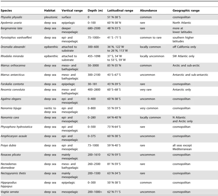
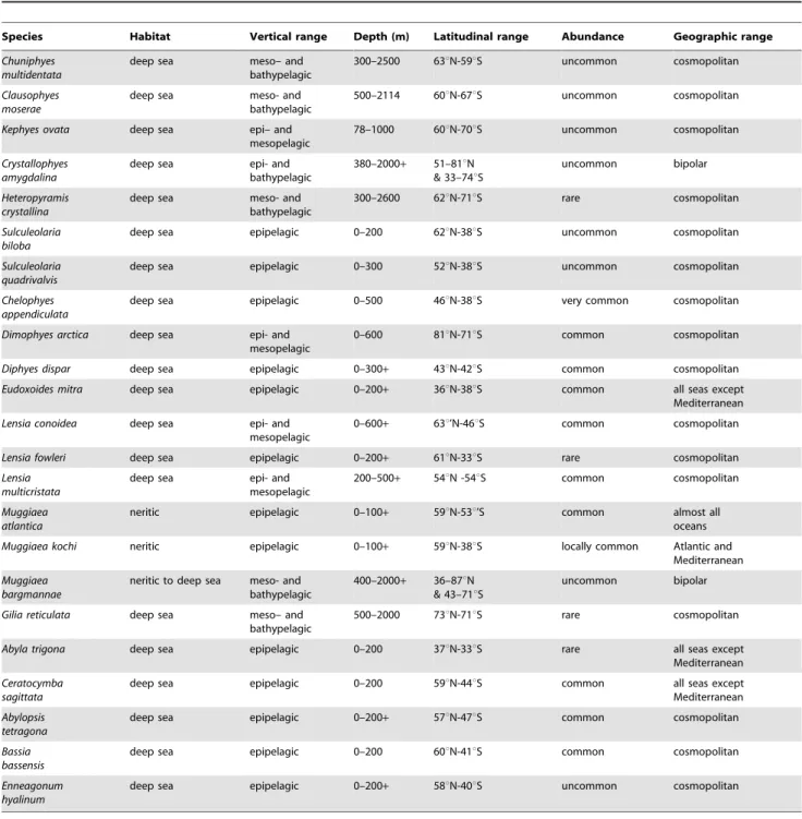
Biogeography. Almost all siphonophores are deep sea pelagic organisms and the majority exhibit a cosmopolitan distribution; that is species present in all three great oceans and the Mediterranean. Siphonophore distribution was well covered in the last review [2], so is only summarized here for 44 selected siphonophore species (Tables 1 and 2).
The majority of siphonophores are deemed cosmopolitan in this paper if their geographical ranges encircle the globe within their preferred latitudinal bands. Such bands are dependent on both water temperature and ocean currents. Warm water siphono-phores such asForskalia contortaandHippopodius hippopus(in Table 1), as well asSulculeolaria biloba,S. quadrivalvis,Diphyes dispar,Eudoxoides mitra and the abylids Abyla trigona, Ceratocymba sagittata, Abylopsis tetragona,Bassia bassensisandEnneagonum hyalinum(in Table 2) mostly inhabit shallow epipelagic layers at tropical latitudes. Other species such as Agalma elegans, Physophora hydrostatica, Vogtia serrata (in Table 1), the clausophyids Chuniphyes multidentata, Clausophyes moserae,Kephyes ovata,Heteropyramis crystallinaand the diphyidsLensia conoidea, L. multicristata and Gilia reticulata (in Table 2) occupy a broader latitudinal range in either epipelagic layers at higher latitudes or deeper mesopelagic layers at lower latitudes. A few species are restricted to deep horizons throughout their ranges (eg
Table 1.Distribution and abundance of selected cystonect, physonect, and prayomorph species.
Species Habitat Vertical range Depth (m) Latitudinal range Abundance Geographic range
Physalia physalis pleustonic surface 0 51uN-38uS common cosmopolitan
Apolemia uvaria deep sea epipelagic 0–100 60uN-38uN rare North Atlantic
Bargmannia lata deep sea deeper mesopelagic
680–2500 48uN-33uS rare more at
lower latitudes
Pyrostephos vanhoeffeni deep sea epi- and mesopelagic
75–1000+ 41uS -71uS common to rare southern higher latitudes
Dromalia alexandri epibenthic attached to substrate
300–600 36uN, 122uW to 26uN, 113uW
locally common off California only
Rhodalia miranda epibenthic attached to substrate
455–1098 37uS, 54uW to 53uS, 59uW
locally uncommon SW Atlantic only
Marrus orthocanna deep sea meso– and bathypelagic
50–3000 85uN-35uN common Arctic and sub-arctic
Marrus antarcticus deep sea meso- and bathypelagic
300–2100 43uS–67uS uncommon Antarctic and sub-antarctic
Forskalia contorta deep sea epipelagic 30–181 43uN-39uS rare cosmopolitan
Resomia convoluta deep sea meso- and bathypelagic
400–2800 60uS–68uS very rare Antarctic only
Agalma elegans deep sea epi- and mesopelagic
0–400 60uN-38uS uncommon cosmopolitan
Nanomia bijuga neritic to deep sea
epi- and mesopelagic
0–800 55uN-59uS very common cosmopolitan
Nanomia cara deep sea epi- and mesopelagic
0–280 64uN-40uN locally common N Atlantic and Arctic only
Physophora hydrostatica deep sea epi- and mesopelagic
0–500 73uN-64uS rare cosmopolitan
Amphicaryon acaule deep sea epi- and mesopelagic
0–375 60uN-38uS uncommon cosmopolitan
Praya dubia deep sea epi- and
mesopelagic
73–1000 59uN-40uS rare all seas except
Mediterranean
Rosacea plicata deep sea mainly mesopelagic
200–1610 62uN-59uS uncommon cosmopolitan
Nectadamas diomedeae
deep sea meso- and bathypelagic
260–2500 81uN-59uS rare cosmopolitan
Nectopyramis thetis deep sea mainly mesopelagic
200–1500 63uN-34uS rare cosmopolitan
Hippopodius hippopus
deep sea epipelagic 0–300 50uN-38uS common cosmopolitan
Vogtia serrata deep sea mesopelagic 200–1000+ 62uN-71uS uncommon cosmopolitan
Key: epipelagic, 0- ca. 300 m; mesopelagic, 300–1000 m; bathypelagic, 1000 m and below. Abundance scale: very common, common, uncommon, rare, very rare. Cosmopolitan refers to species present in all three great oceans and the Mediterranean. For primary literature used to construct this table, see [1].
Bargmannia lata,Resomia convoluta,Nectadamas diomedeaeand Nectopyr-amis thetis), others are bipolar (Crystallophyes amygdalina, Muggiaea bargmannae) or restricted to just one polar region (Marrus orthocanna, M. antarcticus). A number of oceanic species do not occur in the Mediterranean (Tables 1, 2). A few species are neritic (for example Muggiaea species, Table 2), and species of the physonect family Rhodaliidae (Dromalia alexandriand Rhodalia mirandaTable 1) are epibenthic, found only in specific areas of the continental shelf surrounding the major continents [27]. One species, Dimophyes arctica(Table 2) inhabits all latitudes.
Species from the neritic calycophoran family Sphaeronectidae are omitted because a recent review [18] indicates that most species ofSphaeronecteshave been incorrectly identified in the past. Other records of certain species noted by particular authors are also omitted due to suspect identifications. This problem and others associated with estimating the worldwide distribution of siphonophores was reviewed recently by Mapstone [6, section 5.2], to which the reader is referred for further information. Primary data used to construct Tables 1 and 2 is available from the WoRMS Siphonophora List [1], and updated as new reliable records become available.
Table 2.Distribution and abundance of selected diphyomorph species.
Species Habitat Vertical range Depth (m) Latitudinal range Abundance Geographic range Chuniphyes
multidentata
deep sea meso– and
bathypelagic
300–2500 63uN-59uS uncommon cosmopolitan
Clausophyes moserae
deep sea meso- and
bathypelagic
500–2114 60uN-67uS uncommon cosmopolitan
Kephyes ovata deep sea epi– and
mesopelagic
78–1000 60uN-70uS uncommon cosmopolitan
Crystallophyes amygdalina
deep sea epi- and
bathypelagic
380–2000+ 51–81uN & 33–74uS
uncommon bipolar
Heteropyramis crystallina
deep sea meso- and
bathypelagic
300–2600 62uN-71uS rare cosmopolitan
Sulculeolaria biloba
deep sea epipelagic 0–200 62uN-38uS uncommon cosmopolitan
Sulculeolaria quadrivalvis
deep sea epipelagic 0–300 52uN-38uS uncommon cosmopolitan
Chelophyes appendiculata
deep sea epipelagic 0–500 46uN-38uS very common cosmopolitan
Dimophyes arctica deep sea epi- and mesopelagic
0–600 81uN-71uS common cosmopolitan
Diphyes dispar deep sea epipelagic 0–300+ 43uN-42uS common cosmopolitan
Eudoxoides mitra deep sea epipelagic 0–200+ 36uN-38uS common all seas except
Mediterranean
Lensia conoidea deep sea epi- and mesopelagic
0–600+ 63u’N-46uS common cosmopolitan
Lensia fowleri deep sea epipelagic 0–200+ 61uN-33uS rare cosmopolitan
Lensia multicristata
deep sea epi- and
mesopelagic
200–500+ 54uN -54uS common cosmopolitan
Muggiaea atlantica
neritic epipelagic 0–100+ 59uN-53u’S common almost all
oceans
Muggiaea kochi neritic epipelagic 0–100+ 59uN-38uS locally common Atlantic and
Mediterranean
Muggiaea bargmannae
neritic to deep sea meso- and bathypelagic
400–2000+ 36–87uN & 43–71uS
uncommon bipolar
Gilia reticulata deep sea meso– and bathypelagic
500–2000 73uN-71uS rare cosmopolitan
Abyla trigona deep sea epipelagic 0–200 37uN-33uS rare all seas except
Mediterranean
Ceratocymba sagittata
deep sea epipelagic 0–200 59uN-44uS common all seas except
Mediterranean
Abylopsis tetragona
deep sea epipelagic 0–200+ 57uN-47uS common cosmopolitan
Bassia bassensis
deep sea epipelagic 0–200 60uN-41uS common cosmopolitan
Enneagonum hyalinum
deep sea epipelagic 0–200+ 58uN-40uS uncommon cosmopolitan
Key: epipelagic, 0- ca. 300 m; mesopelagic, 300–1000 m; bathypelagic, 1000 m and below. Abundance scale: very common, common, uncommon, rare, very rare. Cosmopolitan refers to species present in all three great oceans and the Mediterranean. For primary literature used to construct this table, see [1].
doi:10.1371/journal.pone.0087737.t002
Body Plans and General Morphology
Siphonophores vary greatly in size and shape, and are polymorphic individuals composed of a number of polypoid and medusoid zooids which together function as an integrated organism. Most siphonophores conform to one of three body plans, representing the three main types Cystonecta, Physonectae and Calycophorae (Figure 3A–C). A typical long-stemmed cystonect (Figure 3A Rhizophysa eysenhardti) has a pneumatophore (float) at the anterior end, followed by an elongate stem bearing groups of iterative (repeated) zooids specialized for different functions. The stem and zooid groups are collectively termed the siphosome, and each zooid group (in the cystonect species shown in Fig. 3A) comprises a gastrozooid with tentacle (for capture, ingestion and digestion of prey items) and a gonodendron bearing several gonophores for reproduction (of one sex only in each individual). Long-stemmed cystonects lack prominent swimming bells and instead, in a calm sea, may drift peacefully at the surface and writhe by contracting the stem muscles [9]. A typical long-stemmed physonect (Figure 3BNanomia bijuga), in contrast, has a pneumatophore and an extra portion of stem interpolated between the pneumatophore and siphosome, termed the nectosome, which bears many nectophores (swimming bells). The nectophores contract in a co-ordinated pumping manner and direct water backwards, moving the animal forward by ‘‘jet propulsion’’. In Nanomia bijugaiterative units are spread out along the siphosomal stem, as in a long-stemmed cystonects, and are termed cormidia because each originates from a single probud (as noted above). A cormidium of N. bijuga comprises a gastrozooid with tentacle (branched in most physonects), several smaller palpons, each with a palpacle, gelatinous bracts of two sizes (for extra buoyancy), and gonodendra (with gonophores of both sexes in each individual). A typical calycophoran (Figure 3C, Lensia conoidea) has two nectophores but no pneumatophore, and an elongate siphosomal stem with many closely spaced and reduced cormidia, each comprising a gastrozooid with a prominent elongate tentacle, one bract and gonophores; the latter start to develop while the cormidium is still attached to the stem, and at maturity the cormidium detaches from the end of the stem to become a free-living eudoxid.
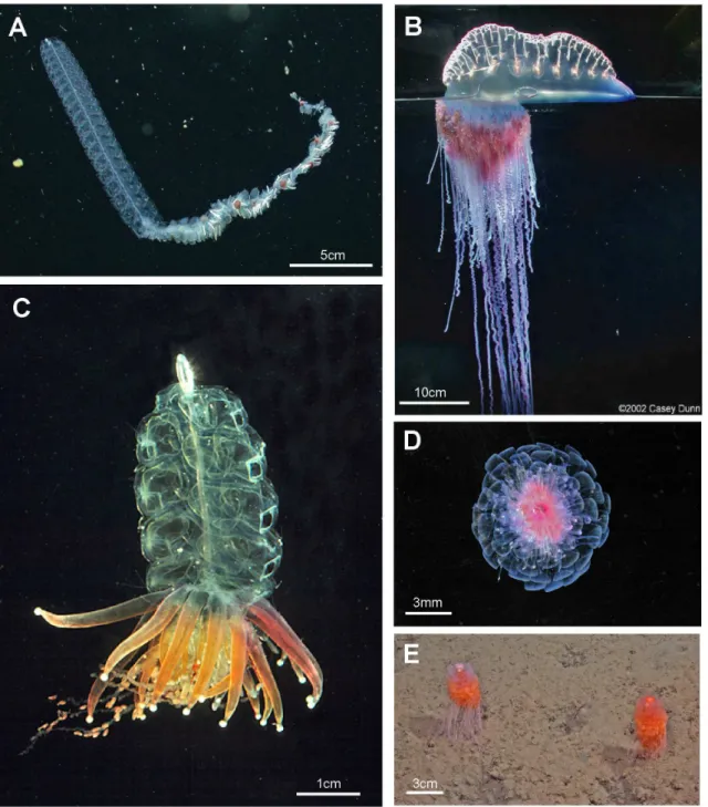
A range of typical and atypical cystonect and physonect body plans are shown in Figure 4.Bargmanniais a typical long-stemmed physonect (Figure 4A), and is larger than theNanomia bijugacolony shown in Figure 3B; the specimen photographed has possibly lost some of its siphosome. The cystonectPhysalia physalis(Figure 4B) is unusual and differs from the more usual cystonects colony shown in Figure 3A because the former has a much larger pneumato-phore, which lies on the sea surface, and no stem. Cormidial siphosomal zooids in P. physalis hang down directly from the underside of the pneumatophore at the ‘oral’, or posterior, end. The physonectPhysophora hydrostatica(Figure 4C) is also somewhat atypical with an intermediate-sized pneumatophore and typical nectophores on an elongate nectosome, but the siphosome is reduced to a swollen corm and surrounded by a ring of prominent enlarged palpons. The physonect Athorybia rosacea has an even more reduced stem (Figure 4D), comprising only a swollen red-tipped pneumatophore and adjacent siphosomal protuberance where enlarged bracts form; these encircle the pneumatophore in rings, and are capable of limited ‘paddling’ locomotion [38]. Rhodaliids are another unusual family of shortened siphono-phores, which, unlike most other families are epibenthic, so live attached to the bottom by their long tentacles. In the rhodaliid Dromalia alexandri(Figure 4E), the pneumatophore is relatively large compared to that of a typical long-stemmed physonect and gives sufficient lift to maintain the animal just above the sea bed [4]; it
can also use the ring of small weak nectophores to swim short distances.
A range of calycophoran body plans are shown in Figure 5 and two main types are distinguished: prayomorphs, with a pair of rounded and opposed swimming bells and an extended siphosome (Figure 5A) and diphyomorphs with a pair of more streamlined bells attached in a linear arrangement one behind the other (Figure 5C). The siphosomal stem of diphyomorphs can be completely withdrawn into the hydroecium for greater protection (Figure 5E). Unusual calycophoran body plans include hippopo-diids with several typically facetted swimming bells arising on pedicels one from another, which enclose a cavity into which the stem can be completely withdrawn (Figure 5B); and sphaeronec-tids in which a single rounded larval swimming bell is retained throughout life (Figure 5G). Swimming bells of tropical abylid diphyomorphs are also arranged linearly (Figure 5F) and their surfaces are also facetted, whereas clausophyid diphyomorphs typically have two staggered bells (Figure 5D) in an arrangement intermediate between the apposed bells of prayomorphs and the linearly aligned bells of diphyomorphs.
The pneumatophore (float) is unique to siphonophores, and a ‘neoformation’ (p. 103 [2]; p. 125 [70]), not a modified medusoid zooid as originally concluded by certain nineteenth century authors [26]. Embryological work by Danielle Carre´ [71,72,73] demonstrated pneumatophore formation in three physonect species. Each pneumatophore comprises a gas gland (pneumade-nia) and a central chitin-lined gas chamber (pneumatosaccus), with a second cavity (the pericystic cavity) typically subdivided by septa which surrounds the gas cavity and is confluent with the gastrovascular cavity of the main stem. Carbon monoxide is secreted into the gas cavity by the gas gland and the pneumat-ophore acts as a hydrostatic organ (reviewed by Mackie et al. (p. 194–196 [2]). For example, as the physonectNanomia bijugarises in the water column, bubbles of expanding gas are released via an apical pore surrounded by a sphincter muscle [74]. The importance of the pneumatophore for buoyancy varies in different species. In cystonects it is the only structure providing lift for the heavy stem and attached zooids. In physonects the pneumato-phore is small, whilst bracts are present that increase buoyancy by the replacement of 44% of the heavy sulphate ions in the mesogloea by lighter chloride ions. Calycophorans lack a pneumatophore, and up to 75% of the sulphate ions in each bract are replaced to provide buoyancy [75].
Nectophores are asexual medusoid structures that contain a muscular nectosac opening via an ostium. Strong contraction of this nectosac forces water out of the bell and propels the siphonophore forwards, or in some cases the ostia are directed forwards to achieve backward swimming [76]. During swimming the stem of physonects shortens to improve streamlining. In many calycophorans streamlining is taken a stage further by contraction of the stem into an additional external hollowed out chamber known as the hydroecium (as noted above). In addition, many calycophoran nectophores contain an extra structure in the mesogloea adjacent to the nectosac termed the somatocyst; this often contains oil globules which can both act as a food store and give extra lift.
(derived from photo image by Rob Sherlock - shown in Fig. 5C): inset Cc shows two tentilla attached to one tentacle (derived from [69] pl. 11, fig. 2). Labels: b - bract; c – cormidium; gd - gonodendron; gz - gastrozooid; h – hydroecium; n – nectophore (swimming bell); nb – nematocyst battery (a coiled cnidoband); np – nematocyst pad; p - pedicel; pn – pneumatophore (float); s – stem; sh – siphosomal horn; so – somatocyst; t – tentacle; tf – terminal filament.
doi:10.1371/journal.pone.0087737.g003
Figure 4. Cystonects and physonects. A. Typical long-stemmed physonect Bargmannia sp., with small anterior pneumatophore, many nectophores on an elongate nectosome and iterative cormidia on an elongate siphosome (MBARI); B. Atypical cystonectPhysalia physalis, pleustonic (lives at surface), with much enlarged pneumatophore, no stem, cormidia arising directly from underside of pneumatophore (Casey Dunnß2002); C. Atypical physonectPhysophora hydrostatica,with pneumatophore, nectophores on an elongate nectosome and cormidia on a short-stemmed corm-like siphosome (Larry MadinßWHOI); D. Atypical physonectAthorybia rosacea, with rose-pink pneumatophore surrounded by rings of large bracts from cormidia on short-stemmed corm-like siphosome; no nectosome (Larry MadinßWHOI); E. Atypical physonectDromalia alexandri,with enlarged penumatophore, ring of nectophores on short nectosome and whorls of iterative cormidia spiralling around corm from growth zone to corm base on short-stemmed siphosome (MBARI). Scale bars approximate.
buoyancy and protection (bracts are absent in cystonects). Tentacles have side branches in most siphonophores, bearing either ‘pads’ of nematocysts (cystonects, Figure 3A inset) or complex nematocyst batteries (physonects and calycophorans, Figure 3C inset) here termed ‘tentilla’. Physonect cormidia also contain one or more reduced gastrozooids called palpons, which have a chemosensory or excretory function (Figure 3B); each palpon bears a reduced tentacle, the palpacle.
Cormidia can be pedunculate (attached at one point on the siphosome), as in calycophorans (Figure 3C) or dispersed along the length of the siphosome, as in long-stemmed cystonects and physonects (Figure 3A–B). In many calycophorans, mature cormidia detach as they reach the end of the stem to become free-living eudoxids, (the sexual stage in the life cycle) in the plankton. In other calycophorans cormidia are retained on the stem throughout life. Free-living eudoxids comprise a single bract (conical buoyant zooid) covering a gastrozooid with tentacle and a gonophore (see below). More gonophores form after the first is released and production may continue for several months.
Example cormidia from a range of physonects are shown in Figure 6, covering typical long-stemmed as well as short-stemmed types. A cormidium from the typical long-stemmed physonect Nanomia bijuga comprises several palpon-gonodendra-bract com-plexes and large posterior gastrozooid with an associated elongate bract (Figure 6A). The palpon complexes become progressively older and larger posteriorly, and all elements of each cormidium originally arose from a pro-bud (as noted earlier) on the siphosomal horn at the anterior end of the siphosome [8] (Figure 3B). One of 10 cormidia from the physonect Physophora hydrostatica occupies a compact segment of the siphosomal corm, and includes three enlarged lateral palpons, an associated hermaphrodite gonodendron of male and female gonophores, with a gastrozooid and tentacle on the posterior surface, but no bracts (Figure 6B a–b). In the rhodaliid Dromalia alexandri (Figure 6C) cormidia are borne on branched cormidial units away from the corm surface, and these units spiral around the inflated corm to the posterior under-surface [4]. Cormidial units originate continuously on a siphosomal horn between the nectophores (swimming bells), on the ventral surface just below the pneumatophore, and each mature unit typically carries three cormidia. A single cormidium includes a gastrozooid, several palpons and many gonophores in a gonodendron [4]. Dendritic growth of the cormidial units enables a large number of cormidia to be carried on a single rotundD. alexandriindividual. Cormidia on the enlarged float ofAthorybia rosacea(Figure 6D) originate from a siphosomal horn adjacent to the float apex, and each includes a group of typically four large larval bracts, an associated branched hermaphrodite gonodendron with small palpons below, and a larger gastrozooid on the posterior corm surface.
Figure 7A illustrates the complexity of a mature Portuguese Man O’WarPhysalia physalisviewed from above and ‘sailing’ with the wind, with many long tentacles extending from the cormidia and streaming out from the windward side. The cormidia ofP.
physalisare shown diagrammatically in Figure 7A, and numbered 1–5 and I –VII; they originate directly from the underside of the float (pneumatophore) in this species and develop in a particularly complex pattern, as described and illustrated in a seminal paper by A.K. Totton [39]. Cormidia bud one from another in a series, and each such series is termed a cormidial complex. There are twelve cormidial complexes in a matureP. physalia, which are attached in two groups separated by a small gap; the oldest complex in each group, (which forms first) lies closest to the anterior (or aboral) end of the animal (Figure 7A). The smaller oral group of complexes (1– 5) lies just posterior of the first gastrozooid to form in the larva, the protozooid, and one cormidial complex from this region is shown in Figure 7B. It bears c. 13 cormidia, on two branches: a smaller oral branch above which is directed towards the oral end of the float, and a larger aboral branch below which is directed towards the aboral, or posterior, end of the animal. Almost all the cormidia ofP. physalis comprise three zooids: a gastrozooid, gonodendron and a separate tentacle with ampulla (where the nematocysts are formed), which together form a tripartite group (Figure 7C). As growth proceeds more tripartite groups develop on lateral branches from the cormidial complex, filling every available space (Figure 7B). Indeed, no other siphonophore buds so prolifically as P. physalis[39]. As sexual maturity is reached, the gonodendra of each cormidial complex sub-branch many times, and detach. The largest such gonodendral ‘sphere’ found by Totton (from a female) measured ,5 cm across, and bore 2400 gonophores on seven main branches, plus 224 very small medusoid bells, an extra zooid present in the cormidial complexes of matureP. physalis.
Cormidia are discrete in calycophorans, and, with one exception, lack the palpons present in physonect cormidia. In many calycophoran cormidia, the bract wraps around the stem in a cloak-like manner and gives protection to the underlying gastrozooid and gonophores (Figure 8A, C). As already noted above, when the cormidium of most diphyomorphs reaches maturity, it detaches and becomes a free-living eudoxid (Figure 8E). In some calycophorans, however, cormidia remain attached to the stem throughout life (prayine prayids and sulculeolariine diphyids). A few groups lack bracts, including members of the prayomorph family Hippopodiidae (see above), andClausophyesspecies of the diphyomorph family Clausophyidae, both of which also probably retain their cormidia on the stem. In hippopodiids, a number of bells remain joined together when mature, forming a hollow cylinder from which the siphosomal stem emerges at the posterior end (Figure 8B). This stem originates between the two youngest nectophores but only the bottom two bells are functional in hippopodiids; their mesogloea, together with that of the other smaller bells, give buoyancy to compensate for the absence of bracts in the cormidia (Figure 8D). Cormidia arise from a siphosomal horn and are small, allowing the stem to be completely withdrawn into the cylindrical chamber when not feeding, as already noted above (Figure 8B).
Figure 5. Calycophorans.A. Typical prayomorphPrayasp., with two rounded bells and a very long siphosome bearing over 100 cormidia; tentacles are extended for feeding, each bearing 80–90 nematocyst batteries, giving,9000+batteries in all (Steven HaddockßMBARI); B. Atypical prayomorphHippopodius hippopuswith several facetted nectophores enclosing central chamber; latter contains short stem with cormidia which lack bracts to facilitate complete stem withdrawal (Russ Hopcroft, UAF); C. Typical diphyid diphyomorphLensia conoideawith two angular linearly aligned bells; stem extended for feeding and with many closely spaced cormidia; each has an elongate tentacle with 15+tentilla (better shown in Figure 3C)
Figure 6. Physonect cormidia.A:Nanomia bijugacormidium (derived from [68] pl. 7, fig. 10); B:Physophora hydrostaticaa. diagram of posterior view of corm surface bearing 10 cormidia (derived from [77] figs. 12a, 16a); b. one cormidium exploded (derived from [26] pl. 20, fig. 18 with two additional palpons added); C:Dromalia alexandridorsal view of corm with many spirally arranged cormidial units, dorsal view (GMM); D:Athorybia rosacealateral view of float with siphosomal horn and attached cormidia (derived from [50] txt fig. 45). Labels: b – bract; bl – bracteal lamella; cu – cormidial unit; gdf – female gonodendron; gdm – male gonodendron; gz – gastrozooid; p – palpon; pl – palpacle; pn – pneumatophore (float); sh – siphosomal horn; t – tentacle with tentilla; te - tentillum.
doi:10.1371/journal.pone.0087737.g006
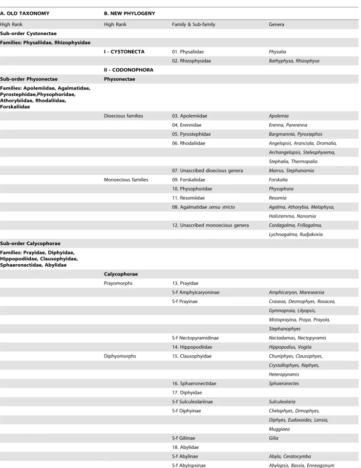
Old and New Phylogenies
The first detailed molecular study of a large range of Siphonophora [10] identified important morphological characters associated with their evolution not previously considered signifi-cant; it is reproduced here as Figure 9. A more recent study [79] used the barcoding gene mtCOI to generate a phylogeny for 95 medusozoan species (including 61 siphonophores), though this gene is more appropriate for phylogenetic characters at family level or below. Analysis of a third gene 28S is unresolved for the clade Codonophora [55], and further siphonophore taxa analyses and application of whole genome sequencing to the group are awaited for more clarification of this clade. The study of Dunn et al. [10] led to further changes in physonect systematics by Pugh [17] as discussed below (Figure 10). The old and new phylogenies are compared in Table 3, from 15 families recognized in 1987 and 16 different families and 67 genera recognized today.
The consensus tree from the molecular study of Dunn et al. (see fig. 6 [10]) is based on data from two genes: the nuclear gene 18S and mitochondrial gene 16S, and is figured here as Figure 9. It concludes that cystonects are sister to all other siphonophores, with the remainder ranked together in a new clade Codonophora, meaning ‘bell bearers’. Within the Codonophora clade, the traditional grouping ‘Physonectae’ are paraphyletic, with the ‘physonect’ family Apolemiidae sister to all other taxa. Clades for the physonect families Forskaliidae and Agalmatidae sensu stricto are well supported, although resolution for taxa representing rhodaliids, erennids, pyrostephids and physophorids is poor. The
traditional group Calycophorae are nested within the non-apolemiid Codonophora and form a monophyletic clade. Within the Calycophorae, prayomorphs are paraphyletic, based on taxa and genes sampled in 2005. Hippopodiid prayomorphs form a distinct clade, and diphyomorphs, together with Sphaeronectes (ignoring one undescribed clausophyid species) form another distinct clade. Intraspecific variation is also demonstrated in multiple individuals of Hippopodius hippopus and Sulculeolaria quadrivalviscollected in the Atlantic and Pacific Oceans. The final important finding of Dunn et al. [10] places abylids within a clade containing the five diphyids tested. Five cryptic species pairs are also identified amongst the Atlantic and Pacific ‘physonects’ analysed (Figure 9).
The new phylogeny shows that character evolution within the Siphonophora is related to reproductive state (figs. 7–8 [10]). Separately sexed individuals are dioecious, whereas monoecious siphonophores bear differentially maturing male and female gonophores on the same individual. Zooid types scored by Dunn et al. [10] include nectosomal nectophores, siphosomal bracts, gastrozooids and palpons, as well as the number of types of each zooid present in each taxon. They found that cystonects, apolemiids, pyrostephids, erennids and rhodaliids, are all dioe-cious, and, surprisingly, all lack a descending ‘pallial canal’ (‘descending surface diverticulum’ of Mapstone [6]) on the proximal surface of the nectophore. In contrast, all remaining codonophorans are monoecious, and in taxa tested from the families Agalmatidaesensu stricto, Forskaliidae and Physophoridae
Figure 7. Cystonect cormidia as exhibited byPhysalia physalis.A: Left-handed drifting specimen viewed from above (derived with minor modification from [39] fig. 5) – added numbers 1–5 identify oral cormidial groups while numbers I–VI identify main cormidial groups – note how Physalia’s surface float drifts to starboard with the wind on a broad reach; B: Oral cormidial complex number 2 viewed from inside the float – note groups 3 to 8 are tripartite, with more tripartite groups on oral and aboral side branches (adapted from [39] txt fig. 12D) – numbers in brackets added to identify tripartite groups; C: A developing tripartite group from main cormidial complex number VI (derived from [39] txt fig. 14B, in part only). Labels: a – ampulla (basigaster); fw – float wall; gd – gonodendron; gz – gastrozooid; pn – pneumatophore (float); ta – tentacle with ampulla (basigaster); T – tentacle; tg – tripartite group.
Figure 8. Calycophoran cormidia.A:Rosacea cymbiformiscormidium (after [6] fig. 2D); B.Hippopodius hippopussection through colony (adapted from [31] fig. 11, [78] txt fig. 13 and [27] fig. 44b); C:Chelophyes appendiculatacormidium (from [34] pl. 11, fig. 1); D.Hippopodius hippopuscormidium; note, no bracts (from [26] pl. 29, fig. 1 in part); E.Dimophyes arcticaeudoxid (Russ Hopcroft, UAF). Labels: b – bract, c – cormidium; go – gonophore; gof – female gonophore; gom – male gonophore; gz – gastrozooid; n – nectophore; nl – nectophoral lamella; o – oil globule (in phyllocyst); ph – phyllocyst; sh – siphosomal horn; ss – siphosomal stem; t – tentacle with tentilla.
doi:10.1371/journal.pone.0087737.g008
Figure 9. Molecular phylogeny of siphonophores from Dunn et al. (fig. 6[10]).Consensus tree of all trees for the Bayesian analysis of the combined data set (from an initial 20 million trees). The left score above the branch is the Bayesian posterior probability (%), the right score above the branch is the ML bootstrap support value (%), and the score below the branch is the MP bootstrap support value (%). The bars to the right of the species names indicate clades and grade taxa. Abbreviations: Atl – Atlantic; Med – Mediterranean; Pac – Pacific. For full details of analyses and consensus tree computations refer to Dunn et al. [10].
Figure 10. Possible phylogeny of the Siphonophora (derived from[17], fig. 21, and[11]).MFZ – muscle-free zone on nectophore; * - dorsal nectosome; ** - one species monoecious.
doi:10.1371/journal.pone.0087737.g010
(exceptAthorybia rosaceawhich lacks nectophores) this condition is coincident with the presence of a descending ‘pallial canal’ on the proximal surface of the nectophore.
Nectosomal nectophores are an apomorphy of the Codono-phora and may have been derived from retained reproductive medusae [10]. Their presence together with the presence or absence of a descending pallial canal, suggests these two characters might have pleiotropic links [10]. Many of one type of nectophore (homomorphic) were found in all the ‘physonects’ tested except Athorybia rosacea, which lacks nectophores. Amongst the Calyco-phorae, nectophores are reduced to two of one type in most prayomorphs tested, except for the two nectopyramidines which had only one of one type, and hippopodiids which, as Dunn et al. [10] conclude, have developed several nectophores of one type from an ancestor which probably had only two of one type (see their fig. 8a). Most diphyomorph calycophorans, in contrast, have two nectophores of two types (an anterior and a posterior: heteromorphic), with one nectophore lost inMuggiaea atlantica, and only a single larval nectophore retained in Sphaeronectes gracilis (fig. 8a [10]).
Palpons are another character found in almost all ‘physonects’, but absent from all the calycophorans tested by Dunn et al. (fig. 8b [10]). Parsimony indicates that palpons were probably present in the ancestral siphonophore and have been lost one or two times, while bracts appeared first in the Codonophora, and might have developed into two or more types several times and at several different specific locations during siphonophore evolution [10]. Bracts, however, which are also characteristic of the Codono-phora, are all of one type in apolemiids and also in all calycophorans which possess them, as well as in some Agalmatidae sensu stricto (Agalma and Athorybia). In erennids and other Agalmatidae sensu stricto (Nanomia and Halistemma species) two types of bracts develop, and four types are found in Forskalia species (see fig. 8b in [10]). Thus, as Dunn et al. [10] conclude, there has been both gain and loss of zooids during siphonophore evolution.
The importance of these characters in shaping siphonophore evolution is reflected in the higher rankings given in Table 3B. A new hypothesis for character evolution given by Pugh [17], which is shown here in Figure 10, proposes a dioecious ancestral siphonophore with pneumatophore and siphosome, but maybe not a nectosome. Such an ancestor may have given rise to two clades: the dioecious cystonects with a pneumatophore and siphosome but no nectosome, and a dioecious ancestral codonophoran with a pneumatophore, nectosome and siphosome. Nectophores of the latter have only an ascending ‘pallial canal’ on their proximal surfaces. The first nectosome to evolve is thought to have had all nectophores attached on the same side of the stem as the siphosomal zooids, which is, by convention, the ventral surface of the stem (p. 931 [10]). A similar condition is found in most families and genera of physonects today.
Apolemiids are also dioecious, with nectosomal palpons between the nectophores [11,80], and were the first group to split from the other Codonophora, with both lineages evolving simultaneously and independently thereafter. The ancestral sister group to the Apolemiidae could have been another clade that lacked nectosomal palpons (Figure 10) and from which, perhaps later in time, a monoecious ancestor emerged. Dioecy could have persisted in a group of physonects which lacked a descending ‘pallial canal’ on the proximal surface of their nectophores, including three extant families and two unascribed dioecious genera (see Table 3B). In one of these families, the Pyrostephidae, a twist may have occurred at the junction between the nectosome and siphosome resulting in nectophores arising on the dorsal
surface (‘dorsal nectosome’) and siphosomal cormidia on the ventral surface. The first monoecious siphonophores could have been physonects with a descending ‘pallial canal’ on the proximal surface of their nectophores, a new diagnostic character. From this clade Pugh [17] proposes a split into the Family Agalmatidaesensu stricto with a dorsal nectosome, and a non-agalmatid clade including the families Forskaliidae, Physophoridae and Resomii-dae together with the unascribed monoecious generaCordagalma, FrillagalmaandLychnagalma(Table 3B) which all exhibit a ventral nectosome (Figure 10). Pugh [17] also suggests that a further monoecious group of siphonophores, the Calycophorae, appeared at some point during the evolution of these various physonect families, (Figure 10). In calycophorans the pneumatophore is lost and the nectosome typically reduced to just two nectophores.
Systematics 1987 to Present
This section summarizes the changes in siphonophore system-atics since the last review in 1987 and is based on the new phylogenies as outlined above [10,17], together with details of families that have been revised or newly introduced, and new genera and species added, moved or now considered invalid. Most of this information for cystonects and physonects is given in Table 4, and for calycophorans in Table 5. Ongoing debates about the validity of certain species, and other systematic information too extensive for inclusion in the tables, is briefly summarized below.
Apolemiidae. Unique nectosomal palpons (previously necto-somal tentacles or polyps) are probably a synapomorphy of the Codonophora, being greatly reduced or absent in other codono-phorans [10]. These zooids arise on the nectosome from the posterior ends of the nectophoral muscular lamellae, either singly or in bunches [85], and are identified as small buds on the nectosome of some other long-stemmed physonects [8]. Other important specific characters include the presence or absence of diverticula penetrating into the mesogloea from the lateral radial canals of the nectophores, the relative size of the siphosomal horn, the type of siphosomal cormidia present (pedunculate or dispersed), and the number of palpon types on the siphosome (one or two) [11]. In older cormidia, secondary gastrozooids may form independent of the growth zone, directly on the siphosome [11], as also shown in the agalmatidNanomia bijuga[8] (see above). Pedunculate cormidia may be either ancestral or derived for the Codonophora [11], but further work and denser sampling of siphonophore phylogeny is needed to resolve this question [11].
Currently, the family is monotypic forApolemia,and includesA. uvaria(Lesueur, 1815),A. vitiazi(Stepanjants, 1967) andA. contorta (Margulis, 1976) [1], together with two newly described speciesA. lanosaandA. rubriversa[11] and a third species not yet described (A. trinegra[84]). Two types of siphosomal palpons are exhibited byA. uvaria(shorter red/brown type and longer opaque type [85,95]), but may also be characteristic of other species, together with pigment distribution in the siphosomal palpons [84]. Apolemiids can reach more than 30 m in length, and the recent paper by Siebert et al. forms the foundation for descriptions of up to 15 further new species [11]. Apolemiids frequently undergo autotomy [6,95], releasing many lengths of siphosome which float freely in the water without nectophores, while the latter swim off or drifted away in a different direction.
Table 3.Old and new classification of the Siphonophora.
A. OLD TAXONOMY B. NEW PHYLOGENY
High Rank High Rank Family & Sub-family Genera
Sub-order Cystonectae
Families: Physaliidae, Rhizophysidae
I - CYSTONECTA 01. Physaliidae Physalia
02. Rhizophysidae Bathyphysa, Rhizophysa
II - CODONOPHORA Sub-order Physonectae Physonectae Families: Apolemiidae, Agalmatidae,
Pyrostephidae,Physophoridae, Athorybiidae, Rhodaliidae, Forskaliidae
Dioecious families 03. Apolemiidae Apolemia
04. Erennidae Erenna, Parerenna
05. Pyrostephidae Bargmannia, Pyrostephos
06. Rhodaliidae Angelopsis, Aranciala, Dromalia,
Archangelopsis, Steleophysema,
Stephalia, Thermopalia
07. Unascribed dioecious genera Marrus, Stephanomia
Monoecious families 09. Forskaliidae Forskalia
10. Physophoridae Physophora
11. Resomiidae Resomia
08. Agalmatidaesensu stricto Agalma, Athorybia, Melophysa,
Halistemma, Nanomia
12. Unascribed monoecious genera Cordagalma, Frillagalma,
Lychnagalma, Rudjakovia
Sub-order Calycophorae Families: Prayidae, Diphyidae, Hippopodiidae, Clausophyidae, Sphaeronectidae, Abylidae
Calycophorae
Prayomorphs 13. Prayidae
S-f Amphyicaryoninae Amphicaryon, Maresearsia
S-f Prayinae Craseoa, Desmophyes, Rosacea,
Gymnopraia, Lilyopsis,
Mistoprayina, Praya, Prayola,
Stephanophyes
S-f Nectopyramidinae Nectadamas, Nectopyramis
14. Hippopodiidae Hippopodius, Vogtia
Diphyomorphs 15. Clausophyidae Chuniphyes, Clausophyes,
Crystallophyes, Kephyes,
Heteropyramis
16. Sphaeronectidae Sphaeronectes
17. Diphyidae
S-f Sulculeolariinae Sulculeolaria
S-f Diphyinae Chelophyes, Dimophyes,
Diphyes, Eudoxoides, Lensia,
Muggiaea
S-f Giliinae Gilia
18. Abylidae
S-f Abylinae Abyla, Ceratocymba
S-f Abylopsinae Abylopsis, Bassia, Enneagonum
doi:10.1371/journal.pone.0087737.t003
Pyrostephidae. This family has been properly diagnosed for the first time and three new species introduced [14]. A likely pyrostephid post-larva has also been described (Table 4), and a comprehensive study of the organisation of siphosomal zooids in Bargmannia elongata shows that new cormidia are formed on a protrusion from the stem termed the ‘‘horn’’ [80]. Here a series of ‘‘probuds’’ form, which each subdivides a number of times to form eight zooids and together these form a single cormidium (see above).
Rhodaliidae. Four new species have been added to this epibenthic family in recent years (Table 4 and [45,89]).Dromalia alexandri has been re-described including the first figures of a rhodaliid siphosomal horn, mature cormidial units and a mature bract, together with a more comprehensive distribution map including both range and density [4]. Herein the doubtful species Steleophysema aurophora Moser, 1924 [27], is re-validated from observations made by Dhugal Lindsay (pers. comm.) of new specimens collected off Japan, and as a resultSagamalia hinomaruis reduced to a junior synonym [1]. The genusTridensaHissmann, 2005, is also reduced to a junior synonym ofSteleophysema, based on the shape and attachment point of its bracts (at base of each
cormidial unit), attachment of the gonophores (with egg pouch) directly to the thin polygastric cormidia just distal of each cormidial gastrozooid, and attachment of the gonopalpons just distal of the gonophores. The two species T. sulawensis and T. rotundabecome junior synonyms ofS. sulawensisandS. rotunda[1]. A full re-description of S. aurophora is underway (D. Lindsay, pers. comm.).
Unascribed dioecious physonects. The generaMarrusand Stephanomia perhaps diverged early from other codonophorans (Figure 10). Marrus orthocannoides may not belong to the genus Marrus, because it has a fully muscular nectosac, whereas those of otherMarrusspecies have a proximal muscle-free zone [90]. The genus nameStephanomiahas been applied to many species in the past (p. 102 [6]), but is herein restricted to the large species Stephanomia amphytridisof Lesueur and Petit, 1807 [1] as applied by Dunn et al. [10] and mentioned on p. 103 of Mapstone [6]. This species has been collected recently in both the Atlantic and Pacific [17], sequenced for 16S and 18S genes [10] and a morphological description is underway.
Forskaliidae. The fragile and often snake-like colonies of this monoecious family have a spiral stem with diffusely attached Table 4.New systematics for cystonect and physonect siphonophore families.
Family Comments
01. Physaliidae Monotypic forPhysalia physalis(P. utriculusconsidered a junior synonym [81]
02. Rhizophysidae Long-stemmed;Bathyphysa japonicaa junior synonym ofB. conifera;SEM studies of budding sequences described forB. sibogae, Rhizophysa filiformisandR. eysenhardti[8]
03. Apolemiidae Long stemmed; monophyletic and sister to all other Codonophora, with unique nectophoral palpons on the nectosome. Nectophores distinctive and ridge-less, cormidia dispersed or discrete; gastrozooids with simple tentacles (no tentilla) resembling palpacles of palpons. Monogeneric forApolemia. Two new species includeA. lanosaandA. rubriversa[11] and three older species A. contorta, A. uvariaandA. vitiazi(Tottonia contortasensu Mapstone 2003 now referable toA. lanosa). A number of other species are known to exist [2,10,11,52,82,83,84,85], and await full description.
04. Erennidae Long-stemmed family erected for 4 species with large prominent straight tentilla, no involucrum and a rigid terminal process lacking nematocysts [15]. Two genera:Erenna(3 species) andParerenna(1 species).E. richardiBedot, 1904, and a new speciesE. laciniatahave large flattened nectophores and large tentilla held close to body and vibrate to attract prey; two further new species E. cornutaandParerenna emilyaehave different and also unique tentilla and gastrozooids [15].
05. Pyrostephidae Long-stemmed family reviewed and revised [14], with 3 new species ofBargmannia:B. amoena, B. gigas, B. lata[14,86]; alsoMica micula, the putative post-larva of a pyrostephid [87,88]. Nectophores with unique lower-lateral wings and much enlarged triangular thrust block; inB. elongatatwo growth zones on stem and composition of the cormidia studied using SEM [80]; pyrostephid cormidia either have oleocysts (modified tentacle-less palpons) (inPyrostephos) or none (inBargmannia) [14].
06. Rhodaliidae Short-stemmed family of 8 genera, with 4 new species includingArchangelopsis jagoa,Arancialia captonia[45,89], and two others herein referred toSteleophysemaMoser, 1924, includingS. sulawensisandS. rotunda.Sagamalia hinomarureduced to a junior synonym ofSteleophysema aurophora[1,89]. Firstin situfeeding observations on four species [89].Dromalia alexandrire-described [4].
07. Unascribed dioecious genera
Long-stemmed generaMarrusTotton, 1954 [90] andStephanomiaLesueur & Petit, 1807 [10] both with muscle-free zones on nectosac and other characters (Fig. 10). A new speciesM. claudanielisdescribed [90] and new specimens of an old speciesS. amphytridis[10] await re-description.
08. Forskaliidae Long stemmed and delicate monotypic family, probably sister to the Physophoridae [10]. Recently revised [16] with two new species added (Forskalia asymmetrica, F. saccula) and one reduced to a Species Inquirenda [1].
09. Physophoridae Family with long nectosome but short corm-like siphosome; previously monotypic forPhysophora hydrostaticabract present only in larva; now a new smaller and less colourful second speciesP. gilmeri, is added, with bracts retained on adult colony [77]; unique tentilla in both species.
10. Resomiidae Long-stemmed family newly introduced for two species previously referred to the Agalmatidae (Moseria convoluta,M. similis) and now transferred to a new monotypic genusResomia[17]; two tentilla types uniquely present on each tentacle. Three new species R. dunni,R. ornicephala,R. persicadescribed in 2010 [91].
11. Agalmatidaesensu stricto Mostly long-stemmed and recently restricted to genera with dorsal nectosome (see above) and involucrate tricornuate tentilla with tightly coiled cnidoband (see below). Now includes two short-stemmed genera (Athorybia,Melophysa) [17]. New species added (Halistemma transliratum) [92] and another re-described (H. foliacea, asH. amphytridis) [17,93].
12. Unascribed monoecious genera
Long-stemmed monotypic generaCordagalma, FrillagalmaandLychnagalmawith ventral nectosomes have been removed from the Agalmatidae [17] and a new speciesC. tottonidescribed [94].Rudjakovia plicataconsidered a valid species [1] and may be transferred to Agalmatidae when more characters are elucidated [17].
![Figure 3. Three typical siphonophore body plans. A. Long-stemmed cystonect Rhizophysa eysenhardti (derived from [66] pl](https://thumb-eu.123doks.com/thumbv2/123dok_br/18408009.359441/9.918.91.842.82.1025/figure-typical-siphonophore-stemmed-cystonect-rhizophysa-eysenhardti-derived.webp)
![Figure 6. Physonect cormidia. A: Nanomia bijuga cormidium (derived from [68] pl. 7, fig](https://thumb-eu.123doks.com/thumbv2/123dok_br/18408009.359441/13.918.94.825.79.966/figure-physonect-cormidia-nanomia-bijuga-cormidium-derived-fig.webp)
![Figure 8. Calycophoran cormidia. A: Rosacea cymbiformis cormidium (after [6] fig. 2D); B](https://thumb-eu.123doks.com/thumbv2/123dok_br/18408009.359441/15.918.81.833.89.946/figure-calycophoran-cormidia-rosacea-cymbiformis-cormidium-fig-d.webp)
![Figure 9. Molecular phylogeny of siphonophores from Dunn et al. (fig. 6 [10]). Consensus tree of all trees for the Bayesian analysis of the combined data set (from an initial 20 million trees)](https://thumb-eu.123doks.com/thumbv2/123dok_br/18408009.359441/16.918.92.813.95.982/figure-molecular-phylogeny-siphonophores-consensus-bayesian-analysis-combined.webp)
![Figure 10. Possible phylogeny of the Siphonophora (derived from [17], fig. 21, and [11])](https://thumb-eu.123doks.com/thumbv2/123dok_br/18408009.359441/17.918.84.811.114.1043/figure-possible-phylogeny-siphonophora-derived-fig.webp)