Histological evaluation of the phenobarbital
(Gardenal™) influence on orthodontic
movement: a study in rabbits
Matheus Melo Pithon*, Antônio Carlos de Oliveira Ruellas**
Introduction: The purpose of this study was to histologically evaluate the influence of
phenobarbital on orthodontic tooth movement. Methods: Twenty-two New Zealand
rab-bits (Oryctolagus cuniculus) were divided into three groups: normal or non-tested (N), control (C), and experimental (E). In Group N (n = 2) no procedure was carried out, except to verify the condition of normality before treatment. In Groups C (n = 10) and E (n = 10) an orthodontic appliance was inserted between the first molars and lower incisors in order to promote a mesial molar movement. In Group E phenobarbital was adminis-tered during the course of the experiment, which differentiates it from the group C. The animals were sacrificed on days 7 and 14 so that anatomical sections could be prepared for further histological analysis. Results: Histologically no difference was observed between
normal and experimental groups. Conclusions: Phenobarbital does not interfere with the
orthodontic tooth movement. Abstract
Keywords: Tooth movement. Pharmaceuticals. Orthodontics.
* Professor, Southwest Bahia University UESB, Bahia, Brazil. MSc and PhD in Orthodontics, School of Dentistry, Federal University of Rio de Janeiro – UFRJ, Brazil. Diplomate of Brazilian Board of Orthodontics and Dentofacial Orthopedics - BBO.
** Professor, Federal University of Rio de Janeiro – UFRJ, Rio de Janeiro, Brazil. MSc and PhD in Orthodontics, School of Dentistry, Federal University of Rio de Janeiro – UFRJ, Brazil.
IntRoduCtIon
Orthodontic tooth movement occurs when forces are applied to teeth and are transmitted to the supporting periodontium, so as to change their position in relation to the surrounding struc-tures, through alveolar bone remodeling.19 That is to say, bone apposition in the area submitted to tension and bone resorption in the area under pressure.17,19
Although the histological mechanisms of this process have been studied for years, the mediators that initiate or facilitate them are not yet com-pletely understood. As a result of this, several au-thors have found it necessary1,5,19 to evaluate the interaction of medications which, acting at cellu-lar level, could interfere with these mediators, and consequently interfere in the process of orthodon-tic movement.
» The authors report no commercial, proprietary, or inancial interest in the
products or companies described in this article.
Studies have shown that many patients
un-der dental treatment make use of medications.12
Some of these patients may be under orthodontic treatment, and some of the medications used by
them could influence orthodontic movement.1,5
Several medications have been studied with regard to the possibility of interfering in orth-odontic tooth movement such as acetylsalicylic acid,15 diazepam,13 acetaminophen,17 contra-ceptives,18 corticosteroids,21 indomethacin24 and bisphosphonates.5,8
Phenobarbital is a recognized anxiolytic and an-ticonvulsant agent according to Davidovitch et al.6 Treatment with anxiolytic and anticonvusant drugs in ambulatory patients (not only during the epileptic state) may affect the action of several hormones, such as inhibition of antidiuretic hor-mone secretion, calcitonin, insulin, adrenocorti-cotropic hormone and PTH. One of the most re-markable side effects with this class of drugs is the alteration in calcium metabolism, which results in depletion of the serum level, in addition to induc-ing a condition of osteomalacia or rickets.7
Since anxiolytic and anticonvulsant drugs af-fect bone remodeling, they may also be capable of affecting orthodontic procedures due to the alter-ation in tissue reaction, and consequently, tooth movement.6 Based on this premise the aim of this study was to perform a histological evaluation of
the influence of phenobarbital (Gardenal™) on
induced orthodontic tooth movement in rabbits.
MAtERIAL And MEtHodS
Twenty-two New Zealand (Oryctolagus cu-niculus) rabbits were used, 11 males and 11 fe-males, divided into the following groups: non-tested, control and experimental. The animals were healthy, ranging from 10 to 14 months of age, which corresponds to the young adult stage. They weighed on average 3 Kg, were pro-vided and kept at the Pharmacy and Nutrition Institute phytopharmacology animal lab of the “José do Rosário Velano University – Unifenas”
during the course of the experiment.
The animals were divided into five groups, named and characterized as follows:
- Non-tested Group: Represented by animals N1 and N2, which were not submitted to any orthodontic procedure or phenobarbital admin-istration. They served as parameter for compari-son with the other animals, and were sacrificed on the first day of the experiment.
- 7-day Control Group: Represented by ani-mals C1, C2, C3, C4 and C5, which were sub-mitted to the orthodontic movement, did not receive phenobarbital administration and were sacrificed after seven days of tooth movement.
- 14-day Control Group: Represented by
animals C6, C7, C8, C9 and C10, which were submitted to orthodontic movement, did not receive phenobarbital administration, and were sacrificed after seven days of tooth movement.
- 7-day Experimental Group: Represented by animals E1, E2, E3, E4 and E5, which in addi-tion to being submitted to orthodontic move-ment, received phenobarbital (Gardenal Sanofi Aventis, São Paulo, Brazil Lot 042389), and were sacrificed 7 days after tooth movement.
- 14-day Experimental Group: Represented by animals E6, E7, E8, E9 and E10, which in ad-dition to being submitted to orthodontic move-ment, received phenobarbital, and were sacri-ficed 14 days after tooth movement.
In the Experimental Groups the medication was administered orally, once a day. Each animal received 30 International Units of medication per kilo of weight; on average, each animal re-ceived 90 International Units.
A
C
B
D
After euthanasia, the mandibles were sepa-rated from the soft tissues, and prepared for his-tological sections.
Histological evaluation
The structures were histologically evalu-ated with an optical microscope (Nikon Eclipse E 600,Tokyo, Japan) and the regions of inter-est photographed with a digital camera (Nikon Coolpix 4500, Tokyo, Japan).
RESuLtS
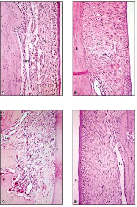
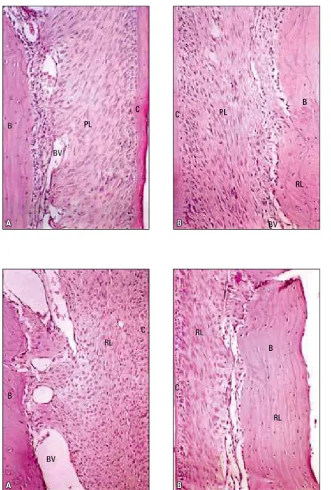
The results demonstrated that animals not sub-mitted to orthodontic movement, they presented periodontal support with normal characteristics. Animals subjected to orthodontic movement, both control and experimental groups presented with areas of pressure and tension (Figs 2-5).
Tissue reactions in the pressure and tension sides of the periodontal ligament were similar in both control and experimental groups at 7 and 14 day period (Figs 2-5).
A
A
B
B
FIGURE 2 - A) Area of inter-radicular pressure, Group C, after 7 days of orthodontic move-ment. Note the narrowing of the periodon-tal ligament space, disorganized fibers and cells, discrete inflammatory infiltrate, dilated vessels, bone surface without resorption lacunae, partially covered with osteoblasts. B) Area of inter-radicular pressure, Group C, after 7 days of orthodontic movement. Note the discrete increase in the ligament thick-ness, stretched fibers, discrete inflammatory infiltrate, irregular bone surface with areas of active resorption. Normal cement surface.
FIGURE 3 - A) Area of inter-radicular pres-sure, Group E, after 7 days of orthodontic movement. Note the narrowing of periodontal ligament space, disorganized fibers and cells, moderate inflammatory infiltrate, dilated and hyperemic vessels, area of intense maxillary bone resorption. B) Area of inter-radicular traction, Group E, after 7 days of orthodon-tic movement. Note the discrete increase in ligament thickness, stretched fibers, discrete inflammatory infiltrate, irregular bone surface without active resorption. Normal cement surface.
dISCuSSIon
Phenobarbital is a derivative of barbituric acid, and was synthesized for the first time in Germany, in 1912, and patented under the brand name of Luminal.7 It acts on the GABA receptor, blocking the entry of calcium into the presynaptic terminals,
inhibiting the transmission of the neurotransmitter glutamate.16 As all barbiturates, chemically pheno-barbital is a cyclic diamide of six carbons.
According to the literature, the treatment with drugs that have an anxiolytic and anticonvulsant ac-tion may affect the acac-tion of various hormones.7,16
B
B
B
B PL
PL
C C
BV BV
BV
BV PL
PL
C
A
A
B
B
One of the most remarkable side effects in therapy with this class of drugs is the alteration in calcium metabolism, which results in depletion of the se-rum level, in addition to inducing a condition of osteomalacia or rickets.7,16 Since it alters the me-tabolism of calcium, phenobarbital may have an in-fluence on tooth movement. Therefore, the present article proposed to evaluate the influence of this
medication on induced tooth movement.
For this purpose, a New Zealand (Oryctolagus cuniculus) breed of rabbits, frequently used in sev-eral studies on the effects of drugs on orthodontic tooth movement was used.18,21,20 In order to in-duce the tooth movement similar to what would be seen clinically, a coil spring was extended from the maxillary incisors to the molars.
FIGURE 4 - A) Area of inter-radicular pressure, Group C, after 14 days of orthodontic move-ment. Note discrete narrowing of periodontal ligament space, with slightly compressed cells, there is rich cellularity and there are dilated blood vessels. Note discrete mono-nuclear leukocytes. B) Area of inter-radicular tension, Group C, after 14 days of orthodontic movement. Note discrete increase in ligament thickness, rich cellularity and few blood ves-sels. Predominant fusiform cells, cement sur-face preserved and covered with cemento-blasts, notable reversal lines.
FIGURE 5 - A) Area of inter-radicular pressure, Group E, after 14 days of orthodontic move-ment. Note discrete narrowing of periodon-tal ligament space, with slightly compressed cells, there is rich cellularity with ovoid and irregular cells. Well dilated and non hyper-emic blood vessels. B) Area of inter-radicular traction, Group E, after 14 days of orthodontic movement. Note discrete increase in ligament thickness, rich cellularity with fusiform cells, without organization and stretching of colla-gen fiber bundles, cement surfaces without resorption lacunae.
B
B
B B
PL
PL
RL
RL RL
RL C
C
C
C BV
BV
Tooth movement was evaluated after 7 and 14 days which reflects the intermediate and final periods of tooth movement in the rabbit
periodontium.18
When the histological preparations from the animals not submitted to orthodontic tooth movement were analysed, the supporting peri-odontium presented characteristics of normal-ity. There was a uniformity in the periodontal ligament width along the roots of the teeth, intense vascularization, high cellularity with various types of cells, particularly fibroblasts, in addition to lymphocytes, undifferentiated mesenchymal cells, osteoblasts and cemento-blasts distributed in the amorphous fundamen-tal substance, as referred to by Junqueira and Carneiro9 and Ten Cate.22
The bony tissue was shown to be of the lamel-lar type, covered by a non lamellamel-lar bone layer. Superficially to the lamellar bone, in some areas, there was deposition of a thin layer of osteoid tis-sue covered by osteoblasts aligned along it. The presence of osteocytes distributed throughout the bone was also observed. This condition of normal-ity demonstrated in the normal animals was men-tioned by Junqueira and Carneiro.9
In the histological preparations of the animals submitted to orthodontic movement, both in the control and experimental groups, the presence of area of pressure in the periodontal ligament re-gion corresponding to the mesial surface of the first permanent molar and in the area under ten-sion on the distal surface was characterized, in agreement with findings of other experimental studies on orthodontic tooth movement.11 Tissue reactions on the pressure and tension sides of the periodontal ligament were similar in the control and experimental groups both at 7 and 14 days, and were characterized as described below.
the pressure side in animals of the 7-day Con-trol and Experimental Groups
Seven days after force application, a reduction
in the width of the periodontal ligament space was observed, in addition to a discrete inflamma-tory infiltrate next to the bone surface, discrete hyaline zones, fibers and disorganized cells. Ab-sence of alterations at the tooth margin (cement) and a discrete bone resorption with an irregular surface were also observed. Another condition found was lacunae without clasts in the major-ity of the laminae while in others, a discrete dis-tant resorption with clasts in lacunae could be observed (Figs 2 and 3).
the tension side in animals of the 7-day Control and Experimental Groups
On the tension side, the presence of inflam-matory infiltrate next to the bone was observed, as well as hyaline zones, increased periodontal ligament space, well distended periodontal liga-ment fibers, dilated vessels and discrete hyper-emia. Some empty lacunae and distant clasts were also observed, as well as absence of bone and cement resorption, demonstrating a normal tooth surface and presence of bone formation with reversal lines (Figs 2 and 3).
These characteristics observed at 7 days are in agreement with those that presented in the literature by Ruellas,20 demonstrating that there are no differences between the normal and ex-perimental groups.
the pressure side in animals of the 14-day Control and Experimental Groups
Areas of cement resorption occurs even with the use of light orthodontic forces, according to
Kurol and Owman-Moll,11 which explain all the
histological characteristics observed on the pres-sure side at the 14-day period.
the tension side in animals of the 14-day Con-trol and Experimental Groups
The space of the periodontal ligament pre-sented with rich cellularity and a few blood vessels. Cells were predominantly fusiform and the fiber bundles were distended. The cement surface was preserved and covered with cementoblasts. The bony surface was slightly cut, without resorption lacunae, and reversal lines indicative of bone de-position were noted. No inflammatory infiltrate or hyaline zones were noted (Figs 4 and 5).
It is important to point out that Group E pre-sented characteristics similar to those of Group C at 14-day period. However, a lower quality in the healing process in Group E could be noted as com-pared to Group C. These characteristics observed on the pressure and tension sides are corroborated by previous findings,19,20 therefore demonstrating no difference between the control and experimen-tal groups after 14 days.
Histological differences at 7 and 14-day period, were found by Chao et al3 and Tenshin,23 when prostaglandins were used. The authors found an in-crease in vascularization and significant changes in its morphology, representing greater bone resorp-tion, thus obtaining double the movement, with-out promoting damage to the periodontal tissues.
Other medications have also been tested such as parathyroid hormone,10 vitamin D metabolites,4 indomethacin,24 aspirin,2 ibuprofen,
acetamino-phen14 and bisphosphonates14 and have
demon-strated histological differences between control and experimental groups.
Although no differences were found in the periodontium of animals treated with pheno-barbital, it is important for the clinician to be aware of patients being treated with this kind of drug particularly because there is no refer-ence in the literature on the long-term effect on tooth movement.
The results of this study were based on sub-jective and overall analysis by an experienced and trained examiner involving various parameters described for optical microscopy. The use of op-tical microscopy and hematoxylin-eosin staining was shown to be practical, low cost and allowed overall analysis of all tissues, with a high degree of fidelity, without the need of any special and spe-cific analytic or staining technique.
ConCLuSIon
1. Brudvik P, Rygh P. Non-clast cells start orthodontic root resorption in the periphery of hyalinized zones. Eur J Orthod. 1994;15(6):467-80.
2. Carter-Bartlett P, Dersot JME, Saffar JL. Periodontal and femoral bone status in periodontitis-affected hamsters receiving a high dose indomethacin treatment. J Biol Buccale. 1989;17(2):93-101.
3. Chao CF, Shih CE, Wang TM. Effects of prostaglandin E2 on alveolar bone resorption during orthodontic tooth movement. Acta Anat (Basel). 1988;132(4):304-9.
4. Collins MK, Sinclair PM. The local use of vitamin D to increase the rate of orthodontic tooth movement. Am J Orthod Dentofacial Orthop. 1988;94(4):278-84.
5. Damian MA. Inluência do bisfosfonato alendronato de sódio (Fosamax®) no movimento ortodôntico em ratos “Wistar”
[tese]. Rio de Janeiro (RJ): Universidade Federal do Rio de Janeiro; 2003. 134 p.
6. Davidovitch Z, Finkelson MD, Steigman S, Shanfeld JL, Montgomery PC, Korostoff E. Electric currents, bone remodeling, and orthodontic tooth movement. II. Increase in rate of tooth movement and periodontal cyclic nucleotide levels by combined force and electric current. Am J Orthod. 1980;77(1):33-47.
7. Hahn TJ, Birge SJ, Scharp CR. Phenobarbital-induced alterations in vitamin D metabolism. J Clin Invest. 1972;51(4):741-8.
8. Igarashi K, Mitani H, Adachi H., Shinoda H. Anchorage and retentive effects of a bisphosphonate (AHBuBP) on tooth movements in rats. Am J Orthod Dentofacial Orthop. 1994;106(3):279-89.
9. Junqueira LC, Carneiro J. Histologia básica. 4ª ed. Rio de Janeiro: Guanabara Koogan; 1995.
10. Kajiyama K, Murakami T, Yokota S. Gingival reactions after experimentally induced extrusion of the upper incisors in monkeys. Am J Orthod Dentofacial Orthop. 1993;104(1):36-47. 11. Kurol J, Owman-Moll P. Hyalinization and root resorption
during early orthodontic tooth movement in adolescents. Angle Orthod. 1998;68(2):161-5.
12. Miller CS, Kaplan AL, Guest GF. Documenting medication use in adult dental patients: 1987-1991. J Am Dent Assoc. 1992;123(11):40-8.
REfEREnCES
13. Paiva DCB. Inluência clínica e tecidual do diazepam no periodonto de sustentação durante o movimento ortodôntico [dissertação]. Rio de Janeiro (RJ): Universidade Federal do Rio de Janeiro; 2001. 154 p.
14. Ramos LVT, Furquim LZ, Consolaro A. A inluência de medicamentos na movimentação ortodôntica: uma análise crítica da literatura. Rev Dental Press Ortod Ortop Facial. 2005;10(1):122-30.
15. Resende AC. A inluência do ácido acetilsalicílico no movimento dentário ortodôntico [dissertação]. Rio de Janeiro (RJ): Universidade Federal do Rio de Janeiro; 2000. 132 p. 16. Rho JM, Sankar R. The pharmacologic basis of antiepilect drug
action. Epilepsia. 1999;40(11):1471-83.
17. Roche JJ, Cisneros GJ. The effect of acetaminophen on tooth movement in rabbits. Angle Orthod. 1997;67(3):231-6. 18. Ruellas ACO. Inluência do uso de anovulatórios na
movimentação ortodôntica: estudo em coelhos [tese]. Rio de Janeiro (RJ): Universidade Federal do Rio de Janeiro; 1999. 157 p.
19. Ruellas ACO, Bolognese AM. Mola de níquel-titânio x mola de aço inoxidável. Comparação do movimento dentário. J Bras Ortodon Ortop Facial. 2000;5(27):26-50.
20. Ruellas ACO, Oliveira AM, Nishioka MH, Tavares AFT. Movimento dentário ortodôntico sob inluência de dipirona sistêmica. J Bras Ortodon Ortop Facial. 2002;7(38):142-7. 21. Sobral MC. Avaliação do movimento dentário em coelhos
com osteosporose induzida por costicosteróide [dissertação]. Rio de Janeiro (RJ): Universidade Federal do Rio de Janeiro; 1999. 100 p.
22. Ten Cate AR. Oral histology: development, structure and function. St. Louis: Mosby; 1994. Year Book.
23. Tenshin S. Remodeling mechanisms of transeptal ibers during and after tooth movement. Angle Orthod. 1995;65(2):141-50. 24. Zhou D, Hughes B, King GJ. Histomorphometric and
biochemical study of osteoclasts at orthodontic compression sites in the rat during indomethacin inhibition. Arch Oral Biol. 1997;42(10-11):717-26.
Contact address
Matheus Melo Pithon
Av. Otávio Santos, 395, sala 705 Centro Odontomédico CEP: 45.020-750 – Vitória da Conquista/BA, Brazil E-mail: [email protected]
Submitted: January 22, 2007