Mutation analysis of
NANOS3
in Brazilian women with
primary ovarian failure
Braian Lucas A. Sousa, Mirian Yumie Nishi, Mariza Gerdulo Santos, Vinicius Nahime Brito, Sorahia Domenice, Berenice B. Mendonca*
Hospital das Clı´nicas da Faculdade de Medicina da Universidade de Sa˜o Paulo, Disciplina de Endocrinologia, Unidade de Endocrinologia do Desenvolvimento, Laborato´rio de Hormoˆnios e Gene´tica Molecular LIM/42, Sa˜o Paulo/SP, Brazil.
OBJECTIVES: Primary ovarian failure is a rare disorder, and approximately 90% of cases are of unknown etiology. The aim of this study was to search for mutations inNANOS3,a gene that was recently related to the etiology of primary ovarian failure, in a group of Brazilian women.
METHODS:We screened forNANOS3DNA variants in 30 consecutive women who were previously diagnosed with primary ovarian failure, of unknown etiology and compared the results with those from 185 women with normal fertility. TheNANOS3gene was amplified by polymerase chain reaction using pairs of specific primers and then sequenced. The resulting sequences were compared with control sequences available in the National Center for Biotechnology and Information database.
RESULTS: No mutations in NANOS3 were found in primary ovarian failure patients, but four previously described polymorphisms were identified at a similar frequency in the control and primary ovarian failure groups.
CONCLUSIONS:Mutations inNANOS3were not associated with primary ovarian failure in the present cohort.
KEYWORDS: Primary Amenorrhea;NANOS3; Gonadal Dysgenesis; Primary Ovarian Failure; Sexual Differentiation.
Sousa BL, Nishi MY, Santos MG, Brito VN, Domenice S, Mendonc¸a BB. Mutation analysis ofNANOS3in Brazilian women with primary ovarian failure. Clinics. 2016;71(12):695-698
Received for publication onMay 3, 2016;First review completed onMay 23, 2016;Accepted for publication onSeptember 2, 2016 *Corresponding author. E-mail: [email protected]
’ INTRODUCTION
Primary ovarian failure (POF), also known as premature ovarian failure, premature ovarian insufficiency, premature menopause, or hypergonadotropic hypogonadism, is defined as the loss of function of the ovaries before age 40 (1). POF is phenotypically characterized by the triad of primary or secondary amenorrhea, hypergonadotropic hypogonadism and hypoestrogenism.
There are several causes of POF, and the genetic causes are associated with a defect in ovarian development.
Sex determination in mammals is a dynamic and complex process that requires the interaction of multiple factors, and both primordial germ cells (PGCs) and somatic cells acquire sex-specific characteristics.
PGCs are essential for the differentiation and maintenance of the fetal ovary; when few germ cells enter the genital ridge during embryogenesis, the supporting lineage cells do not differentiate, generating strike ovaries (2-4). Because the
differentiation of ovarian somatic cells relies on PGCs, genes involved in the differentiation and subsequent maintenance of PGCs are potential candidates for the etiology of 46,XX gonadal development disorders (5).
Nanos homolog 3was identified in PGCs ofDrosophila, and the NANOS3 protein has anti-apoptotic activity during the migration of PGCs toward the gonad (6). Male and female mice that do not expressNanos3experience a complete loss of PGCs during gonadal development (7). The human NANOS3
gene is located on the short arm of chromosome 19 (region 13.13) and has two exons.
A preliminary study in a Brazilian population identified aNANOS3c.385G4A homozygous mutation in two sisters
with POF.In vitroanalysis showed that the NANOS3 protein in these two patients had an impaired ability to prevent the apoptosis of PGCs, suggesting a possible etiology for the disorder (8).
Our objective was to search for inactivatingNANOS3 muta-tions in an additional group of 30 women with POF without apparent cause.
’ MATERIALS AND METHODS
This was a cohort study performed in accordance with the ethical standards of the Ethics Committee of Hospital das Clínicas, University of São Paulo School of Medicine, Brazil (Protocol number 1226/07).
DOI:10.6061/clinics/2016(12)03
Copyright&2016CLINICS–This is an Open Access article distributed under the terms of the Creative Commons License (http://creativecommons.org/licenses/by/ 4.0/) which permits unrestricted use, distribution, and reproduction in any medium or format, provided the original work is properly cited.
No potential conflict of interest was reported.
695
POF was defined as the loss of function of the ovaries before age 40 (1). We invited 30 consecutive patients with the diagnosis of POF of unknown etiology who were followed in the Developmental Endocrinology Unit of Hospital das Clínicas, São Paulo, Brazil, to undergo screening forNANOS3
mutations. Thyroid, adrenal and ovarian autoimmune dis-orders, as well as mutations inFSHR,NR5A1,BMP15,GDF9,
GATA4,DMRT1,FOXL2,LHCGR, andSTRA8and premutation of FMR1, had been ruled out in all these patients. All the patients had a normal 46,XX karyotype obtained by analysis of 50 metaphases.
Hormonal tests required for the POF diagnosis were performed at the Laboratory of Hormones and Molecular Genetics, LIM/42 of the HCFMUSP, using immunofluoro-metric assays (Delfia, Turku, Finland).
Basal gonadotropin levels were elevated in all patients, with predominantly high serum FSH levels ranging from 32 to 111 U/L and serum LH levels ranging from 13 to 107 U/L. Serum estradiol levels were low or within the follicular phase range in all patients.
The control group was composed of 185 women aged 18 to 65 years, all of whom had normal menstrual cycles and no history of menopause before 40 years of age or treatment for subfertility. The control group was screened for theNANOS3
allelic variants found in the POF group.
Genomic DNA was extracted from peripheral blood leucocytes using the salting out procedure (9). The DNA concentration and purity were determined using the NanoPhotometer (Implen GmbH, Schatzbogen, Germany), considering an ideal 260/280 nm ratio of 1.75, and DNA integrity was assessed by 1% agarose gel electrophoresis, staining with SYBR Safe (Invitrogen Life Technologies Cor-poration, CA, USA) and visualization with ultraviolet light.
NANOS3was amplified by PCR using specific pairs of primers located in the intronic regions of the gene (Table 1), and the PCR products were subjected to direct automated sequencing. We performed PCR amplifications in a total volume of 25ml (Table 2) as follows:
Exon 1: (94o
C - 5 min) x 1+(94o
C - 30 sec; 58o
C - 30 sec; 72o
C - 45 sec) x 35+(72o
C - 10 min) x 1. Exon 2: (94o
C - 5 min) x 1+(94o
C - 30 sec; 57o
C - 30 sec; 72o
C - 45 sec) x 35+(72o
C - 10 min) x 1.
The PCR products were analyzed by electrophoresis on 1% agarose gels stained with SYBR Safe (Invitrogen Life Technologies Corporation, CA, USA) and visualized with ultraviolet light. To assess the DNA concentration of the PCR products, the intensity of the signal emitted by the bands was
compared with the Low DNA Mass molecular weight marker (Invitrogen, Life Technologies Corporation, CA, USA). After amplification, PCR products were purified by incubation with ExoSAP-IT (GE Healthcare Life Sciences, Buckinghamshire, United Kingdom) for 15 min at 37o
C and then 15 min at 80o
C. DNA sequencing was performed according the protocol of the ABI Prism BigDye Terminator Cycle Sequencing Ready Reaction Kit (Life Technologies Cor-poration, CA, USA).
The samples were subjected to direct automated sequen-cing using the ABI Prism Genetic Analyzer 3130XL (Life Tech-nologies Corporation, CA, USA). The sequences obtained were compared to those in the National Center for Biotechnology Information database(NCBIhttp://www.ncbi.nlm.nih.gov/). We performedin silicoanalysis of the identified variants using Mutation Taster (available online at http://www.mutationta-ster.org).
Comparisons between unrelated patients with POF and controls were performed using contingency tables and w2
analysis for each polymorphism in SigmaStat for Windows v.3.5.p valueso0.05 were considered statistically significant.
’ RESULTS
NANOS3mutation screening in 30 unrelated women with POF of unknown etiology identified the rs897790, rs2016163, rs369192674 and rs371590850 variants in 18 patients.
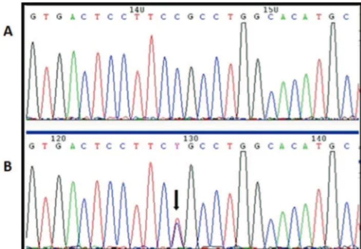
The rs897790 variant is located in the 5’ untranslated region of exon 1, where it caused the replacement of a cytosine with a thymine at position -23 (c.-23C4T); this
variant was found in 6 patients in the heterozygous state (Figure 1). In the control group, the rs897790 variant was identified in 59 women, of whom 54 were heterozygous and 5 were homozygous.
The rs2016163 polymorphism is located in exon 1 and causes the substitution of an adenine with a guanine at
Table 1-Sequences of the primers used to amplifyNANOS3exons.
Exon Primer sequence
1 Forward 50CTG CTC CTC CCT CTT CAC AC 30 Reverse 50GTC TTC CCC TAA CCC TTG GA 30 2 Forward 50GTC ACG GGG TCG CTG TCT 3’
Reverse 50AGT GGG GGC AGT CGT CAT AG 30
Table 2-PCR amplification protocol for theNANOS3gene.
Exon H2O (ml) 5X Buffer (ml) dNTP (mM) Forward Primer (pmol) Reverse Primer (pmol) Taq DNA Polymerase (U)
1 16.3 5 0.2 10 10 0.75
2 16.3 5 0.2 10 10 0.75
dNTP: deoxynucleoside triphosphate;ml: microliters; mM: millimolar; pmol: picomoles; U: units.
Figure 1 - A, Electropherogram of a patient with wild type
NANOS3. B, Electropherogram of a patient with the
hetero-zygous rs897790 (c.23C4T) variant, indicated by a black arrow.
696 Mutation Analysis ofNANOS3in POF
position 354 (c.354A4G). This substitution does not change the amino acid sequence. Ten of the 30 patients harbored this polymorphism, one in the homozygous state and 9 in the heterozygous state (Figure 2). This variant was present in 70 individuals in the control group, of whom 58 were heterozygous and 12 were homozygous.
Six patients presented with both variants. These pre-viously identified variants are reported in control databases, such as the 1000 Genomes Project, at a frequency of greater than 1%, which classifies them as polymorphisms.
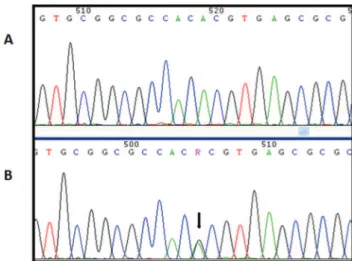
Two patients harbored a GAG duplication at position 512-514 (c.512_514dupGAG) in the transcribed region of exon 1 (rs369192674). This trinucleotide duplication led to the addition of a glycine between positions p.171 and p.172 (p.Gly171dup) (Figure 3). In silico analysis of this variant classified it as not deleterious. This variant is described in the NCBI database, although its population frequency is not known. This variant was found in three women in the control group in the heterozygous state.
Another patient harbored a heterozygous insertion of 18 nucleotides between positions c.134 and c.135 (c.134_
135insGCCGGAGCCGGTGTCAGC) in the transcribed region of exon 1 (rs371590850). This insertion leads to the addition of six amino acids between positions p.45 and p.46 (p.Ala45_ Leu46insProGluProValSerAla) (Figure 4). In silico analysis classified this variant as not harmful. This variant is described in the NCBI database, although its population frequency is not known. In the control group, this variant was identified as heterozygous in 6 women.
The screening results for the rs897790, rs2016163, rs3691 92674 and rs371590850 variants in the control and POF groups are summarized in Table 3.
The allele distributions of the rs897790, rs2016163, rs369192674 and rs371590850 variants were not significantly different between the POF patients and the control group (p=0.271,p=0.787,p=0.295 andp=0.597, respectively) (Table 3). We did not identify any deleteriousNANOS3 variants in the studied population.
’ DISCUSSION
In the present study, we screened for NANOS3 muta-tions in a cohort of women with POF and identified only four previously described variants, which were detected at a similar frequency in the control group.
Although most POF cases are sporadic, 10-15% are familial, corroborating the hypothesis that this disorder has a genetic component. However, despite recent advances in molecular biology, approximately 90% of POF cases are of unknown etiology (1,10). The role of NANOS3 in inhibit-ing the apoptosis of PGCs and thus contributinhibit-ing to their maintenance makes this protein a natural candidate for the etiology of POF in humans (11). Loss of NANOS3 function may lead to PGC death:Nanos3knockout mice do not have PGCs (7). Based on this finding, three groups used Sanger sequencing to screen women with POF forNANOS3 muta-tions (8,12-14). Qin et al. studied samples from 80 Chinese
Figure 2 - A, Electropherogram of a patient with wild type
NANOS3. B, Electropherogram of a patient with the
hetero-zygous rs2016163 (c.354A4G) variant, indicated by a black arrow.
Figure 3 - A, Electropherogram of a patient with wild type
NANOS3. B/C, Electropherogram of a patient heterozygous for
the rs369192674 (c.512_514dupGAG) variant. The overlapping sequences of the two alleles, wild type and variant, are shown beginning at the black arrow.
Figure 4 - A, Electropherogram of a patient with wild type
NANOS3. B, Electropherogram of the patient with the
hetero-zygous rs371590850 (c.134_135insGCCGGAGCCGGTGTCAGC) var-iant. The overlapping sequences of the two alleles, wild type and variant, are shown beginning at the black arrow.
Table 3-Distribution ofNANOS3variants in the control and patient groups.
Allelic Variant POF Group Control Group p#
rs897790 6*/30** 59*/185** 0.271
rs2016163 10*/30** 70*/185** 0.787
rs369192674 2*/30** 3*/185** 0.295
rs371590850 1*/30** 6*/185** 0.597
POF: Premature Ovarian Failure.
* : number of individuals with the variant; **: total number of individuals; #: Chi-square test;po0.05 indicated significance.
697
and 88 Caucasian women by denaturing high-performance liquid chromatography (DHPLC), followed by Sanger seq-uencing, and did not identify any causative mutations in
NANOS3(12). Wu et al. evaluated 100 Chinese patients with POF and reported a heterozygous p.Arg153Trp mutation in
NANOS3in a 23-year-old patient born from consanguineous parents. Transfection assays in HEK293 cells showed that the half-life of the mutant p.Arg153Trp protein was significantly reduced. Using an animal model, these researchers also showed that the PGC population was directly related to NANOS3 protein levels. Together, these two findings impli-cate the c.457C4T mutation (p.Arg153Trp) in the etiology of
POF in this patient (13).
Finally, a previous study in a Brazilian cohort evaluated 85 patients with primary or secondary ovarian failure and reported the novel homozygous c.358G4A mutation
(p.Glu120Lys) in NANOS3in two sisters with POF.In vitro
analysis showed a significant loss of PGC viability, and
in silico modeling of mutant NANOS3 suggested that this amino acid change might destabilize the protein-RNA inter-action. This evidence supports the hypothesis that the c.358G4A mutation, which increases the apoptosis rate of
PGCs during the embryonic period, is the etiological factor for POF in these two patients (8).
Considering these three studies involving the index case of 343 families, theNANOS3mutation frequency in POF is less than 0.5% (8,12-14).
Our study included 30 additional Brazilian women with POF, but no mutations were found in theNANOS3gene. The four variants that were identified in the present study have already been described, but only the rs897790 and rs2016163 variants were previously reported in POF women (8,12,13). The rs2016163 variant was also identified in normal women by Qin et al. (12). No significant difference was observed in the allele frequencies of the rs897790, rs2016163, rs369192674 and rs371590850 variants between POF patients and the control group. Our negative results should be interpreted cautiously due to the small number of patients; POF is a rare disorder. In addition, only theNANOS3coding regions and exon boundaries, not the intronic and regulatory regions, were sequenced.
In conclusion, despite the prominent role ofNANOS3in ovarian development, our findings suggest that NANOS3
mutations were not associated with POF in the present cohort.
’ ACKNOWLEDGMENTS
The authors thank the staff of the Laboratory of Hormones and Molecular Biology (LIM/42), whose dedication and effort made this study possible. This study was partially supported by grants from Conselho Nacional de Desenvolvimento Cientifico e Tecnológico–CNPq (301339/2008-9 to
B.B.M. and 149339/2013-5 to B.L.A.S.). Berenice B. Mendonc¸a was
parti-ally supported by grants from the Conselho Nacional de Desenvolvimento Científico e Tecnológico – CNPq (3057-43/2011-2). This work was
partially supported by grants from Fundac¸ão de Amparo a Pesquisa do
estado de São Paulo FAPESP 2013/02162-8.
’ AUTHOR CONTRIBUTIONS
Sousa BL, Santos MG and Nishi MY performed and analyzed the experi-mental assays. Brito VN performed the statistical analysis. Sousa BL was responsible for writing the manuscript. Domenice S followed and selected the patients. Mendonca BB designed the project, supervised the study execution and wrote the manuscript.
’ REFERENCES
1. Nelson LM. Clinical practice. Primary ovary insufficiency. N Engl J Med. 2009;360(6):606-14, http://dx.doi.org/10.1056/NEJMcp0808697. 2. MacLaren A. Germ and somatic cell lineages in the developing gonad.
Mol Cell Endocrinol. 2000;163(1-2):3-9, http://dx.doi.org/10.1016/S0303-7207(99)00234-8.
3. Kocer A, Reichmann J, Best D, Adams IR. Germ cell sex determination in mammals. Mol Hum Reprod. 2009;15(4):205-13, http://dx.doi.org/ 10.1093/molehr/gap008.
4. Menke DB, Koubova J, Page DC. Sexual differentiation of germ cells in XX mouse gonads occurs in an anterior-to-posterior wave. Dev Biol. 2003; 262(2):303–12, http://dx.doi.org/10.1016/S0012-1606(03)00391-9. 5. Matzuk MM, Lamb DJ. The biology of infertility: research advances and
clinical challenges. Nat Med. 2008;14(11):1197-213, http://dx.doi.org/ 10.1038/nm.f.1895.
6. Suzuki H, Tsuda M, Kiso M, Saga Y. Nanos3 maintains the germ cell lineage in the mouse by suppressing both Bax-dependent and -indepen-dent apoptotic pathways. Dev Biol. 2008;318(1):133-42, http://dx.doi. org/10.1016/j.ydbio.2008.03.020.
7. Tsuda M, Sasaoka Y, Kiso M, Abe K, Haraguchi S, Kobayashi S, et al. Conserved role of nanos proteins in germ cell development. Science. 2003;301(5637):1239-41, http://dx.doi.org/10.1126/science.1085222. 8. Santos MG, Machado AZ, Martins CN, Domenice S, Costa EM, Nishi MY,
et al. Homozygous inactivating mutation in NANOS3 in two sisters with primary ovarian insufficiency. Biomed Res Int. 2014;787465, http://dx. doi.org/10.1155/2014/787465.
9. Miller SA, Dykes DD, Polesky HF. A simple salting out procedure for extracting DNA from human nucleated cells. Nucleic Acids Res. 1988; 16(3):1215, http://dx.doi.org/10.1093/nar/16.3.1215.
10. Caburet S, Zavadakova P, Ben-Neriah Z, Bouhali K, Dipietromaria A, Charon C, et al. Genome-wide linkage in a highly consanguineous ped-igree reveals two novel loci on chromosome 7 for non-syndromic familial premature ovarian failure. PLoS One. 2012;7(3):e33412, http://dx.doi. org/10.1371/journal.pone.0033412.
11. Julaton VT, Reijo Pera RA. NANOS3 function in human germ cell development. Hum Mol Genet. 2011;20(11):2238-50, http://dx.doi.org/ 10.1093/hmg/ddr114.
12. Qin Y, Zhao H, Kovanci E, Simpson JL, Chen ZJ, Rajkovic A. Mutation analysis of NANOS3 in 80 Chinese and 88 Caucasian women with pre-mature ovarian failure. Fertil Steril. 2007;88(5):1465–7, http://dx.doi.org/ 10.1016/j.fertnstert.2007.01.020.
13. Wu X, Wang B, Dong Z, Zhou S, Liu Z, Shi G, et al. A NANOS3 mutation linked to protein degradation causes premature ovarian insufficiency. Cell Death Dis. 2013;4:e825, http://dx.doi.org/10.1038/cddis.2013.368. 14. Qin Y, Jiao X, Simpson JL, Chen ZJ. Genetics of primary ovarian
insuffi-ciency: new developments and opportunities. Hum Reprod Update. 2015;21(6):787-808, http://dx.doi.org/10.1093/humupd/dmv036.
698 Mutation Analysis ofNANOS3in POF