R
E
V
IS
Ã
O
R
E
V
IE
W
1 Neurotoxicology and Molecular Imaging Laboratory, Department of Environmental Health Sciences, Johns Hopkins Bloomberg School of Public Health, Baltimore, Maryland, USA 60 Haven Ave., B1-108. 10032 New York NY USA. [email protected]
Manganese and Parkinson’s Disease:
A Critical Review and New Findings
Manganês e Doença de Parkinson:
Uma revisão crítica e novas descobertas
Resumo O objetivo desta revisão foi examinar se
a exposição crônica ao Mn produz degeneração do neurônio pela dopamina e DP ou se é apenas uma apresentação neuropatológica e clínica dife-rente. Foram revisados estudos clínicos, de neu-roimagens e neuropatológicos disponíveis sobre humanos e primatas expostos ao Mn ou outras condições humanas que resultam em concentra-ções elevadas de Mn no cérebro. Foi examinada a literatura sobre humanos e primatas e compara-das as mudanças clínicas de neuroimagem e neu-ropatológicas associadas com o “parkinsonimo” induzido por Mn, envolvendo a degeneração do sistema dopaminérgico nigro-estriatal como no caso da DP. as evidências decisivas mostram que o “parkinsonismo” induzido pelo Mn não envolve a degeneração dos neurônios de dopamina do mesencéfalo e que o dopa-1 não é uma terapia eficaz. Novas evidências estão presentes em um mecanismo putativo pelo qual o Mn pode produ-zir anormalidades de movimento. A confirmação desta hipótese em humanos é essencial para to-mar decisões adequadas sobre o tratamento, pla-nejar estratégias terapêuticas eficazes e estabele-cer guias regulatórios.
Palavras-chave Gânglio basal, Dopamina,
Hu-manos, Manganês, Desordens de movimento, Neuroimagem, Neurotoxicidade, Primatas, Do-ença de Parkinson, Estriatal
Abstract The goal of this review was to examine
whether chronic Mn exposure produces dopam-ine neuron degeneration and PD or whether it has a distinct neuropathology and clinical pre-sentation. I reviewed available clinical, neuroim-aging, and neuropathological studies in humans and nonhuman primates exposed to Mn or other human conditions that result in elevated brain Mn concentrations. Human and nonhuman pri-mate literature was examined to compare clini-cal, neuroimaging, and neuropathological changes associated with Mn-induced parkinsonism. Clin-ical, neuroimaging, and neuropathological evi-dence was used to examine whether Mn-induced parkinsonism involves degeneration of the nigros-triatal dopaminergic system as is the case in PD. The overwhelming evidence shows that Mn-in-duced parkinsonism does not involve degenera-tion of midbrain dopamine neurons and that l-dopa is not an effective therapy. New evidence is presented on a putative mechanism by which Mn may produce movement abnormalities. Confir-mation of this hypothesis in humans is essential to make rational decisions about treatment, de-vise effective therapeutic strategies, and set regu-latory guidelines.
Key words Basal ganglia, Dopamine, Humans,
Manganese, Movement disorder, Neuroimaging, Neurotoxicity, Nonhuman primates, Parkinson’s disease, Striatum
G
Idiopathic Parkinson’s disease (PD) is a progres-sive neurodegenerative disorder with a slow on-set, and compared with the familial forms of the disease, it is associated with advanced age (> 55 years of age). The four cardinal signs of idio-pathic PD are tremor at rest, bradykinesia (hy-pokinesia and akinesia), rigidity, and postural instability (Jankovic 2008; Lees et al. 2009; Tolosa et al. 2006). The diagnosis of idiopathic PD is typically based on the presence of two or more of the four cardinal signs and a response to l-dopa therapy. Unilateral tremor of the hand at rest with a frequency of 4–6 Hz is the earliest and most easily recognized symptom of PD. Autopsy-based studies have shown that the percentage of pa-tients with confirmed PD diagnosis who exhibit-ed resting tremor ranges from 76–100% (Hugh-es et al. 1993; Louis et al. 1997; Rajput et al. 1991). Bradykinesia is defined by slowness of move-ments or difficulty in initiating and executing movement, and it is one of the essential signs used to diagnose idiopathic PD (Lees et al. 2009). Bradykinesia appears to correlate with the de-gree of dopamine deficiency in the caudate and putamen (striatum) (Vingerhoets et al. 1997). Ri-gidity is characterized by increased resistance of the limbs and is the result of the muscles becom-ing tensed and contracted so that the person feels stiff and weak. Postural instability is the loss of postural reflexes, and this occurs in the late stag-es of the disease. Postural instability causstag-es pa-tients to develop a forward or backward lean that causes them to fall. As the disease progresses, walking is affected, and patients walk in quick, small steps like they are hurrying forward in or-der to maintain balance.
Although the clinical diagnosis of PD is based on a combination of the four cardinal motor signs, other parkinsonian disorders also express many of these signs (Tolosa et al. 2006), and a definite diagnosis of PD requires neuropatho-logical confirmation (Gelb et al. 1999). It has been estimated that > 10% of PD cases can be diag-nosed incorrectly by movement disorder special-ists when clinical signs are the only basis for di-agnosis (Hughes et al. 2002). Consistent with the known deficiency of dopamine in the striatum, the clinical symptoms of PD are significantly al-leviated with l-dopa therapy, the precursor sub-strate for the synthesis of dopamine, the chemi-cal that is decreased in PD (Savitt et al. 2006). This is important information when one exam-ines the hypothesis that manganese (Mn)-induced
parkinsonism involves nigrostriatal dopamine neuron degeneration.
Molecular imaging is a useful strategy for the diagnosis of PD and has provided extensive evi-dence that PD patients exhibit decreased levels of presynaptic dopamine neuron terminal markers in the striatum (Felicio et al. 2009). This is consis-tent with the loss of dopamine terminals as a re-sult of degeneration of neuronal cell bodies in the substantia nigra pars compacta (SNpc) (Figure 1). Single photon emission computed phy (SPECT) and positron emission tomogra-phy (PET) studies have shown that PD patients exhibit decreased levels of dopamine transporters (DATs) and vesicular monoamine transporter type 2 (VMAT2) and reduced activity of dopa decar-boxylase as measured by the conversion of l-dopa to dopamine in the striatum using [18F]-fluoro-dopa PET (Felicio et al. 2009). Postsynaptic D2-dopamine receptors (D2Rs) either are not affect-ed or are increasaffect-ed in the striatum of untreataffect-ed PD patients (Antonini et al. 1994; Brooks et al. 1992; Rinne et al. 1990). Similar changes in these markers have been documented in a nonhuman primate model of PD (Chen et al. 2008).
A major discovery in understanding the mo-lecular basis of PD came about when a deficit in the concentration of the neurotransmitter dopamine was discovered in the striatum of brain samples from PD patients (Ehringer and Hornyk-iewicz 1960; HornykHornyk-iewicz 2006). Scientists now know that a marked loss of dopamine-contain-ing neurons in the SNpc results in decreased dopamine levels in the caudate and putamen (Sav-itt et al. 2006). A hallmark neuropathological fea-ture of SNpc dopamine neurons in PD is pro-teinacious intraneuronal aggregations called Lewy bodies that seem to be associated with dopamine neuron degeneration (Dauer and Przedborski 2003; Wakabayashi et al. 2007).
d
e C
o
le
tiv
a, 16(
11)
:4549-4566,
2011
Idiopathic PD and Mn-Induced Parkinsonism: The Early Writings of James Parkinson and John Couper
In 1817, the physician James Parkinson published the first description of a neurological disorder that is now recognized by his name, Parkinson’s disease (Parkinson 1817). His monograph, titled An Essay on the Shaking Palsy, describes five cas-es with shaking palsy, a term that was vaguely employed by medical writers at the time. He com-mented that
[T]he first symptoms perceived are, a slight sense of weakness, with a proneness to trembling in some particular part; sometimes in the head, but most commonly in one of the hands and arms. These symptoms gradually increase in the part first af-fected; and at an uncertain period, but seldom in less than twelve months or more, the morbid in-fluence is felt in some other part. Thus assuming one of the hands and arms to be first attacked, the other, at this period becomes similarly affected.
Parkinson went on to say that as the disease proceeds, “walking becomes a task which cannot be performed without considerable attention.” As the malady proceeds,
The propensity to lean forward becomes invin-cible, and the patient is thereby forced to step on the toes and fore part of the feet, whilst the upper part of the body is thrown so far forward as to render it difficult to avoid falling on the face . . . irresistibly impelled to take much quicker and shorter steps, and thereby to adopt unwillingly a running pace.
He then described the later stages,
As the disease proceeds toward its last stage, the trunk is almost permanently bowed, the muscular power is more decidedly diminished, and the trem-ulous agitation becomes violent. . . His words are now scarcely intelligible,
and he is no longer able to feed himself. “The chin is now almost immoveably bent down upon the sternum” . . . with the saliva continually trick-ling from the mouth. “The power of articulation is lost.”
Twenty years after the essay by James Parkin-son, John Couper described the first symptoms of Mn toxicity in humans (Couper 1837). The observations made in this brief essay are impor-tant in order to understand the working condi-tions and the symptoms of Mn intoxication in humans. A comparison of the two essays makes it clear how the two conditions are similar and how they are different. Couper wrote,
In the chemical works of Charles Tennant and Co., a considerable number of men are employed in
grinding the black oxide of manganese, to be em-ployed in the manufacture of bleaching powder. The surface of their bodies is of course constantly cov-ered with the manganese; the air which they breathe is loaded with it in the form of fine powder, and they are ever exposed, from neglect of cleanliness, to swallow portions of it along with their food.
It is clear from this description that working conditions were very hazardous and personal hygiene was poor. As a result, these workers were exposed to extremely high concentrations of Mn on a daily basis.
In the description of the workers who were affected by Mn poisoning, Couper went on to say that,
The loss of power is most apparent in the lower extremities, which are so considerably affected that the patient staggers, and inclines to run forward when he attempts to walk. The arms are also weak-ened, but only to a small extent. The patient com-plains that in speaking he cannot make himself heard by persons at a moderate distance, as for-merly; and the inability seems to depend, not on any defect of articulation, but on weakness of voice. There is no deficiency of sensibility in any part of the body; the intellect and external senses are un-impaired; but there is an obvious expression of vacancy in the countenance, apparently from the paralyzed state of the muscles of the face. From the same cause the saliva is apt to escape from the mouth, especially during speaking. There is no tremor in any part of the body.
A comparison of the two essays indicates that certain symptoms are similar and some are dif-ferent from those in PD. The Mn-exposed work-ers exhibited gait disturbance, a propensity to fall, masked face, hypophonia and dysphonia, and drooling. The absence of resting tremor, a prominent and early cardinal sign of PD patients, as initially described by James Parkinson, is a dis-tinguishing observation that is different from PD in these workers heavily exposed to Mn.
G
Reference
Jankovic 2008 Lee 2000
Yamada et al. 1986 Cook et al. 1974 Mena et al. 1967
Huang 2007 (summary of Taiwan cohort) Kessler et al. 2003
Kenangil et al. 2006 Bowler et al. 2006 Josephs et al. 2005 Racette et al. 2001 Sadek et al. 2003 Meral et al. 2007 Sikk et al. 2007 de Bie et al. 2007 Sanotsky et al. 2007 Varlibas et al. 2009 Stepens et al. 2008 Selikhova et al. 2008 Colosimo and Guidi 2009 Nagatomo et al. 1999 Burkhard et al. 2003 Klos et al. 2006 Klos et al. 2005 Faviani et al. 2007 Kim et al. 2007 Schaumberg et al. 2006
Category IPD Mn-O Mn-O Mn-O Mn-O Mn-O Mn-O Welding Welding Welding Welding Welding Ephedron Ephedron Ephedron Ephedron Ephedron Ephedron Ephedron Ephedron PN LD LD LD LD LD Mn-O/LD Resting tremor + -+ -+ Some -Some -Action/ postural tremor + + + + + + + + + + + + + + + + + Bradykinesia + + + + + + + + + + + + + + + + + + + + + + + -Rigidity + + + + + + + + + + + + + + + + + + + + + -Postural instability + + + + + + + + + + + + + + + + + + + + + + + +
Table 1. Clinical symptoms of parkinsonism in idiopathic PD and in human conditions with elevated brain Mn concentrations.
Reference
Jankovic 2008 Lee 2000
Yamada et al. 1986 Cook et al. 1974 Mena et al. 1967
Huang 2007 (summary of Taiwan cohort) Kessler et al. 2003
Kenangil et al. 2006 Bowler et al. 2006 Josephs et al. 2005 Racette et al. 2001 Sadek et al. 2003 Meral et al. 2007 Sikk et al. 2007 de Bie et al. 2007 Sanotsky et al. 2007 Varlibas et al. 2009 Stepens et al. 2008 Selikhova et al. 2008 Colosimo and Guidi 2009 Nagatomo et al. 1999 Burkhard et al. 2003 Klos et al. 2006 Klos et al. 2005 Faviani et al. 2007 Kim et al. 2007 Schaumberg et al. 2006
Gait disorder + + + + + + + + + + + + + + + + + + + + + + + + Dystonia + Secondary + + + + + + + + + + + + + + + + Micrographia + + + + + + + + + + Difficulty walking backward, turning, pull-test + + + + + + + + + + + + + + + + + + Hypophonia, dysphonia/ dysarthria + + + + + + + + + + + + + + + + + + Falls + + + + + + + + + + l-Dopa response Excellent Minimal
Minimal to none None None None Minimal Excellent None None None None Minimal to none
None None None Minimal None Minimal Partial None Not tested
d
e C
o
le
tiv
a, 16(
11)
:4549-4566,
2011
which Mn levels are markedly increased in the brain, and the motor symptoms in idiopathic PD. The conclusion from these reports is that, although several of the clinical signs between PD and Mn-induced parkinsonism are similar, in Mn-induced parkinsonism, unlike in PD, there is an absence of resting tremor and the lack of a response to l-dopa. Also, the progression of Mn-induced parkinsonism appears to be a gait dis-order of early onset with dystonia that occurs much later in the slow progression of the move-ment abnormalities in PD.
Mn-Induced Parkinsonism
from Human Conditions Not Related to Occupational Exposures
Besides occupational exposures, other conditions have been shown to increase accumulation of Mn in the brain and have been valuable sources of information in understanding Mn-induced neu-rological dysfunction, including parkinsonism.
Psychostimulant drug abusers: ephedron.
Re-cently, there have been a number of reports, pri-marily originating from Eastern European coun-tries and Russia, of young addicts afflicted with an atypical form of parkinsonism (de Bie et al. 2007; Meral et al. 2007; Sanotsky et al. 2007; Se-likhova et al. 2008; Sikk et al. 2007; Stepens et al. 2008; Varlibas et al. 2009) (Table 1). This form of
parkinsonism is the result of intravenous injec-tions of a psychostimulant drug called “ephe-drone” or “Russian cocktail,” a drug in which ephedrine is oxidized using potassium perman-ganate and acetic acid (Sanotsky et al. 2007; Ste-pens et al. 2008). Typically, this homemade chem-ical mixture is not purified before intravenous injection, so milligram to gram amounts of Mn are injected with multiple injections occurring during the course of weeks and months. Normal Mn concentrations in whole blood are d” 10–12 ìg/L, but the blood Mn concentrations in these young addicts have been measured at levels as high as 2,000–3,000 ìg/L (Stepens et al. 2008; Varli-bas et al. 2009). Consistent with the fact that high concentrations of Mn are injected, most of these individuals exhibit bilateral T1-weighted magnet-ic resonance imaging (MRI) hyperintensive sig-nals in the basal ganglia and other brain regions reflecting Mn accumulation (Figure 1, Table 2).
The parkinsonian signs in these young drug-abuse subjects are consistent with those in occu-pationally exposed Mn workers (Table 1). An important observation on the etiological role of Mn in the parkinsonism in these young addicts is based on the fact that movement abnormalities are observed in ephedrone users in Eastern Eu-rope and Russia where the chemical preparation uses potassium permanganate as the oxidizing agent. However, a parkinsonian syndrome has not been observed in North America, where
G
mate is used as the oxidizing agent rather than potassium permanganate (Selikhova et al. 2008; Stepens et al. 2008). The most parsimonious ex-planation is that Mn is the causative agent in this atypical form of parkinsonism.
Patients with liver disease. There is evidence of
parkinsonism associated with chronic liver dis-ease. Patients with advanced cirrhosis have been documented with a form of parkinsonism with clinical symptoms similar to Mn-induced
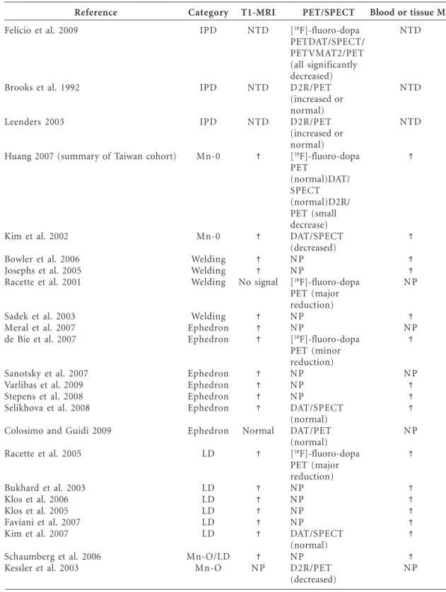
parkin-Table 2. Brain imaging studies in idiopathic PD and in human conditions that show elevated
concentrations of Mn in the brain.
Reference
Felicio et al. 2009
Brooks et al. 1992
Leenders 2003
Huang 2007 (summary of Taiwan cohort)
Kim et al. 2002
Bowler et al. 2006 Josephs et al. 2005 Racette et al. 2001
Sadek et al. 2003 Meral et al. 2007 de Bie et al. 2007
Sanotsky et al. 2007 Varlibas et al. 2009 Stepens et al. 2008 Selikhova et al. 2008
Colosimo and Guidi 2009
Racette et al. 2005
Bukhard et al. 2003 Klos et al. 2006 Klos et al. 2005 Faviani et al. 2007 Kim et al. 2007
Schaumberg et al. 2006 Kessler et al. 2003
Category
IPD
IPD
IPD
Mn-0
Mn-0
Welding Welding Welding
Welding Ephedron Ephedron
Ephedron Ephedron Ephedron Ephedron
Ephedron
LD
LD LD LD LD LD
Mn-O/LD Mn-O
Abbreviations: IPD, idiopathic PD; LD, liver disease; Mn-O, occupational Mn exposure; NP, not performed; NTD, not typically done. PET/SPECT studies were done in the striatum (caudate and putamen). Arrows indicate increased T1-weighted MRI signal in the globus pallidus and other basal ganglia structures.
T1-MRI
NTD
NTD
NTD
No signal
Normal
NP
PET/SPECT
[18F]-fluoro-dopa
PETDAT/SPECT/ PETVMAT2/PET (all significantly decreased) D2R/PET (increased or normal) D2R/PET (increased or normal) [18
F]-fluoro-dopa PET
(normal)DAT/ SPECT (normal)D2R/ PET (small decrease) DAT/SPECT (decreased) NP NP
[18F]-fluoro-dopa
PET (major reduction) NP NP
[18F]-fluoro-dopa
PET (minor reduction) NP NP NP
DAT/SPECT (normal) DAT/PET (normal) [18
F]-fluoro-dopa PET (major reduction) NP NP NP NP
DAT/SPECT (normal) NP D2R/PET (decreased)
Blood or tissue Mn
NTD
NTD
NTD
NP
NP
NP
NP
d
e C
o
le
tiv
a, 16(
11)
:4549-4566,
2011
sonism. This finding is likely because Mn is ex-creted in the bile, and in persons with chronic liver disease, the excretion of Mn is markedly impaired, with subsequent accumulation in the brain. The clinical symptoms associated with idiopathic PD, chronic liver disease, ephedrone abuse, and occu-pational exposures to Mn are described in Table 1. Some of the symptoms are common to those in PD, but there are significant differences. In con-ditions in which Mn is the most likely etiological agent for the parkinsonism, there is a rapid pro-gression of the motor symptoms and early gait and postural impairment with focal dystonia (cock gait in worst cases). Further, there is an ab-sence of resting tremor but an expression of ac-tion or postural tremor and no consistent asym-metry (Table 1). Elevated concentrations of Mn in basal ganglia structures have been measured in liver disease patients (Klos et al. 2006) and are consistent with basal ganglia hyperintensive sig-nals in T1-weighted MRI. Increased T1-weighted MRI hyperintensive signal is not observed in PD patients. From a clinical perspective, most per-sons who were occupationally exposed to Mn, users of ephedron, and patients with liver disease and parkinsonism are not responsive or are min-imally responsive to l-dopa therapy, the mainstay therapy that ameliorates the early movement ab-normalities in PD (Table 1). Racette et al. (2001) noted an exception to this general observation; they found that welders with movement abnor-malities (presumably from the Mn in the welding fume) had an excellent response to l-dopa py. However, a positive response to l-dopa thera-py is not typical of Mn-induced parkinsonism but is representative of PD (Table 1). Further, their study has been criticized by several investigators (Ravina et al. 2001; Sadak and Schulz 2001).
Neuroimaging Studies in Idiopathic PD and Mn-Induced Parkinsonism
With the advent of molecular imaging techniques in the 1980s, a number of neuroimaging modal-ities have been used to understand the structural, cellular, and molecular changes that occur in neu-rological and neurodegenerative diseases. In PD and conditions associated with Mn-induced par-kinsonism, three principal neuroimaging modal-ities have been used: T1-weighted MRI, SPECT, and PET. Structural MRI and magnetic resonance spectroscopy have also been used, but to a lesser extent, and are not discussed in this review.
In human conditions where Mn is the most
likely etiological agent for the parkinsonism, a high percentage of the subjects exhibited a hyperinten-sive signal in T1-weighted MRI that is typically first observed in the globus pallidus and in other basal ganglia structures such as the substantia nigra, caudate, and putamen (Figure 1, Table 2). This finding is because Mn is a paramagnetic metal that decreases the relaxation time in a T1-weight-ed MRI, which makes the signal hyperintensive. It should be noted that the basal ganglia T1-weight-ed MRI was normal in a small number of persons who had been occupationally exposed to Mn, who had injected the ephedron preparation, or patients who had liver disease. This obsesrvation is most likely due to the fact that the T1-weighted MRI study was performed after a significant amount of time had passed from the time of exposure to the point when the Mn had been eliminated from the brain. Importantly, for patients with liver dis-ease, the hyperintensive T1-weighted MRI signal in the basal ganglia is normalized after liver trans-plantation (Aggarwal et al. 2006) that corrects the impairment in Mn excretion.
Mn-G
induced parkinsonism, Huang (2007) observed small but significantly decreased D2R signals, which is the opposite of what is observed in PD. These findings have provided evidence for a lack of nigrostriatal dopamine neuron degeneration in workers occupationally exposed to Mn who exhibit parkinsonism.
For the most part, available evidence from SPECT and PET studies of dopamine neuron ter-minal markers in ephedron addicts and in liver disease patients have confirmed the lack of an effect of elevated brain Mn on dopa decarboxy-lase activity using [18F]-l-dopa PET and on DAT levels in the striatum (Colosimo and Guidi 2009; de Bie et al. 2007; Kim et al. 2007; Selikhova et al. 2008; see Table 2). In one study, Kim et al. (2002) observed significant reductions in DAT levels measured by SPECT in two workers who had been occupationally exposed to Mn (Table 2). However, both subjects exhibited an excellent re-sponse to l-dopa therapy, which is typically not observed in Mn-induced parkinsonism (Table 1). Therefore, the possibility that these two subjects were PD cases with concurrent Mn exposure is high. Also, scientists generally assumed that de-creased levels of DAT as measured by PET or SPECT are representative of dopamine terminal loss in the striatum. Although this is clearly the case in PD patients where the neuropathology is well documented, this cannot be assumed in Mn-exposed subjects because studies have shown that Mn directly interacts with DAT. Chen et al. (2006) have shown that an acute dose of Mn produces a transient increase in DAT levels in the nonhuman primate striatum. This transient increase is most likely due to acute inhibition of DAT, which pro-duces an up-regulation of the protein. Consis-tent with this hypothesis, a direct inhibitory ef-fect of Mn on radioligand binding to DAT was demonstrated in the same study. That is, Mn in-hibited [3H]-WIN 35,428 binding to DAT in neu-ronal membranes from rat striatum. Thus, in the study by Kim et al. (2002) and in other SPECT and PET DAT imaging studies, it is possible that the decrease in DAT measured by SPECT may not be representative of DAT loss in the striatum (or, by inference, of dopamine terminal degener-ation); rather, the decrease in DAT may reflect the ability of Mn to interfere with radioligand bind-ing to DAT. For both of the Mn-exposed cases in the Kim et al. (2002) study, there had been a long history of Mn exposure. Thus, it is possible that a progressive accumulation of Mn occurred in the striatum that reached concentrations suffi-cient to interfere with radioligand binding to DAT.
This possibility can be explored in future studies because other dopamine terminal markers such as VMAT2 can be imaged and are less likely to be influenced by elevated levels of Mn in the stria-tum. In two studies, Racette et al. described re-duced [18F]-fluoro-dopa PET signals in two welders (2001) and in a patient with liver disease (2005). These findings suggest the possibility of nigrostriatal dopamine neuron degeneration. However, other factors need to be considered. First, in the 2001 study, 15 career welders were studied, and [18F]-fluoro-dopa PET was per-formed in only two of them. Second, all of the welders were responsive to l-dopa therapy, and this response is not consistent with Mn-induced parkinsonism. Third, none of the welders tested with T1-weighted MRI scans (8 of 15) exhibited a hyperintensive signal in the basal ganglia de-spite being career welders. Further, one of the two welders that had reduced [18F]-fluoro-dopa uptake in the posterior putamen had a positive family history of PD, which suggests the possi-bility of a familial form of the disease. Taking all of the information together, the most parsimo-nious explanation is that the parkinsonism in these welders was not likely associated with the Mn in the welding fumes. The second study (Rac-ette et al. 2005) was a case report of a patient with alcoholic cirrhosis. This single subject had par-kinsonian symptoms and elevated Mn in the blood and was responsive to l-dopa therapy, a clinical response that has not been observed in Mn-induced parkinsonism. This case had a re-duction in [18F]-fluoro-dopa uptake through-out the caudate and putamen; however, the cau-date and posterior putamen ratio was more sim-ilar to the control subjects than to the PD con-trols. In summary, the evidence from neuroim-aging studies have indicated that in different hu-man conditions where Mn concentrations mark-edly increase in the brain to produce movement abnormalities, there is a lack of degeneration of the nigrostriatal dopaminergic terminals.
Neurochemical and Neuropathological Studies in PD and Mn-Induced Parkinsonism
associat-d
e C
o
le
tiv
a, 16(
11)
:4549-4566,
2011
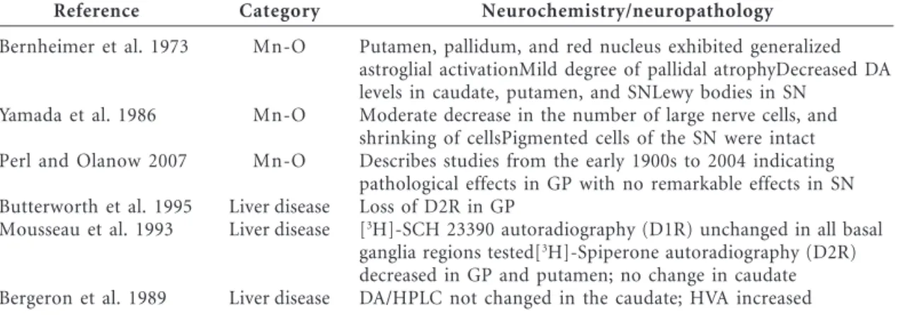
ed with occupational Mn exposures were per-formed in the early 1900s and lacked detailed ex-amination of brain tissue. Neurochemical meth-ods used in the early studies did not have the sensitivity and specificity available today. Never-theless, most studies described marked changes in the globus pallidus of Mn-exposed individu-als, with no remarkable effects on pigmented cells of the substantia nigra, which indicates a lack of an effect on dopamine cell bodies, the only mid-brain neurons that contain melanin (Table 3). Neurochemical studies of the brains of patients with liver disease have confirmed for the most part the results from neuroimaging studies: a sig-nificant decrease in D2R levels in the basal gan-glia and no observed decrease in dopamine levels (Table 1). In one study, Mousseau et al. (1993) used in vitro autoradiography and found de-creased D2R levels in the globus pallidus and putamen with no change in D1-dopamine recep-tors (D1Rs) in the basal ganglia of patients with liver disease (Table 3).
In general, the available neurochemical and neuropathological evidence from subjects with increased Mn concentrations in the brain suggests primary involvement of the globus pallidus, ex-pressing the loss or shrinkage of neurons and glial cell activation. Consistent evidence has shown that D2R levels are decreased, and one study indicated no change in D1R. Most studies have indicated a lack of degeneration of midbrain dopamine neu-rons, which is consistent with the majority of the neuroimaging studies. Parenthetically, in the stud-ies that indicated a loss or a shrinking of neurons in the globus pallidus or striatum of Mn-exposed subjects, no attempt has been made to identify the neuronal phenotype affected.
Behavioral, Neuroimaging,
Neurochemical, and Neuropathological Evidence of Mn Neurotoxicity
in Nonhuman Primates
Behavior. Experimental animals have been used to advance the understanding of Mn neu-rotoxicity. Rodent studies are not described in this review because, unlike nonhuman primates, they lack behavioral similarities to humans and are less sensitive to Mn than are humans and nonhuman primates. Nonhuman primates have improved our understanding of the effects of Mn on motor function, in vivo brain chemistry, and neuropathology (Table 4). Historically, nonhu-man primate studies can be divided into two cat-egories. The studies from the 1960s to the early 1990s used Mn doses that were considerably high-er than those used in studies from 1995 to 2008 (Table 4). Most studies before 1995 used cumu-lative doses > 300 mg Mn/kg body weight. These early studies did not use neuroimaging techniques because of their more recent availability, with the exception of Newland and Weiss (1992) (Table 4), who administered very low doses of Mn and used T1-weighted MRI. Another important dif-ference between the early studies and more re-cent ones has been the use of quantitative, ad-vanced histological, neurochemical, and neuro-pathological techniques to analyze brain tissue. For example, some of the early studies used gross anatomical examination of brain tissue, and the analysis of neurotransmitter levels used colori-metric reactions that were prone to interference and lacked sensitivity compared with current state-of-the-art techniques (Table 4).
Table 3. Neurochemical studies in human postmortem brain tissue.
Reference
Bernheimer et al. 1973
Yamada et al. 1986
Perl and Olanow 2007
Butterworth et al. 1995 Mousseau et al. 1993
Bergeron et al. 1989
Category
Mn-O
Mn-O
Mn-O
Liver disease Liver disease
Liver disease
Neurochemistry/neuropathology
Putamen, pallidum, and red nucleus exhibited generalized astroglial activationMild degree of pallidal atrophyDecreased DA levels in caudate, putamen, and SNLewy bodies in SN
Moderate decrease in the number of large nerve cells, and shrinking of cellsPigmented cells of the SN were intact Describes studies from the early 1900s to 2004 indicating pathological effects in GP with no remarkable effects in SN Loss of D2R in GP
[3
H]-SCH 23390 autoradiography (D1R) unchanged in all basal ganglia regions tested[3
H]-Spiperone autoradiography (D2R) decreased in GP and putamen; no change in caudate DA/HPLC not changed in the caudate; HVA increased
G
Table 4. Behavioral, neuroimaging, and neurochemical studies in Mn-exposed nonhuman primates.
Reference
Pentschew et al. 1963
Neff et al. 1969
Chandra et al. 1979
Bird et al. 1984
Eriksson et al. 1987
Newland et al. 1989
Newland and Weiss 1992
Species
Rhesus (5 monkeys, only 1
described)
Squirrel monkey (control = 4; Mn-group A = 5; Mn-group B
= 6)
Rhesus (4)
Rhesus (4)
Rhesus (4)
Long-tailed macaque (3)
Cebus (3)
Route
im (Mn dioxide)
sc (Mn dioxide)
Oral
Inhalation (Mn oxide)
sc (Mn oxide)
iv and inhalation
(MnCl2)
iv (MnCl2)
ΣMn dose
695 mg Mn/kg (calculated)
379 mg Mn/kg in highest dose
group (calculated)
23,580 mg Mn/ kg (MnCl2)
(calculated)
30 mg Mn/m3
air (exposure rate)
1,543 mg Mn/kg (calculated)
30–50 mg Mn/kg (injection)
40–60 mg Mn/kg
Behavior
Excitability Loss of postural
stability Falling upon
jumping Clumsiness No cogwheel,
tremor, or involuntary movements Muscular rigidity
Impaired equilibrium Tremors on intension Impaired equilibrium
ND
No behavioral or neurological abnormalities
Hyperactive tending to fall then hypoactive
Unsteady gait Action tremor Loss of power in
limbs Clumsy hands
and feet movement
ND
Action tremor
Neuroimaging
ND
ND
ND
ND
ND
T1-MRI hyperintensity
basal ganglia
T1-MRI hyperintensity
basal ganglia
Neuropathology/ neurochemistry
Neuronal loss and gliosis in subthalamic nucleus and medial segment of
GPGliosis in SN (?)Atrophic nerve
cells in lateral pallidum
Decreased DA and NE in caudate
Decreased DA in striatum and
midbrain Decreased DA in
caudate and GP but not in putamen or SN Severe neuronal loss and gliosis in
GP, rest of brain appears normal Decreased DA and
DOPAC in putamen but not
changed in the caudate HVA levels normal
in most animals Decreased dopa decarboxylase
activity in putamen of
two-thirds of the animals ChAT activity reduced in GP of
all animals GAD activity
unaffected Glutathione levels
decreased in the striatum, GP, and SN of one third of
the animals ND
ND
d
e C
o
le
tiv
a, 16(
11)
:4549-4566,
2011
Table 4. continuation
Reference
Eriksson et al. 1992a
Eriksson et al. 1992b
Shinotoh et al. 1995
Olanow et al. 1996
Dorman et al. 2006
Chen et al. 2006 Guilarte et
al. 2006a
Guilarte et al. 2006b
Species
Long-tailed macaque (3)
Long-tailed macaque (2)
Rhesus (3)
Rhesus (3) (same animals as Shinotoh et
al. 1995)
Rhesus (20)
Baboon (2)
Cynomolgus (3–5)
Cynomolgus (4)
Route
sc (Mn oxide)
sc (Mn oxide)
iv (MnCl2)
iv (MnCl2)
Inhalation
sc and iv
iv
iv
ΣMn dose
333–444 mg Mn/kg (calculated)
680 mg Mn/kg (calculated)
71–87 mg Mn/kg
71–87 mg Mn/kg
Various levels of exposure
10–100 mg Mn/kg (acute) 165.5 ± 4.7 mg Mn/kg (range
= 152–174)
165.5 ± 4.7 mg Mn/kg (range
= 152–174)
Behavior
Decreased activity in 1/2 of the Mn-exposed
animals
Unsteady gait Hypoactivity Clumsiness of hands and feet
Two animals hypoactive, one
normal Not responsive
to l-dopa
Two animals hypoactive, one
normal Not responsive
to l-dopa
ND
ND
Subtle deficits in fine motor
control Small decrease
in activity
See Guilarte et al. 2006a
Neuroimaging
ND
DAT/PET (60% decrease) D2R/PET (40%
decrease then normalized) l-Dopa/PET (normal)
T1-MRI
hyperintensity in basal ganglia
D2R/PET l-Dopa/PET
(normal) FDG/PET (normal)
T1-MRI hyperintensity basal
ganglia See Shinotoh et al.
(1995)
T1-MRI
hyperintensity in basal ganglia
DAT/PET (transient increase)
DAR/PET (significantly
decreased) DAT/PET (normal) D2R/PET (normal)
T1-MRI
hyperintensity in basal ganglia and
other brain regionsMRS changes in regions
outside basal ganglia
Neuropathology/ neurochemistry
DAT/autorad (decreased in caudate
and putamen, no change in GP)
D1R/autorad (decreased in caudate
and putamen, no change in GP) D2R/autorad (no change in any region)
mAChR/autorad (no change in any region) GABAaR/autorad (no change in any region)
ND
Minimal cell loss and prominent gliosis in GP and lesser degree in
SNpr (normal) SNpc relatively spared
Minimal cell loss and prominent gliosis in GP and to lesser degree
in SNprS Npc appeared normal Striatal DA and HVA levels are normal in two affected animals
ND
ND
DAT/autorad (normal) D2R/autorad (normal)
TH/immuno histochemistry
(normal) DA-HVA/HPLC
(normal) See Guilarte et al.
(2006a)
G
Table 4. continuation
Reference
Struve et al. 2007 Guilarte et
al. 2008
Species
Rhesus (20)
Cynomolgus (13; includes four animals
from Guilarte et
al. 2006a)
Route
Inhalation
iv (Mn sulfate)
ΣMn dose
Various levels of exposure 68–250 mg
Mn/kg
Behavior
ND
See Schneider et al. 2006, 2009
Neuroimaging
ND
DAR/PET (significantly
decreased) DAT/PET (normal)
D2R/PET (small decrease)
Neuropathology/ neurochemistry
DA-DOPAC-HVA/ HPLC (normal) DAT/autorad (normal) D2R/autorad (normal) D1R/autorad (normal) CB1/autorad (normal)
TH/immuno histochemistry
(normal) DA-HVA/HPLC
(normal)
Abbreviations: autorad, autoradiography; CB1, cannabinoid receptor 1; ChAT, choline acetyltransferase; DA, dopamine; DAR, dopamine release; DOPAC, 3,4-dihydroxyphenylacetic acid; D2R, dopamine receptor; FDG, fluorodeoxyglucose; GABAaR, γ-aminobutyric acid A receptor; GAD, glutamic acid decarboxylase; GP, globus pallidus; im, intramuscular; iv, intravenous; mAChR, muscarinic acetylcholine receptor; MRS, magnetic resonance spectroscopy; ND, not determined; NE, norepinephrine; sc, subcutaneous; SN, substantia nigra; SNpr, substantia nigra pars reticulata; TH, tyrosine hydroxylase. Arrows indicate increased T1-weighted MRI signal in the globus pallidus and other basal ganglia structures.
From a behavioral perspective, early nonhu-man primate studies examined the effects of Mn on movement and coordination. It is clear from these studies that nonhuman primates exhibited movement abnormalities similar to those in Mn-exposed humans (compare Tables 1 and 4), in-cluding loss of postural stability, excitability, hy-poactivity, falling, muscular rigidity, tremors of intension or action tremor, unsteady gait, loss of power in limbs, and clumsy foot movement (Ta-ble 4). From 1995, studies used cumulative Mn concentrations < 300 mg Mn/kg body weight and found more subtle movement abnormalities, in-cluding hypoactivity, deficits in fine motor con-trol, action tremor, and, more recently, deficits in working memory (Table 4). Consistent with the human literature, the Mn-induced motor abnor-malities in nonhuman primates were not respon-sive to l-dopa therapy (Shinotoh et al. 1995).
Neurochemistry. The early nonhuman
pri-mate studies of Mn exposure described decreased levels of dopamine in the caudate (Neff et al. 1969), striatum and midbrain (Chandra et al. 1979), caudate and globus pallidus but not in the putamen or substantia nigra (Bird et al. 1984), and putamen but no change in the caudate (Eriks-son et al. 1987). Some relevant comments are important to put these results in perspective. First, in the study by Neff et al. (1969), the monkeys in group A were injected with single doses of 200 mg Mn oxide on two different occasions. The report indicated that “2 weeks following the first injection, 1 control and 4 MnO2 treated
mon-keys died.” This was a highly unusual event and makes one question the health status of these animals prior and during treatment. They report-ed a significant decrease of dopamine in the cau-date of group A monkeys, and a less severe dopamine deficit but still a significant one in an-other group of Mn-exposed animals that received a third injection (group B).
d
e C
o
le
tiv
a, 16(
11)
:4549-4566,
2011
Mn-exposed animals that received a higher cu-mulative Mn dose exhibited less dopamine loss than did animals with a lower dose whose tissues were stored for a longer time.
Eriksson et al. (1987) measured reduced con-centrations of dopamine in the putamen and glo-bus pallidus but not in the caudate, although one of the three animals analyzed did express a de-crease in dopamine content in the caudate. An observation in the Eriksson et al. (1987) study that has relevance to a mechanism of action of Mn is that in the brains of these monkeys, in which they measured significant decrements in tissue dopamine concentration, they found no significant effect on glutathione concentrations in two of the three monkeys examined. In gener-al, these early studies demonstrate differences in basal ganglia regions where dopamine concen-trations appear to be decreased and the regional pattern does not follow the well-characterized loss of dopamine in idiopathic PD (Dauer and Przedborski 2003; Felicio et al. 2009).
The most consistent observation of these early studies was that Mn produced morphological changes in the globus pallidus, subthalamic nu-clei, and substantia nigra pars reticulata, wheas the SNpc remained intact (Table 4). More re-cent studies (after 1995) that have used cumula-tive Mn doses < 300 mg/kg and state-of-the-art high-performance liquid chromatography (HPLC) with electrochemical detection analytical methods have provided no evidence of changes in dopamine concentrations in the caudate and puta-men (Guilarte et al. 2006a, 2008; Olanow et al. 1996; Struve et al. 2007) (Table 4).
In vitro autoradiography. In vitro quantita-tive receptor autoradiography has been used to examine dopamine neuron markers in the brain of Mn-exposed nonhuman primates (Table 4). The groups headed by Eriksson in Sweden (1992a) and by Guilarte in the United States (2006a, 2008), have used this approach exten-sively because not only is it quantitative, it also offers exquisite anatomical information with high resolution. Eriksson et al. (1992a) performed re-ceptor autoradiography studies in the basal gan-glia of monkeys exposed to 0.1 g Mn/month for 26 months. They indicated that this dosing regi-men was comparable with what workers might inhale in dusty environments. They found that the binding of [3H]-mazindol to DAT (the pre-sumed target, but see below) was reduced by 75% in the caudate and putamen of Mn-exposed an-imals. However, there were technical problems that need to be discussed: a) The authors stated
that the level of nonspecific [3H]-mazindol bind-ing in the caudate and putamen was 50% and 60%, respectively. This level of nonspecific bind-ing was very high, so the specific bindbind-ing signal-to-noise (nonspecific binding) ratio was very low. Typically in this type of assay, one wants a low level of nonspecific binding, in the range of 10– 15% of total binding. b) [3H]-Mazindol is known to bind to other monoaminergic uptake sites be-sides DAT (May et al. 1994). To ensure that one is selectively measuring DAT, an antagonist for the other monoaminergic uptake sites should be in-cluded in the assay. This procedure was not per-formed by Eriksson et al. (1992a); thus, the effect of Mn on the [3H]-mazindol autoradiography results were a combination of DAT and other monoaminergic uptake sites. c) As pointed out, even if [3H]-mazindol was made selective for DAT by including antagonists for other monoamin-ergic transporters, a reduction in [3H]-mazin-dol binding to DAT would not necessarily reflect dopamine terminal loss, but it could represent a competitive inhibition of Mn with radioligand binding to DAT as shown by Chen et al. (2006). Parenthetically, Eriksson et al. (1992a) also found a significant decrease in [3H]-SCH 23390 bind-ing to D1R in the caudate and putamen with no change in [3H]-spiperone binding to D2R in the same regions. These results were opposite to what Eriksson et al. (1992b) and Guilarte et al. (2008) reported with in vivo PET imaging and in vitro autoradiography—that is, decreased D2R and no effect on D1R.
G
basal ganglia. This latter study not only included a naive control group in which the animals did not receive Mn or neuroimaging studies, but it also included an imaged control group in which the neuroimaging studies were done without Mn exposure. The inclusion of an imaged control group was essential because one of the longitudi-nal PET studies included the administration of amphetamine (Guilarte et al. 2006a, 2008), a psy-chostimulant that depletes dopamine levels and down-regulates DAT. This control group provid-ed valuable information for assessing the effect of amphetamine alone. Although several of the dopamine neuron markers that were analyzed postmortem, such as dopamine and DOPAC (3,4-dihydroxyphenylacetic acid) in the putamen, DAT and VMAT2 in the caudate and putamen and D2R in the putamen were significantly lower in Mn-exposed animals than in naive controls; they were not different from all controls when the imaged control group was included. The values for these parameters in the imaged control animals were actually lower than those for the Mn-exposed animals. The fact that studies of Mn-exposed an-imals that received amphetamine with PET had higher levels of these dopamine markers than did imaged controls that received the same amphet-amine with PET but no Mn suggests that Mn has an “antagonistic effect” on the action of amphet-amine on these dopamphet-aminergic markers. Because this action on dopaminergic markers is mediated by DAT, these results provide evidence that Mn interacts directly with DAT and are consistent with other studies that indicate an interaction of Mn with DAT (Anderson et al. 2007; Chen et al. 2006; Ingersoll et al. 1999).
Neuroimaging studies: MRI and PET.
Simi-lar to the studies involving humans exposed to Mn, several investigators have performed neu-roimaging studies of nonhuman primates to ex-amine the distribution of Mn in the brain by T1-weighted MRI and brain chemistry changes us-ing PET. The T1-weighted MRI studies (New-land et al. 1989; New(New-land and Weiss 1992) (Table 4) showed that Mn first accumulates in the glo-bus pallidus and produces a hyperintensive sig-nal in T1-weighted MRI even at relatively low doses of Mn. Thus, it is a sensitive method for detecting small increases in brain Mn concentra-tions. These findings have been confirmed by other investigators, and subsequent studies have shown that although the basal ganglia is one of the first to accumulate Mn, other brain struc-tures also accumulate the metal, albeit to a lesser extent (Dorman et al. 2006; Ingersoll et al. 2006b).
PET imaging in nonhuman primates exposed to Mn was first performed by Eriksson (1992b) (Table 4). They studied two monkeys exposed to Mn for 16 months that received [11C]-nomifensine PET to measure DAT (although this ligand also recognizes other monoaminergic transporters), [11C]-raclopride PET for D2R, and [11C]-l-dopa for l-dopa decarboxylase ac-tivity. They found a progressive decrease in [11C]-nomifensine binding as a function of cumulative dose in both animals. One of the animals had a transient decrease in [11C]-raclopride binding to D2R that returned to baseline by the end of the exposure period, and no effect of Mn treatment on [11C]-l-dopa PET was observed. These stud-ies indicate that dopamine neurons are not de-generating, but there is a potential loss of dopam-ine terminals based on the [11C]-nomifensdopam-ine PET. However, a firm conclusion that chronic Mn results in the loss of DAT and presumably dopam-ine neuron terminals in the striatum cannot be made because [11C]-nomifensine is not a selec-tive DAT ligand and because Mn can directly in-hibit ligand binding to DAT.
Shinotoh et al. (1995) performed PET studies in three monkeys (not all monkeys received the three different types of PET studies) that were chronically treated with Mn. These investigators found no significant effect of Mn on fluo-ro-l-dopa PET, [11C]-raclopride PET, or [18F]-deoxyglucose PET despite the fact that the ani-mals exhibited hypoactivity (Table 4). Thus, their work is also consistent with the lack of degenera-tion of nigrostriatal dopaminergic neurons.
Neuroimaging studies: new PET findings.
d
e C
o
le
tiv
a, 16(
11)
:4549-4566,
2011
the ability of dopamine neurons to synthesize dopamine, and it may indirectly measure the dopamine-releasing capacity of dopamine neu-rons. Guilarte et al. (2006a, 2008) used a more direct method to assess in vivo dopamine release using a bolus plus continuous infusion of [11C]-raclopride (D2R ligand) with amphetamine chal-lenge (Zhou et al. 2006). This type of PET study is based on the ability of endogenous dopamine (re-leased by the administration of amphetamine) to compete with the binding of [11C]-raclopride to D2R (Laruelle 2000). An animal with a high ca-pacity to release dopamine will produce a large decrease in the amount of [11C]-raclopride up-take in the striatum, whereas an animal with a low capacity will produce a smaller change or no change. These studies showed that the most sig-nificant effect of chronic Mn at all of the cumula-tive doses of Mn administered was a marked in-hibition (> 50% on average) of in vivo dopamine release in the striatum measured by PET (Gui-larte et al. 2006a, 2008) (Table 4). The effect of Mn on in vivo dopamine release was observed in the absence of a change in DAT levels and with a small but significant decrease on D2R levels in the stria-tum (Guilarte et al. 2008). Therefore, consistent with human and other nonhuman primate stud-ies, this work also showed a lack of dopamine neuron degeneration. However, these studies pro-vided new information: although chronic Mn ex-posure did not result in dopamine neuron degen-eration, dopamine neurons were dysfunctional because they had a reduced capacity to release dopamine. Neurochemical studies in the brain of the same animals that received PET studies showed that the marked effect of Mn on in vivo dopamine release measured by PET was not due to a reduction in tissue dopamine levels, because dopamine concentrations were not significantly different from all controls (Guilarte et al. 2006a, 2008). It appears that the deficit of in vivo dopam-ine release is associated with the ability of Mn to disrupt presynaptic release mechanisms.
Conclusions
The available evidence from human and nonhu-man primate studies using behavioral, neuroim-aging, neurochemical, and neuropathological end points provides strong support to the
hypothe-sis that, although excess levels of Mn accumula-tion in the brain results in an atypical form of parkinsonism, this clinical outcome is not asso-ciated with the degeneration of nigrostriatal dopaminergic neurons as is the case in PD. In fact, the new evidence suggests that it may be the dysfunction of this system or the inability to re-lease dopamine that produces the movement abnormalities documented in Mn-exposed sub-jects. This new finding is consistent with the fact that l-dopa therapy does not provide a benefit to the Mn-induced movement abnormalities as it does in PD (Lu et al. 1994) (Table 1). This is be-cause Mn-induced motor dysfunction does not seem to be a problem of decreased synthesis or concentration of dopamine in presynaptic ter-minals, but rather a problem of the ability to release the available dopamine. These findings provide a starting point and a new avenue of re-search to delineate a putative mechanism of Mn-induced movement abnormalities.
G
References
Aggarwal A, Vaidya S, Shah S, Singh J, Desai S, Bhatt M. 2006. Reversible parkinsonism and T1W pallidal hyperintensities in acute liver failure. Mov Disord 21:1986–1990.
Anderson JG, Cooney PT, Erikson KM. 2007. Inhibi-tion of DAT funcInhibi-tion attenuates manganese accu-mulation in the globus pallidus. Environ Toxicol Pharmacol 23:179–184.
Antonini A, Schwarz J, Oertel WH, Beer HF, Madeja UD, Leenders KL. 1994. [11C]-raclopride and positron emission tomography in previously un-treated patients with Parkinson’s disease: influence of L-dopa and lisuride therapy on striatal dopam-ine D2 receptors. Neurology 44:1325–1329. Bergeron M, Reader TA, Layrargues GP, Butterworth
RF. 1989. Monoamines and metabolites in autop-sied brain tissue from cirrhotic patients with hepat-ic encephalopathy. Neurochem Res 14:853–859. Bernheimer H, Birkmayer W, Hornykiewicz O, Jellinger
K, Seitelberger F. 1973. Brain dopamine and the syndromes of Parkinson and Huntington. Clinical, morphological and neurochemical correlations. J Neurol Sci. 20:415–455.
Bird ED, Anton AH, Bullock B. 1984. The effect of man-ganese inhalation on basal ganglia dopamine con-centrations in rhesus monkey. Neurotoxicology 5:59– 65.
Bowler RM, Koller W, Schulz PE. 2006. Parkinsonism due to manganism in a welder: neurological and neuropsychological sequelae. Neurotoxicology 27:327–332.
Brooks DJ, Ibanez V, Sawle GV, Playford ED, Quinn N, Mathias CJ, et al. 1992. Striatal D2 receptor status in patients with Parkinson’s disease, striatonigral de-generation, and progressive supranuclear palsy measured with 11C-raclopride and positron emis-sion tomography. Ann Neurol 31:184–192. Burkhard PR, Delavelle J, Du Pasquier R, Spahr L. 2003.
Chronic parkinsonism associated with cirrhosis: a distinct subset of acquired hepatocerebral degen-eration. Arch Neurol 60:521–528.
Butterworth RF, Spahr L, Fontaine S, Layrargues GP. 1995. Manganese toxicity, dopaminerigc dysfunc-tion and hepatic encephalopathy. Metab Brain Dis 10:259–267.
Chandra SV, Srivastava RS, Shukla GS. 1979. Regional distribution of metals and biogenic amines in the brain of monkeys exposed to manganese. Toxicol Lett 4:189–192.
Chen MK, Kuwabara H, Zhou Y, Adams RJ, Brasic JR, McGlothan JL, et al. 2008. VMAT2 and dopamine neuron loss in a primate model of Parkinson’s dis-ease. J Neurochem 105:78–90.
Chen MK, Lee JS, McGlothan JL, Furukawa E, Adams RJ, Alexander M, et al. 2006. Acute manganese ad-ministration alters dopamine transporter levels in the nonhuman primate striatum. Neurotoxicology 27:229–236.
Colosimo C, Guidi M. 2009. Parkinsonism due to ephe-drone neurotoxicity: a case report. Eur J Neurol 16:e114–e115.
Cook DG, Fahn S, Brait KA. 1974. Chronic manganese intoxication. Arch Neurol 30:59–64.
Couper J. 1837. On the effects of black oxide of manga-nese when inhaled in the lungs. Br Ann Med Phar-macol 1:41–42.
Dauer W, Przedborski S. 2003. Parkinson’s disease: mech-anisms and models. Neuron 39:889–909.
de Bie RM, Gladstone RM, Strafella AP, Ko JH, Lang AE. 2007. Manganese-induced Parkinsonism asso-ciated with methcathinone (Ephedrone) abuse. Arch Neurol 64:886–889.
Dorman DC, Struve MF, Wong BA, Dye JA, Robertson ID. 2006. Correlation of brain magnetic resonance imaging changes with pallidal manganese concen-trations in rhesus monkeys following subchronic manganese inhalation. Toxicol Sci 92:219–227. Ehringer H, Hornykiewicz O. 1960. Verteilung von
No-radrenalin und Dopamin (3-hydroxytyramin) im Gehirn des Menschen und ihr Verhalten bei Erkrankungen des extrapyramidalem Systems [in German]. Klin Wochenschr 38:1236–1239. Eriksson H, Gillberg PG, Aquilonius SM, Hedstrom KG,
Heilbronn E. 1992a. Receptor alterations in manga-nese intoxicated monkeys. Arch Toxicol 66:359–364. Eriksson H, Mägiste K, Plantin LO, Fonnum F, Hedström KG, Theodorsson-Norheim E, et al. 1987. Effects of manganese oxide on monkeys as revealed by a com-bined neurochemical, histological and neurophysi-ological evaluation. Arch Toxicol 61:46–52. Eriksson H, Tedfoff J, Thuomas KA, Aquilonius SM,
Hartvig P, Fasth KJ, et al. 1992b. Manganese in-duced brain lesions in Macaca fascicularis as re-vealed by positron emission tomography and mag-netic resonance imaging. Arch Toxicol 66:403–407. Faviani G, Rogacheski E, Wiederkehr JC, Khouri J, Cian-farano A. 2007. Liver transplantation in a patient with rapid onset parkinsonism-dementia complex induced by manganism secondary to liver failure. Arq Neuropsiquiatr 65:685–688.
Felicio AC, Shih MC, Godeiro-Junior C, Andrade LA, Bressan RA, Ferraz HB. 2009. Molecular imaging studies in Parkinson disease: reducing diagnostic uncertainly. Neurologist 15:6–16.
Gelb DJ, Oliver E, Gilamn S. 1999. Diagnosis criteria for Parkinson disease. Arch Neurol 56:33–39. Guilarte TR, Burton NC, McGlothan JL, Verina T, Zhou
Y, Alexander M, et al. 2008. Impairment of nigros-triatal dopamine neurotransmission by manganese is mediated by pre-synaptic mechanism(s): impli-cations to manganese-induced parkinsonism. J Neu-rochem 107:1236–1247.
Guilarte TR, Chen MK, McGlothan JL, Verina T, Wong DF, Zhou Y, et al. 2006a. Nigrostriatal dopamine system dysfunction and subtle motor deficits in manganese-exposed nonhuman primates. Exp Neu-rol 202:381–390.
Guilarte TR, McGlothan JL, Degaonkar M, Chen MK, Barker PB, Syversen T, et al. 2006b. Evidence for cortical dysfunction and widespread manganese accumulation in the nonhuman primate brain fol-lowing chronic manganese exposure: a 1H-MRS and MRI study. Toxicol Sci 94:351–358.
d
e C
o
le
tiv
a, 16(
11)
:4549-4566,
2011
Huang CC. 2007. Parkinsonism induced by chronic manganese intoxication—an experience in Taiwan. Chang Gung Med J 30:385–395.
Hughes AJ, Daniel SE, Ben Shlomo Y, Lees AJ. 2002. The accuracy of diagnosis of parkinsonian syn-dromes in a specialist movement disorder service. Brain 125:861–870.
Hughes AJ, Daniel SE, Lees AJ. 1993. The clinical fea-tures of Parkinson’s disease in 100 histological prov-en cases. Adv Neurol 60:595–599.
Ingersoll RT, Montgomery EB Jr, Aposhian HV. 1999. Central nervous system toxicity of manganese. II: cocaine or reserpine inhibit manganese concentra-tion in the rat brain. Neurotoxicology 20:467–476. Jankovic J. 2008. Parkinson’s disease: clinical features
and diagnosis. J Neurol Neurosurg Psychiatry 79:368–376.
Josephs KA, Ahlskog JE, Klos KJ, Kumar N, Fealey RD, Trenerry MR, et al. 2005. Neurologic manifesta-tions in welders with pallidal MRI T1 hyperintensi-ty. Neurology 64:2033–2039.
Kenangil G, Ertan S, Sayilir I, Ozekmekçi S. 2006. Pro-gressive motor syndrome in a welder with pallidal T1 hyperintensity on MRI: a two year follow-up. Mov Disord 21:2197–2200.
Kessler KR, Wunderlich G, Hefter H, Seitz RJ. 2003. Secondary progressive chronic manganism associ-ated with markedly decreased striatal D2 receptor density. Mov Disord 18:217–218.
Kim J, Kim J-M, Kim YK, Shin JW, Choi SH, Kim SE, et al. 2007. Dopamine transporter SPECT of a liver cirrhotic with atypical parkinsonism. Ind Health 45:497–500.
Kim Y, Kim JM, Kim JW, Yoo CI, Lee CR, Lee JH, et al. 2002. Dopamine transporter density is decreased in parkinsonian patients with a history of manganese exposure: what does it mean? Mov Disord 17:568– 575.
Klos KJ, Ahlskog JE, Josephs KA, Fealey RD, Cowl CT, Kumar N. 2005. Neurologic spectrum of chronic liver failure and basal ganglia T1 hyperintensity on magnetic resonance imaging: probable manganese neurotoxicity. Arch Neurol 62:1385–1390.
Klos KJ, Ahlskog JE, Kumar N, Cambern S, Butz J, Burr-itt M, et al. 2006. Brain metal concentrations in chronic liver failure patients with pallidal T1 MRI hyperintensity. Neurology 67:1984–1989.
Laruelle M. 2000. Imaging synaptic neurotransmission with in vivo binding competition techniques: a crit-ical review. J Cereb Blood Flow Metab 20:423–451. Lee JW. 2000. Manganese intoxication. Arch Neurol
57:597–599.
Lees AJ, Hardy J, Revesz T. 2009. Parkinson’s disease. Lancet 373:2055–2066.
Leenders KL. 2003. Significance of nonpresynaptic SPECT tracer methods in Parkinson’s disease. Mov Disord 18(suppl 7):S39–S42.
Louis ED, Klatka LA, Liu Y, Fahn S. 1997. Comparison of extrapyramidal features in 31 pathologically con-firmed cases of diffuse Lewy body disease and 34 pathologically confirmed cases of Parkinson’s dis-ease. Neurology 48:376–380.
Lu CS, Huang CC, Chu NS, Calne DB. 1994. Levodopa failure in chronic manganism. Neurology 44:1600– 1602.
May CH, Guilarte TR, Wagner HN Jr, Vogel S. 1994. Intrastriatal infusion of lisuride—a potential treat-ment for Parkinson’s disease? Behavioral and auto-radiographic studies in 6-OHDA lesioned rats. Neu-rodegeneration 3:305–313.
Mena I, Marin O, Fuenzalida S, Cotzias GC. 1967. Chron-ic manganese poisoning. ClinChron-ical pChron-icture and man-ganese turnover. Neurology 17:128–136.
Meral H, Kutukcu Y, Atmaca B, Ozer F, Hamamcioglu K. 2007. Parkinsonism caused by chronic usage of intravenous potassium permanganate. Neurologist 13:92–94.
Mousseau DD, Perney P, Layrargues P, Butterworth RF. 1993. Selective loss of pallidal dopamine D2 recep-tor density in hepatic encephalopathy. Neurosci Lett 162:192–196.
Nagatomo S, Umehara F, Hanada K, Nobuhara Y, Tak-enaga S, Arimura K, et al. 1999. Manganese intoxi-cation during total parenteral nutrition: report of two cases and review of the literature. J Neurol Sci 162:102–105.
Neff NH, Barrett RE, Costa E. 1969. Selective depletion of caudate nucleus dopamine and serotonin during chronic manganese dioxide administration to squir-rel monkeys. Experientia 25:1140–1141.
Newland MC, Ceckler TL, Kordower JH, Weiss B. 1989. Visualizing manganese in the primate basal ganglia with magnetic resonance imaging. Exp Neurol 106:251–258.
Newland MC, Weiss B. 1992. Persistent effects of man-ganese on effortful responding and their relation-ship to manganese accumulation in the primate globus pallidus. Toxicol Appl Pharmacol 113:87–97. Olanow CW, Good PF, Shinotoh H, Hewitt KA, Vinger-hoets F, Snow BJ, et al. 1996. Manganese intoxica-tion in the rhesus monkey: a clinical, imaging, patho-logic, and biochemical study. Neurology 46:492– 498.
Parkinson J. 1817. An Essay on the Shaking Palsy. Avail-able: http://www.gutenberg.org/files/23777/23777-h/ 23777-h.htm [accessed 3 June 2010].
Pentschew A, Ebner FF, Kovatch RM. 1963. Experimen-tal manganese encephalopathy in monkeys. A pre-liminary report. J Neuropathol Exp Neurol 22:488– 499.
Perl DP, Olanow CW. 2007. The neuropathology of manganese-induced Parkinsonism. J Neuropathol Exp Neurol 66:675–682.
Racette BA, Antenor JA, McGee-Minnich L, Moerlein SM, Videen TO, Kotagal V, et al. 2005. [18F]FDOPA PET and clinical features in parkinsonism due to manganism. Mov Disord 20:492–496.
Racette BA, McGee-Minnich L, Moerlein SM, Mink JW, Videen TO, Perlmutter JS. 2001. Welding relat-ed parkinsonism: clinical features, treatment, and pathophysiology. Neurology 56:8–13.
G
Ravina B, Siderowf A, Farrar J, Hurtig H. 2001. Weld-ing-related parkinsonism: clinical features, treat-ment, and pathophysiology. Neurology 57:936–937. Rinne UK, Laithinen A, Rinne JO, Nágren K, Bergman J, Ruotsalainen U. 1990. Positron emission tomog-raphy demonstrates dopamine D2 receptor super-sensitivity in the striatum of patients with early Par-kinson’s disease. Mov Disord 5:55–59.
Sadak AH, Schulz PE. 2001. Welding-related parkin-sonism: clinical features, treatment, and pathophys-iology. Neurology 57:1738–1739.
Sadek AH, Rauch R, Schulz PE. 2003. Parkinsonism due to manganism in a welder. Int J Toxicol 22:393–401. Sanotsky Y, Lesyk R, Fedoryshyn L, Komnatska I, Matviy-enko Y, Fahn S. 2007. Manganic encephalopathy due to “ephedrone” abuse. Mov Disord 22:1337– 1343.
Savitt JM, Dawson VL, Dawson TM. 2006. Diagnosis and treatment of Parkinson disease: molecules to medicine. J Clin Invest 116:1744–1754.
Schaumberg HH, Herskovitz S, Cassano VA. 2006. Oc-cupational manganese neurotoxicity provoked by hepatitis C. Neurology 67:322–323.
Schneider JS, Decamp E, Clark K, Bouquio C, Syversen T, Guilarte TR. 2009. Effects of chronic manganese exposure on working memory in nonhuman pri-mates. Brain Res 1258:86–95.
Schneider JS, Decamp E, Koser AJ, Fritz S, Gonczi H, Syversen T, et al. 2006. Effects of chronic manga-nese exposure on cognitive and motor functioning in nonhuman primates. Brain Res 1118:222–231. Selikhova M, Fedoryshyn L, Matviyenko Y, Komnatska
I, Kyrylchuk M, Krolicki L, et al. 2008. Parkinsonism and dystonia caused by illicit use of ephedrone—a longitudinal study. Mov Disord 23:2224–2231. Shinotoh H, Snow BF, Hewitt KA, Pate BD, Doudet D,
Nugent R, et al. 1995. MRI and PET studies of man-ganese-intoxicated monkeys. Neurology 45:1199– 1204.
Sikk K, Taba P, Haldre S, Bergquist J, Nyholm D, Zjablov G, et al. 2007. Irreversible motor impairment in young addicts—ephedrone, manganism or both? Acta Neurol Scand 115:385–389.
Stepens A, Logina I, Liguts V, Aldins P, Eksteina I, Plat-käjis A, et al. 2008. A Parkinsonian syndrome in methcathinone users and the role of manganese. N Engl J Med 358:1009–1017.
Struve MF, McManus BE, Wong BA, Dorman DC. 2007. Basal ganglia neurotransmitter concentrations in rhesus monkeys following subchronic manganese sulfate inhalation. Am J Ind Med 50:772–778. Tolosa E, Wenning G, Poewe W. 2006. The diagnosis of
Parkinson’s disease. Lancet Neurol 5:75–86. Varlibas F, Delipoyraz I, Yuksel G, Filiz G, Tireli H,
Gecim NO. 2009. Neurotoxicity following chronic intravenous use of “Russian cocktail.” Clin Toxicol (Phila) 47:157–160.
Vingerhoets FJ, Schulzer M, Calne DB, Snow BJ. 1997. Which clinical sign of Parkinson’s disease best re-flects the nigrostriatal lesion? Ann Neurol 41:58–64. Wakabayashi K, Tanji K, Mori F, Takahashi H. 2007. The Lewy body in Parkinson’s disease: molecules implicated in the formation and degradation of al-pha-synuclein aggregates. Neuropathology 27:494– 506.
Yamada M, Ohno S, Okayasu I, Okeda R, Hatakeyama S, Watanabe H, et al. 1986. Chronic manganese poisoning: a neuropathological study with deter-mination of manganese distribution in the brain. Acta Neuropathol 70:273–278.
Zhou Y, Chen M-K, Endres CJ, Ye W, Brasic JR, Alex-ander M, et al. 2006. An extended simplified refer-ence tissue model for the quantification of dynam-ic PET with amphetamine challenge. Neuroimage 33:550–563.