Original Article
Artigo Original
Paulo Fernando Aragon de Macedo1 Esther Mandelbaum Gonçalves Bianchini2
Descritores
Diagnóstico Adulto Deglutição Mastigação
Keywords
Diagnosis Adult Deglutition Mastication
Correspondence address:
Paulo Fernando Aragon de Macedo Rua Jornalista Irineu Marinho, 520/102, Icaraí, Niterói (RJ), Brasil,
CEP: 24230-126.
E-mail: [email protected]
Received: 01/28/2014
Accepted: 09/03/2014
Study carried out at the Professional Masters Program in Speech, Language and Hearing Sciences, Universidade Veiga de Almeida – UVA – Rio de Janeiro (RJ), Brazil.
(1) Universidade Veiga de Almeida – UVA – Rio de Janeiro (RJ); Universidade Federal Fluminense (UFF) – Niterói (RJ), Brazil.
(2) Graduate Program, Universidade Veiga de Almeida – UVA – Rio de Janeiro (RJ), Brazil.
Conlict of interests: nothing to declare.
Myofunctional orofacial examination: comparative
analysis in young adults with and without complaints
Análise comparativa das informações de exame clínico
miofuncional orofacial em adultos jovens com e sem queixas
ABSTRACT
Purpose: To verify myofunctional orofacial characteristics in young adults and to compare data on individuals with and without myofunctional complaints, aiming to identify the main myofunctional problems and differentiating them from characteristics that are common for this population, as well as to list items for myofunctional evaluation in this population. Methods: Cross-sectional study with 85 adult participants, aged between 19 and 39 years, selected through consecutive sampling at the Department of Speech, Language and Hearing Sciences of Universidade Veiga de Almeida. The participants were divided into two groups: G1 (comprising 50 individuals referred for orofacial myofunctional disorders) and G2 (comprising 35 volunteers without complaints). Descriptive evaluation of craniofacial structures of hard and soft tissues, kinesiology and mandible range of motion and functional patterns of breathing, chewing, and swallowing was applied. Three expert Speech-Language pathologists assessed all participants. Statistical analysis was done using χ2-test, Student’s t-test, or Mann–Whitney test. The reliability level was 99%. Results: A predominance of
Angle Class I pattern of occlusions for G2 (p<0.0001) was found. G1 showed (p<0.0001) mandible movements with deviations and joint noises, amplitude reduction in lateral and protrusive movements, unilateral chewing, nonexpected muscle contraction, temporomandibular joint noises, swallowing with excessive contraction of the orbicularis oris muscle, loud noise, and residues (p=0.006). Conclusion: The main myofunctional orofacial alterations in young adults with complaints refer to changes of the mandibular movements and patterns of chewing or of swallowing, relecting the main items of the clinical evaluation. Many items of assessment and characterization do not differ between the groups, and these should be analyzed regarding their relevance.
RESUMO
Objetivo: Veriicar as características miofuncionais orofaciais em adultos jovens e analisar comparativamente dados de indivíduos com e sem queixas miofuncionais, visando apontar os principais problemas miofuncionais e diferenciando-os de características típicas dessa população, além de elencar os itens de avaliação miofuncional que possam merecer maior atenção para essa população. Métodos: Estudo transversal com 85 participantes adultos, 19 a 39 anos, selecionados por amostra consecutiva no Serviço de Fonoaudiologia da Universidade Veiga de Almeida, divididos em dois grupos — G1: 50 indivíduos encaminhados por queixas miofuncionais orofaciais; G2: 35 indivíduos voluntários sem queixas. Todos foram avaliados por três fonoaudiólogos, especialistas. O exame constou de avaliação clínica descritiva quanto às estruturas craniofaciais de tecidos duros e moles, análise dos movimentos mandibulares, veriicação funcional quanto à respiração, mastigação e deglutição. Análise estatística:teste do χ2,
INTRODUCTION
The stomatognathic system presents great adaptive capacity of its components to maintain the functionality and integrity of the structures that compose it. The precision, coordination, range, and eficiency of movements developed by the orofacial muscles and structures deine many of the characteristics of breathing, chewing, and swallowing and depend fundamentally on their anatomy and organization(1-4).
Clinical evaluation of orofacial motricity is a fundamental step in the diagnostic process in this ield, because it allows the under-standing of the anatomical and functional condition of the sto-matognathic system. It thus seeks to guide the therapy, deines the need for interdisciplinary referrals, and points to the prognosis(5-9).
Accordingly, numerous conditions, intrinsic and/or extrinsic, can interfere with responses that are observed in myofunctional evaluation. The age group is one of the aspects to be considered regarding the criteria of what is considered acceptable or an altera-tion when it comes to stomatognathic funcaltera-tions(9-11). Studies with
young adults suggest variations in chewing and swallowing from the analysis of various issues that range from structural features, such as alterations in occlusion curve, to temporomandibular disorders and even the textures of food(4,5,12).
In recent years, many publications containing orofacial motricity assessment protocols are available, showing a concern about the standardization of these instruments for the detection of orofacial myofunctional disorders(6,8,13-18). However, the main
focus points of interpretation of these studies are varied, and many of them apply to any age group. Studies involving adults mainly focus on certain speciicities of that population and propose protocols that are unique to certain problems(16-19). Disorders and
conditions that are speciic to the adult population studies are addressed in studies focusing speciically on young adults, such as respiratory disorders(20), sleep disorders(21), and
temporo-mandibular joint (TMJ) dysfunction(21-23). Breathing, chewing,
swallowing, and speech disorders appear associated with altera-tions of the occlusion and of facial type(1,2,5), imbalances of the
musculature(14,15,18), and temporomandibular dysfunction(16,17,21-23).
Protocols with scores were validated, providing assessment parameters and determining myofunctional diagnosis(6,15,17,18).
Young adults are a population that often seeks or is re-ferred to the Speech-Language Pathologist (SLP) with speciic complaints, be it associated with negative interference in the evolution of orthodontics; the presence of orofacial pain and limitation of mandibular movements; and dificulties in chew-ing, breathchew-ing, and/or sleeping. However, it appears that many adaptations occur and allow the functionality of the stomato-gnathic system, even with present or incipient alterations(1,4,5,7,12),
without those factors or characteristics denoting complaint, discomfort, or reason for referral.
Based on the hypothesis that similar structural and/or myo-functional characteristics are also present in young adults without complaints, which may not cause a signiicant myofunctional impact, the purpose of this study was to investigate the orofacial myofunctional characteristics in young adults and to compare the data obtained from the descriptive evaluation of individuals with and without myofunctional complaints. It seeks, therefore,
to address the main myofunctional problems in this population, differentiating them from typical characteristics of this age group, as well as to list the items for myofunctional evaluation that may require greater attention in this population.
METHODS
This study was approved by the research ethics commit-tee of the Professional Masters Program in Speech-Language Pathology and Audiology, Universidade Veiga de Almeida (under protocol no. 01684312.2.0000.5291).
After relevant ethical procedures, 85 adults of both genders participated in this study. All procedures were performed in the Department of Speech-Language Pathology and Audiology, Hospital Universitário Antonio Pedro (HUAP), Universidade Federal Fluminense (UFF).
A total of 50 individuals, referred from speciic sectors of the hospital on complaints related to orofacial function in breathing, chewing, and swallowing, were evaluated, con-stituting the study group (G1). Also, 35 individuals without complaints related to these functions, who voluntarily agreed to participate in the study, were evaluated, constituting the control group (G2), matched by age (G1: mean age 28.1±6.5 years and G2: mean age 26.4±5.4 years; p=0.07) and by gender (G1: fe-male, 56%; fe-male, 44%; and G2: fefe-male, 68.6%; fe-male, 31.4%; p=0.24), seeking homogeneity between groups.
One of the criteria for inclusion, for both groups, was being in the age range between 19 and 39 years. This range was de-ined with the aim of restricting possible variations arising from the extensive range of age considered adulthood (19–60 years), characterizing the population study as young adults.
Exclusion criteria for both groups were the following: presenting any type of neurological and/or cognitive impair-ment; presenting congenital or acquired structural deformities or alterations in orofacial structures; presenting any moderate or severe hearing impairment; individuals with missing teeth characterized by the presence of edentulous spaces in the arcades; individuals who had undergone previous orofacial myofunctional speech rehabilitation; and individuals with speech alterations of any kind.
To investigate the criteria for inclusion and exclusion of subjects, as well as to conirm the inclusion of subjects in the respective groups, a screening consisting of interviews with all study participants was performed through the application of a semistructured questionnaire(23), containing items
relat-ing to identiication data, age, gender, presence or absence of myofunctional complaint, history and background, problem de-scription, harmful habits, functional dificulties, breathing prob-lems, and sleep problems. Concomitantly to the questionnaire, myofunctional screening was performed to check the other exclusion criteria that were not included in the questionnaire.
to the two Speech-Language pathologists who participated in the analysis. All participants were individually examined by the three Speech-Language pathologists at the same time, but they took notes independently and did not exchange infor-mation during the analysis. All participants were videotaped and photographed during the examination to enable further review, should there be disagreement in the analysis of the results. Compatibility of responses was conducted jointly by the three evaluators, requiring revision of the videos in 8% of cases, leading to consensus.
This examination consisted of the use of descriptive struc-tured orofacial myofunctional evaluation, which included items assessing craniofacial structures of hard and soft tissues; veriication of kinesiology and mandibular movements; func-tional veriication; and description of breathing, chewing, and swallowing. A descriptive evaluation comprised the variables of interest and categories of results that allowed speciic marking of answers, performed according to instructions and explana-tions of the Orofacial Myofunctional Evaluation Explanatory Manual(24) applied in previous training for 4 months.
Data from each patient were duly noted, documented, and archived in a digital spreadsheet(24), consisting of the following
descriptions:
• Examination of craniofacial structures of hard and soft tis -sues using anthropometric measurements by digital caliper (in mm); description of occlusion and dental relationship; and subjective muscle characterization through visual inspection, palpation, request for contraction against the resistance of the examiner’s inger, and veriication of di-rected movements, characterized as satisfactory or altered; • Examination of kinesiology mandibular movements: open -ing, laterality, protrusion, with analysis of the movement observed for the presence or absence of deviations in the path and determination of the amplitude of these movements by measuring with a digital caliper;
• Functional examination and description of breathing, chew -ing, swallow-ing, and its structural correlations. The evalu-ation of chewing and swallowing of solids was performed with bread rolls, with standard analysis of the following variable: crushing, chewing pattern, lip closure, unexpected muscular twitching, presence of noise in the TMJ, and number of chewing cycles. Swallowing was evaluated using liquid (water) in a disposable cup. The variables analyzed were lip and tongue posture, containment of food and liq-uid, contraction of the orbicularis oris and/or chin muscle, auxiliary head movement, noise, and presence or absence of coordination and residue after swallowing, as pointed out in the results tables.
In this study, description was chosen in spite of scores, seeking to pinpoint the characteristics of the data between the groups analyzed.
The following equipment were used for in the register process: digital caliper, Sony DCR-SX20 digital camcorder, and Sony Alpha 1000 digital camera. The participants were photographed and videotaped to allow data review and ar-chiving of the material.
Statistical analysis
The descriptive analysis showed the observed data, ex-pressed by the frequency (n) and percentage (%) for categori-cal data, and mean, standard deviation (SD), and median for numerical data, in the form of tables. The inferential analysis comprised the χ2-test or Fisher’s exact test, the comparison
of categorical data, and the Student’s t-test for independent samples, or the (nonparametric) Mann–Whitney test for com-paring the numerical data between groups, the latter being used for data without normal distribution. A nonparametric method was used, because some variables did not present Gaussian distribution according to the Kolmogorov–Smirnov test.
Considering that several comparisons between the groups were necessary due to the large number of variables analyzed in the application of the assessment protocol (31 tests in total), it was necessary to increase the level of reliability. Once the Bonferroni correction could be too rigid, and signiicant data could be lost, the choice was to adopt the conidence level of 0.01, that is, 99%.
Statistical analysis was performed with SAS® System
sta-tistical software, version 6.11.
RESULTS
Regarding the variables age and gender, according to statistical study, the groups did not differ (p=0.24), allowing comparison of the results of the tests applied.
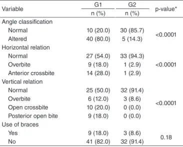
The characterization of the sample in dento-occlusal as-pects and use of braces for the two groups studied are shown in Table 1.
Regarding the occlusal classification, prevalence of occlusion considered normal was found — Angle Class I — for G2. Among the occlusal changes observed, the following were observed: for G2, the presence of Class II, division in 14.5% of participants, whereas for G1, the presence of Class II, first division in 70% of participants; Class II, second division in 4%; and Class III, division in 6% of participants.
In both groups, there were participants with and without braces, with a prevalence of participants without braces, con-stituting a similar sample.
The results regarding the characteristics of execution of mandibular movements (opening and closing), for the two groups, are shown in Table 2. The presence of deviations and noise during both the opening and closing was noteworthy, especially for G1, with differences.
The results of the analysis of mandibular movements on its amplitude are shown in Table 3. Smaller amplitudes in both lateral and protrusive movements can be observed in G1, with differences.
Statistical analysis done using χ2-test or Fisher’s exact test
showed difference (p=0.006) between the groups.
The veriication of symmetry or asymmetry between the nostrils did not show difference between the groups (p=0.30), and, in G1, 70% of participants presented symmetric nos-trils and 80% in G2.
The results and data analysis regarding the characteristics of mastication, for both groups, are shown in Table 4.
Table 1. Sample characterization regarding the classification of the occlusion and use of braces for both groups
Variable G1 G2 p-value*
n (%) n (%)
Angle classification
Normal 10 (20.0) 30 (85.7)
<0.0001
Altered 40 (80.0) 5 (14.3)
Horizontal relation
Normal 27 (54.0) 33 (94.3)
<0.0001
Overbite 9 (18.0) 1 (2.9)
Anterior crossbite 14 (28.0) 1 (2.9)
Vertical relation
Normal 25 (50.0) 32 (91.4)
<0.0001
Overbite 6 (12.0) 3 (8.6)
Open crossbite 10 (20.0) 0 (0.0)
Posterior open bite 9 (18.0) 0 (0.0)
Use of braces
Yes 9 (18.0) 3 (8.6)
0.18
No 41 (82.0) 32 (91.4)
*χ2-test or Fisher’s exact test
Caption: G1 = research group; G2 = control group
Table 2. Characteristics of mandibular movements for both groups studied
Variable
G1 G2
p-value*
n (%) n (%)
Opening (altered)**
Normal 14 (29.2) 23 (67.7)
0.001
Deviation to the right 12 (25.0) 4 (11.8)
Deviation to the left 11 (22.9) 7 (20.6)
Noises 11 (22.9) 0 (0.0)
Closing
Normal 21 (42.0) 30 (85.7)
<0.0001
Deviation to the right 11 (22.0) 5 (14.3)
Deviation to the left 7 (14.0) 0 (0.0)
Noises 11 (22.0) 0 (0.0)
*χ2-test or Fisher’s exact test; **For analytical purposes, three cases were excluded (two with limitations and one with pain)
Caption: G1 = research group; G2 = control group
Table 3. Range of mandibular movements for both groups studied
Variable
G1 Mean±SD
(median)
G2 Mean±SD
(median)
p-value*
Maximum opening (mm) 54.6±10.1 (55.7) 50.5±7.8 (48) 0.046**
Right side (mm) 5.8±2.6 (5.1) 8.2±2.0 (7.7) 0.0001**
Left side (mm) 6.0±2.3 (5.8) 8.3±2.0 (8.0) 0.0001**
Protrusion (mm) 5.9±1.4 (6.0) 8.0±1.0 (7.9) 0.0001**
*χ2-test or Fisher’s exact test; **Data on maximum opening and protrusion were compared by Student’s t-test for independent samples, and data on right and left sides were compared by Mann-Whitney test
Caption: G1 = research group; G2 = control group
Table 4. Results concerning the characteristics of the masticatory function for the groups studied
*χ2-test or Fisher’s exact test; **Data on the number of cycles were compared by Mann-Whitney test
Caption: SD = standard deviation; TMJ = temporomandibular joint; G1 = research group; G2 = control group
Variable G1 G2 p-value*
n (%) n (%)
Crushing
Posterior 35 (71.4) 30 (85.7)
0.12
Anterior 14 (28.6) 5 (14.3)
Chewing pattern
Bilateral 11 (22.0) 24 (68.6)
<0.0001
Unilateral 39 (78.0) 11 (31.4)
Labial closure
Yes 43 (86.0) 35 (100.0)
0.020
No 7 (14.0) 0 (0.0)
Unexpected muscle contraction
Yes 46 (92.0) 10 (28.6)
<0.0001
No 4 (8.0) 25 (71.4)
Noise in the TMJ
Yes 22 (44.0) 2 (5.7)
<0.0001
No 28 (56.0) 33 (94.3)
Number of cycles
Mean±SD (median) 21.8±6.8 (21.2) 22.5±6.0 (22) 0.56**
It was observed that most participants in both groups per-formed the crushing of the food on the posterior teeth. Excessive use of the tongue by pressing the food was previously observed only in one participant from G1 (2%).
There was a predominance of unilateral masticatory characteristic for G1 and bilateral for G2, with differences between the two groups. By analyzing the chewing pattern in detail, a bilateral alternating pattern was observed for in 16% of the sample, and simultaneous bilateral pattern in 6%. For G2, only the standard bilateral alternating pat-tern (68%) was observed. Unilateral chewing was preferred for 70% of the G1 sample, and chronic unilateral for 8%. In G2, there was only a preferential unilateral pattern in 31.4% of participants.
overbite, considering normal overbite, no overbite, open bite, or excessive overbite (p=0.81).
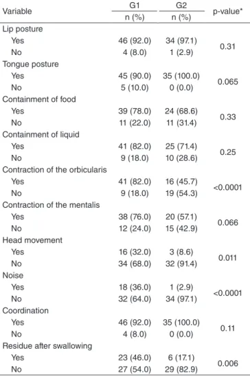
The results and analysis of data on swallowing characteris-tics for the two study groups are shown in Table 5.
movements, and higher percentage of subjects with these movements accompanied by deviations and/or noise during mandibular route. The literature suggests that the organiza-tion of mandibular movements relates to the integrity of the TMJ and the action of skeletal muscles(3,8), and some signs
of temporomandibular disorders refer to alterations in these movements(15-18).
In a related way, the variables of mandibular motion regarding the extent of laterality and maximum mandibular protrusion showed signiicantly lower values in the G1 com-pared to the G2, which are also characteristics that indicate signs of temporomandibular disorders, agreeing with previ-ous studies(14,17,22,23). In this sense, the smaller amplitude of
alterations of mandibular movements found in G1, compared to those in G2, may show the presence of temporomandibular disorders in the irst group. These indings agree with previ-ous studies that indicate the presence of reduced range and deviations in laterality of the mandibular path for individu-als with restriction or limitation of mandibular movements, which are quite frequent in subjects with temporomandibular disorders(16-18,21-23). Thus, it seems important to observe and
analyze the amplitude of mandibular movements obtained for the two groups analyzed in this study. The mean values of the opening movement obtained for both G1 and G2 were within the reference limits reported in the literature(6,8,15),
between 40 and 55 mm. However, for laterality movements and maximum protrusion, the average values obtained for G1 were signiicantly lower than those obtained for G2, falling below the reference values(6,8,15), close to the interval between
7 and 11 mm.
Regarding the occlusal characterization, data from this study showed, as expected, that in the G2, the Angle Class I pattern of occlusions prevailed. In parallel, in G1, the Class II pattern of malocclusion prevailed, as well as alterations in vertical and horizontal occlusal relationships. One study(7) points to
the existence of a relationship between measures of mandibu-lar movements with malocclusions, indicating that these may lead to changes in the position of the condyles, changing the biodynamic and inluencing the performance of laterality and protrusion movements(7).
The use of braces does not seem to interfere with the func-tional results, because the difference between the two groups was not found with regard to this variable. However, these data do not corroborate a previous study(25), in which oral
discom-fort and other dificulties related to oral function are reported, such as dificulty in chewing and swallowing. Different types of braces can justify these discrepant indings, because this study only presented individuals with ixed vestibular braces, in both groups.
With an analysis of orofacial functions, one can note differences between the groups in this study regarding re-spiratory function, with oronasal and oral breathing types being signiicantly more frequent in G1, whereas the nasal type was more frequent for G2. These indings raise agree-ment with studies that show associations between alterations in the breathing pattern and other myofunctional altera-tions(2,5,10,15,18). However, the analysis of the breathing pattern
Table 5. Results concerning the characteristics of the swallowing function for both groups studied
*χ2-test or Fisher’s exact test
Caption: G1 = research group; G2 = control group
Variable G1 G2 p-value*
n (%) n (%)
Lip posture
Yes 46 (92.0) 34 (97.1)
0.31
No 4 (8.0) 1 (2.9)
Tongue posture
Yes 45 (90.0) 35 (100.0)
0.065
No 5 (10.0) 0 (0.0)
Containment of food
Yes 39 (78.0) 24 (68.6)
0.33
No 11 (22.0) 11 (31.4)
Containment of liquid
Yes 41 (82.0) 25 (71.4)
0.25
No 9 (18.0) 10 (28.6)
Contraction of the orbicularis
Yes 41 (82.0) 16 (45.7)
<0.0001
No 9 (18.0) 19 (54.3)
Contraction of the mentalis
Yes 38 (76.0) 20 (57.1)
0.066
No 12 (24.0) 15 (42.9)
Head movement
Yes 16 (32.0) 3 (8.6)
0.011
No 34 (68.0) 32 (91.4)
Noise
Yes 18 (36.0) 1 (2.9)
<0.0001
No 32 (64.0) 34 (97.1)
Coordination
Yes 46 (92.0) 35 (100.0)
0.11
No 4 (8.0) 0 (0.0)
Residue after swallowing
Yes 23 (46.0) 6 (17.1)
0.006
No 27 (54.0) 29 (82.9)
DISCUSSION
This study had a sample of young adults, and those who composed the G1 were referred for orofacial myofunctional problems. Thus, this group was constituted of patients who met the inclusion criteria for the established age range, 19–39 years, according to the interest of the study.
Variations of some characteristics of the stomatognathic functions were also identified in relation to gender(9,10).
Therefore, the two groups were also paired regarding this vari-able, to avoid possible bias in the analyses.
based on observational data on posture, both habitual and in usual activities, represents a dificult conclusive possibil-ity. Although the purpose of this study was the analysis of orofacial myofunctional clinical examination in the chosen population, examinations of breathing can be considered one of the study’s limitations, because the determination of the respiratory pattern raises speciic needs for additional veriication.
By analyzing masticatory function, this study showed that G1 presented a unilateral preferred chewing pattern, unexpected muscle contraction, and noises in the TMJ, with differences from G2. These results corroborate previous studies related to masticatory patterns(1,3,8,14-18).
Chewing seems to be affected by dental morphology and the temporomandibular situation. In general, dental-occlusal disharmony seems to interfere directly in this function and can lead to unilateral chewing patterns. The causes cited refer to the asymmetry of masticatory muscles, temporomandibular disorders, unilateral muscle problem, and occlusal factors, such as premature tooth contacts that would cause deviations in the path of mandibular closure, and the preference for a particular side in chewing seems to be directly related to the better quality of occlusal relationship(4,8,26-28).
Regarding the analysis of the swallowing function, it was found that G1 showed changes such as contraction of the orbi-cularis oris muscle, excessive noise, and waste after swallow-ing, with statistically higher frequency than in G2. These data disagree with the indings in previous studies(1,2,6,8,9,15,18), which
describe these changes related to functional swallowing abnor-malities. It is worth mentioning the importance of checking the volume of the bolus to be swallowed in the analysis of swallowing characteristics, because this produces variations to be considered(9,11,12).
This study fulilled its objective of outlining the main myofunctional changes in the population studied, indicat-ing these as items that require greater attention and detail in the myofunctional evaluation process, corroborating previous studies. Furthermore, considering the variables that showed no statistical differences between groups, we emphasize the need for more research comparing groups of adults with and without myofunctional complaints, seeking to ascertain the items that are common to the two groups, therefore presenting less clinical manifestation. This type of examination can point to the assessment items in which there are similarities in the results, and can be singled out as normal variations between adults.
The inherent dificulty of application of orofacial myo-functional evaluation protocols can also be highlighted, because even those who use scores and are validated always include subjective data analysis, such as those related to stress, appearance, strength, and perception of muscular and/ or functional interference, which depend on the impressions and expertise of the examiners. In this study, this can be considered a limitation, especially because descriptive as-sessment is used, which, although being partially structured and requiring previous training, can present variations in the views of different evaluators.
CONCLUSION
The main orofacial myofunctional disorders in young adults with complaints refer to the limitations and alterations of mandibular movements; unilateral masticatory pattern with unexpected muscle contraction and noise in the TMJ; swallow-ing with excessive contraction of the orbicularis oris, evident noise when swallowing and leftover residues, indicating that these are the most common items in the assessment analysis.
Considering the population studied, several items of evalu-ation and characterizevalu-ation of disturbances were found to not differ between the groups. These should be carefully analyzed.
*PFAM was responsible for bibliographic research, collection and tabulation of data, analysis of results, and drafting of the manuscript; EMGB collaborated in the preparation of the project, supervised the collection and tabulation of data, guided the analysis of the results, and the inal writing of the manuscript.
REFERENCES
1. Pereira AC, Jorge TM, Ribeiro PDJ, Berretin-Felix G. Características das funções orais de indivíduos com má oclusão classe III e diferentes tipos de face. R Dental Press Ortodon Ortop Facial. 2005;10(6):111-19. 2. Ramires RR, Ferreira LP, Marchesan IQ, Cattoni DM, Silva MAA.
Tipologia facial aplicada à Fonoaudiologia: revisão de literatura. Rev Soc Bras Fonoaudiol. 2010;15(1):140-5.
3. Shiga H, Kobayashi Y, Arakawa I, Yokoyama M, Nakajima K. Inluence of two masticating conditions on assessment of movement path stability. J Prosthodont Res. 2012;56(2):125-9.
4. Fueki K, Yoshida E, Okano K, Igarashi Y. Association between occlusal curvature and masticatory movements with different test foods in human young adults with permanent dentitions. Arch Oral Biol. 2013;58(6):674-80.
5. Castro AMA, Teles RP. Inluência do tipo facial no tamanho do espaço aéreo nasofaríngeo. Rev Ortodontia. 2008;41(4):393-8.
6. Genaro KF, Berretin-Felix G, Rehder MIBC, Marchesan IQ. Avaliação miofuncional orofacial – protocolo MBGR. Rev CEFAC. 2009;11(2):237-55.
7. Metzger ALT, Campiotto AR, Muzy PC. Interferência do tipo de má oclusão nas medidas dos movimentos mandibulares: um estudo realizado com o apoio do exército brasileiro. Rev CEFAC. 2009;11(1):78-85. 8. Bianchini EMG. Avaliação da motricidade orofacial. In: Bianchini EMG,
organizadora. Articulação temporomandibular - implicações, limitações e possibilidades fonoaudiológicas. Carapicuíba: Pró-fono; 2010. p. 193-256. 9. Hiss SG, Treole K, Stuart A. Effects of age, gender, bolus volume,
and trial on swallowing apnea duration and swallow/respiratory phase relationships of normal adults. Dysphagia. 2001;16(2):128-35. 10. Martin-Harris B, Brodsky MB, Michel Y, Ford CL, Walters B, Heffner J.
Breathing and swallowing dynamics across the adult lifespan. Arch Otolaryngol Head Neck Surg. 2005;131(9):762-70.
11. Humbert IA, Fitzgerald ME, McLaren DG, Johnson S, Porcaro E, Kosmatka K et al. Neurophysiology of swallowing: effects of age and bolus type. Neuroimage. 2009;44(3):982-91.
12. Peyron MA, Gierczynski I, Hartmann C, Loret C, Dardevet D, Martin N et al. Role of physical bolus properties as sensory inputs in the trigger of swallowing. PLoS One. 2011;6(6):e21167.
13. Paskay LC. Instrumentation and measurement procedures in orofacial myology. Int J Orofacial Myology. 2006;32:37-57.
15. Marchesan IQ, Berretin-Félix G, Genaro KF. MBGR Protocol of Orofacial Myofunctional evaluation with scores. Int J Orofacial Myology. 2012;38:38-77.
16. Felício CM, Mazzetto MO, da Silva MA, Bataglion C, Hotta TH. A preliminary protocol for multi-professional centers for the determination of signs and symptoms of temporomandibular disorders. Cranio. 2006;24(4):258-64.
17. Felício CM, Melchior MO, da Silva MA. Clinical validity of protocol for multi-professional centers for determination of signs and symptoms of temporomandibular disorders. Part II. Cranio. 2009;27(1):62-7. 18. Felício CM, Medeiros APM, Melchior MO. Validity of the protocol
of orofacial myofunctional evaluation with scores for young and adult subjects. J Oral Rehabil. 2012;39(10):744-53.
19. Santoro PP, Furia CL, Forte AP, Lemos EM, Garcia RI, Tavares RA, et al. Otolaryngology and Speech Therapy evaluation in the assessment of oropharyngeal dysphagia: a combined protocol proposal. Braz J Otorhinolaryngol. 2011;77(2):201-13.
20. Lee SM, Ahn JS, Noh CS, Lee SW. Prevalence of allergic diseases and risk factors of wheezing in Korean military personnel. J Korean Med Sci. 2011;26(2):201-6.
21. Smith MT, Wickwire EM, Grace EG, Edwards RR, Buenaver LF, Peterson S et al. Sleep disorders and their association with laboratory pain sensitivity in temporomandibular joint disorder. Sleep. 2009;32(6):779-90.
22. Kitsoulis P, Marini A, Iliou K, Galani V, Zimpis A, Kanavaros P et al. Signs and symptoms of temporomandibular joint disorders related to the degree of mouth opening and hearing loss. BMC Ear Nose Throat Disord. 2011;11:5.
23. Akhter R, Morita M, Ekuni D, Hassan NM, Furuta M, Yamanaka R et al. Self-reported aural symptoms, headache and temporomandibular disorders in Japanese young adults. BMC Musculoskelet Disord. 2013;14:58. 24. Macedo PFA. Protocolo explicativo de exame miofuncional orofacial em
adultos - ferramenta digital de avaliação fonoaudiológica [dissertação]. Rio de Janeiro: Universidade Veiga de Almeida; 2013.
25. Navarro PR, Assis GB, Souza LL, Macluf Filho E, Azenha CR, Tessitori A. Alterações de funções orais na presença de aparelhos o r t o d ô n t i c o s fi x o s c o m r e c u r s o s i n t r a o r a i s . R ev C E FAC . 2013;15(5):1281-91.
26. Camargo MA, Santana AC, Cara AA, Roda MI, Melo RODN, Mandetta S, et al. Lado preferido da mastigação. Acaso ou oclusão? Revista de Odontologia da Universidade Cidade de São Paulo. 2008;20(1):82-6.
27. Pereira CC, Felício CM. Os distúrbios miofuncionais orofaciais na literatura odontológica: revisão crítica. R Dental Press Ortodon Ortop Facial. 2005;10(4):134-42.