Abstract
Submitted: April 10, 2017 Accepted: April 20, 2017
Effects of orofacial myofunctional
therapy on masticatory function in
individuals submitted to orthognathic
surgery: a randomized trial
Objectives: The esthetic and functional results of orthognathic surgery of severe dentofacial deformities are predictable, however there are differences regarding the effects on stomatognathic system. The aim was to investigate the effects of orofacial myofunctional therapy (OMT) on the masticatory function in individuals with dentofacial deformity submitted to orthognathic surgery (OGS). Material and Methods: Forty-eight individuals (18-40 years) were evaluated, 14 undergoing OMT (treated group-TG), 10 without this treatment (untreated group-UTG) and 24 in a control group with normal occlusion; for clinical aspects the data of an individual was missed (n=46). Chewing was performed using the Expanded protocol of orofacial myofunctional evaluation with scores (OMES-E). Muscle tone and mobility were also analyzed before (P0), three (P1) and six months (P2) after OGS. Surface electromyography of the masseter and temporalis muscles was performed, considering the parameters amplitude and duration of act and cycle, and the number of masticatory cycles. The OMT consisted of ten therapeutic sessions along the postoperative period. The results were compared using parametric and non-parametric tests. Results: TG showed higher scores in P1 and P2 than
P0; for the masticatory type the scores in P2 were signiicantly higher than
P0. In addition, the proportion of individuals with adequate tone of lower
lip and adequate tongue mobility for TG increased signiicantly from P1 and
P2 in relation to P0. The EMG results showed a decrease in act and cycle duration in P2 in relation to P0 and P1 for the TG; furthermore the values were close to controls. An increase in the number of cycles from P0 to P2 was also observed, indicating faster chewing, which may be attributed to an improvement of balanced occlusion associated with OMT. Conclusion: There were positive effects of OMT on the clinical and electromyography aspects of chewing in individual submitted to orthognathic surgery.
Keywords: Dentofacial deformities. Orthognathic surgery. Myofunctional
therapy. Mastication. Electromyography. Daniela Galvão de Almeida PRADO1
Giédre BERRETIN-FELIX2
Renata Resina MIGLIORUCCI3
Mariana da Rocha Salles BUENO3
Raquel Rodrigues ROSA2
Marcela POLIZEL4
Isadora Ferraz TEIXEIRA5
Maria Beatriz Duarte GAVIÃO6
http://dx.doi.org/10.1590/1678-7757-2017-0164
1Universidade Estadual de Campinas, Faculdade de Odontologia de Piracicaba, Departamento de
Ciências Fisiológicas, Piracicaba, SP, Brasil.
2Universidade de São Paulo, Faculdade de Odontologia de Bauru, Departamento de Fonoaudiologia,
Bauru, SP, Brasil.
3Clínica particular, Bauru, SP, Brasil. 4Clínica particular, Piracicaba, SP, Brasil.
5Universidade Estadual de Campinas, Faculdade de Odontologia de Piracicaba, Piracicaba, SP,
Brasil.
6Universidade Estadual de Campinas, Faculdade de Odontologia de Piracicaba, Departamento de
Odontologia Infantil, Piracicaba, SP, Brasil. Corresponding address:
Introduction
Individuals with severe dentofacial deformities
(DFD) submitted to orthodontic treatment and
orthognathic surgery (OGS) usually are seeking improvements in facial esthetics and function of the
stomatognathic system; consequently, better occlusal
relations can be achieved4. The esthetic and functional
results are predictable, but there are differences regarding the respective effects23.
Chewing is an important function of the
stomatognathic system; the ideal pattern is bilaterally
alternated, with sealed lips and jaw rotation movements with no movement of the head or other body parts,
enabling the distribution of masticatory forces with
functional and muscular balance, but depending on
factors of occlusal balance25.
Chewing can be altered in individuals with DFD2.
In Class III malocclusion the vertical mandibular
movements are predominant, with utilization of the
tongue dorsum to crush the food against the palate and little or no action of the buccinator muscles.
In Class II malocclusion, usually, the lack of lip
sealing can be observed in the presence of long face,
determining little use of orbicularis oris muscles and buccinators, accompanied by less movement of tongue
lateralization14,24.
Some protocols for clinical evaluation of chewing
have been developed in the area of Orofacial Myology, such as the Expanded protocol of orofacial
myofunctional evaluation with scores (OMES-E)7,8,
which has been proved to be a valid and reliable
instrument for orofacial myofunctional evaluation, allowing grading of the respective conditions within
the limits of selected items7. This protocol comprises
analysis of the posture of components of the
stomatognathic system; mobility of lips, tongue, jaw and cheeks and evaluation of orofacial functions, for
which scores were assigned according to the severity
of change.
An instrumental method to evaluate masticatory function consists in the surface electromyography
(EMG electromyography), which records muscle
activity in microvolts (µV) and in seconds, through
bipolar electrodes. The EMG detects the electric
potential of the muscle ibers and can simultaneously
record the muscles of the craniomandibular region in
both sides. EMG records can provide information about
muscle function in experimental conditions3.
Most studies about masticatory function in
individuals with DFD submitted to orthodontic-surgical treatment showed that the EMG of masticatory
muscles is lower compared to subjects with normal
occlusion16,27. Moreover, changes in masticatory
function or in its components after correction of DFD by OGS are evident. The period of time for occurrence
of changes is controversial and may be related to
differences in evaluation methods and treatment
types21.
Regarding the duration of chewing, Ueki, et al.26
(2009) found no changes in this characteristic after
OGS in Class III malocclusion, and the same was
found by Youssef, et al.28 (1997) in individuals with
Class II and III malocclusion. Conversely, a reduction
was observed in the duration of muscle activity in the
postoperative period compared to the preoperative
in patients with Class III malocclusion15. It is relevant
to consider the methodological differences between
researches, since the knowledge about adaptation
of this function with the correction of form still has
limitations.
A recent research showed increasing trend of the
total number of chewing cycles after 36 months of
orthodontic-surgical treatment in patients with Class
III malocclusion, determining improvement in the balance of the masticatory muscles after surgery19.
Nevertheless, the literature about orofacial
myofunctional therapy (OMT) for patients submitted
to OGS has been controversial, probably due to methodological differences15,17,22. Due to alterations of
the orofacial structures in individuals with DFD after
OGS, a new proprioceptive scheme must be acquired
so the soft structures may satisfactorily perform their functions. Therefore, to complement clinical
evaluation and to understand the functional changes
in DFD, it is important to study the effect of OMT on
the functional aspects of masticatory muscles before and after surgical correction of DFD, to elucidate the
adaptation of these muscles after surgery.
In this context, the eficacy of OMT rehabilitation
in a short time must be more precisely investigated to know if the functionality of the stomatognathic
system and the possible relapses caused by inadequate
maintenance of adaptive patternscould be recovered
early15.
Thus, the aim of this study was to determine the
effects of OMT on the clinical and electromyography
before, three and six months after OGS.
Material and methods
The study was approved by the Institutional Review Board under protocol 074/2012. The registration
number of clinical trial is RBR-4mt6yr.
Sample selection
The study is a randomized longitudinal clinical trial,
parallel with allocation ratio of 1:1. Young adults with DFD, receiving orthodontic treatment before OGS
and attending the Maxillofacial Surgery area of the
University were enrolled, forming the experimental
group. Furthermore, a control group without DFD was obtained, age- and gender-matched with the
individuals undergoing treatment. All individuals
signed a free informed consent form. The procedures
were carried out along 2013 to 2015.
The sample was selected by convenience. The
inclusion criteria of the experimental group were
healthy individuals, aged from 18 to 45 years,
both genders, presenting at least 24 teeth, with skeletal Class II or III malocclusion, diagnosed by
cephalometric radiographs and clinical evaluation
carried out before OGS by the staff of the Maxillofacial
Surgery Area. The control group should present good relation between dental arches; overbite and overjet
ranging from 1 to 3 mm; all natural teeth at least up
to the second molar; nasal breathing; the face height
should be similar to the face width to be classiied into
medium facial type, evaluated using a digital caliper
(Mitutoyo, Santo Amaro-SP, Brazil).
Exclusion criteria for both groups were neurological,
psychiatric or intellectual deicits, partially or totally
edentulous patients and the presence of cleft lip or
palate. The respective information was obtained by
interview and clinical evaluation.
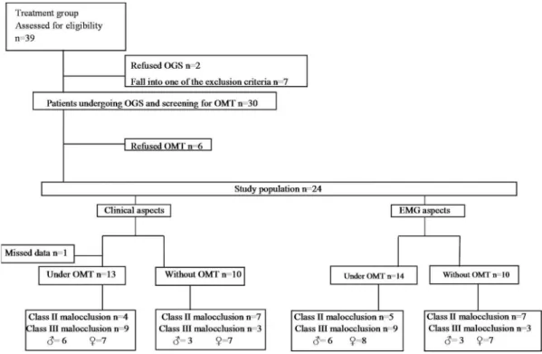
After OGS, the experimental group was composed of 24 individuals allocated in two sub-groups, namely
those who received OMT (Treated group – TG) and
those without OMT (untreated group – UTG) (Figure
1). The allocation was performed by randomization. The numbers 1-24 were randomized on an Excel
worksheet, and the irst 14 numbers drawn were part
of the TG and the last 10 of the UTG. In evaluations
of clinical aspects, data of one individual of TG were missed between the second and third evaluations, who
was excluded from the analysis.
The inal sample of clinical aspects was composed
of 13 individuals (29.31±8.87 years) allocated in TG
and 10 in the UTG (31.20±7.02 years), both with
their corresponding controls (mean age 28.39±7.34
years and mean age 28.10±5.30 years), respectively. For EMG aspects, 14 individuals (29.62±8.78 years)
allocated in TG and 10 in the UTG (31.20±7.02 years),
both with their corresponding controls (mean age
28.38±X years and mean age 28.10±5.30 years), respectively.
After the last evaluation, OMT was offered to the
UTG.
Below, the sample characteristics according to the malocclusion and surgery:
- Class II - Sagittal osteotomy of the mandibular
ramus (TG n=1; UTG n=7) and sagittal osteotomy of
the mandibular ramus with maxilla setback (TG n=3; UTG n=0);
- Class III - Le Fort I osteotomy (TG n=4; UTG
n=1); Le Fort I osteotomy and mandibular setback
(TG n=5; UTG n=1); Mandibular setback (TG n=0; UTG n=1).
One individual with class II malocclusion was
excluded from the clinical analyses due to the missed
data, but included on the instrumental analyses. Individuals with Class II and III malocclusion
were compared by the t test or Mann Whitney test
for all variables according to data normality. Since no
signiicant difference was found, the data were pooled.
TG and UTG were evaluated in three stages: before,
one or two weeks before OGS; and post stages, three
and six months after OGS. The OMT was applied in
the postoperative period, 30 days after surgery, with 10 sessions, one per week. The control group was
evaluated in a single period.
Procedures
Clinical evaluation of chewing
The masticatory function was evaluated using
OMES-E8, considering that the higher the score, the
better the function. The study analyzed the incision,
masticatory type, movements of the head or other body parts, altered head posture and food escape.
These assessments were recorded using a Coolpix
L810 camera (Nikon, São Paulo, SP, Brazil). Three
examiners, professional experts in the area, performed the analysis; the agreement between at least two
of them was taken into account, according to the
Following the protocol, mastication was recorded
with the individual sitting in a chair with a backrest,
the feet resting on the loor at a standardized distance
(1 m) from the camera lens, which was mounted on
a tripod with focus on the face, neck and shoulders.
The individuals chewed one wafer biscuit and in their habitual manner.
The bite was evaluated during ilming and the
scores were attributed as following: 1=when the
individual did not bite the food but broke it into pieces with his hands before bringing it to his mouth; 2=biting
with the molars; 3=biting with the canines and the
premolars; 4=biting with the incisors.
The counting of masticatory strokes for mastication type was made considering the jaw movements of
opening and closing until occurrence of contact of
teeth. The following scores were attributed: 1=when
the patient did not perform the function; 2=when the masticatory strokes occurred on the same side
78–94% of the times; 3=chronic unilateral, when
the masticatory strokes occurred on the same side
95–100% of the time, or anterior when occurred in the region of the incisors and canines; 4=unilateral
preference grade 2 when the masticatory strokes
occurred on the same side 78–94% of the times;
6=unilateral preference grade 1 when the masticatory strokes occurred on the same side 61–77% of the
times; 8=simultaneously bilateral, with the masticatory
strokes occurring on both sides of the oral cavity 95%
of the times; 10=when it was bilateral and alternate,
i.e., the masticatory strokes occurred on each side
50% of the times, or 40% on one side and 60% on the other.
In addition, it was analyzed the movement and/or
altered posture of the head and of other parts of the
body, food escape and uncoordinated jaw movements. Score 1 was attributed to the presence of the alteration
and score 2 to the absence.
Clinical evaluation of tone and mobility
During clinical evaluation, the mobility of the lips and tongue was observed, and the individuals were
asked to perform the following movements: Lips:
protrude closed,retract closed, protrude open, retract
open, protrude closed to the right, protrude closed to the left, pop protracted, pop retracted. Tongue:
protrude and retract, touch right and left commissures
and upper and lower lips sequentially, touch incisive
papilla, touch right cheek, touch left cheek, click tip, suck tongue on palate. If the individual did not perform
one of the tasks, the mobility was considered altered.
The tone of the upper and lower lip was evaluated
and classiied as normal, reduced or increased; both
reduced and increased were considered as altered.
Instrumental examination
Data were collected at the Ultrasonography and
Electromyography Laboratory of the Pediatric Dentistry Department (FOP–UNICAMP), which has proper
environment and conditions for adequate collection of
EMG signal. EMG recordings were obtained from four
channels of the electromyography (EMG SYSTEM, São
José dos Campos-SP, Brazil), model 810c. According to the manufacturer’s recommendation the calibration
used was -2500 to +2500 μV. The instrument was
connected to a computer for data storage and
subsequent analysis.
The evaluations were performed with the individual
sitting on a chair; the surface of the skin over the
muscles was cleaned with alcohol wipes (70th GL)
in order to remove the supericial fat, dead cells,
reduce the skin impedance and thus avoid interference
and ensure signal quality. The muscles evaluated
were: right masseter (RM), left masseter (LM), right
temporalis (RT) and left temporalis (LT).
Disposable surface double Hal electrodes were
used (Miotec Biomedical Equipment, Porto Alegre-RS,
Brazil), placed on the skin with conductive paste and
ixated using micropore®. The electrodes were placed
on the belly of the masseter and anterior temporalis as
follows: masseter - between the level of the zygomatic
arch and gonial angle, close to the occlusal plane level;
anterior temporalis muscle - in front of the hairline, in
the longitudinal direction of the anterior bundle ibers deined by palpation during clenching. The ground electrode was ixated on the right wrist of the patient
after application of conductive paste.
Mastication of a latex rubber with 2.0-cm length and 1.0-cm diameter was carried out for 60 seconds in
the usual manner. In addition, the maximum isometric
voluntary contraction (MIVC) was performed along
20 seconds; the subject was instructed to bite with maximum possible force (teeth clenching) for three
times and the mean of the respective records was
considered for analysis. The results were obtained in µV Root Mean Square (RMS), which gives the number of motor units activated (recruitment) or the
amplitude of the EMG signal. During analysis of the
electromyograms, the irst two seconds were discarded
and 10 subsequent seconds were considered.
The percentage of muscle activity was calculated as
follows: (RMSx100)/MIVC. Additionally, the duration of
chewing act and cycle in seconds were obtained. The
Figure 2- Aspects addressed during therapeutic sessions
masticatory act is the amount of time that the muscle
remains active during the occlusal phase. The chewing cycle involves three phases, namely opening, closing
and occlusal phase (Figure 3).
Analyses of chewing side preference
Furthermore, the chewing side preference was
evaluated to better understand the variations on EMG records along time. The respective task was
video recorded (Nikon Coolpix L810, São Paulo-SP,
Brazil). The subject remained seated on a chair with
a backrest, with their feet resting on the loor at a
standardized distance (1 m) from the camera lens,
which was mounted on a tripod with focus on the face,
neck and shoulders. The subjects chewed one wafer
biscuit as usual. Analysis of the video and classiication
of the preferred side was performed by three expert
examiners in the area; the agreement between at least
two of them was taken into account.
Orofacial myofunctional therapy
In the preoperative period, after completion of clinical assessment, the patient received orientation
and clarification for the orofacial myofunctional
conditions resulting from the DFD and myofunctional
consequences arising from OGS. Guidelines were reported about surgical trauma, facial edema,
decreased sensitivity and facial movements, diet, oral
hygiene and postoperative care.
In the treatment process, the “Post Orthognathic surgery therapy Protocol” was applied, which was
prepared by the project team based on the literature
and effective application in 11 individuals (unpublished
data). The protocol consists of 10 sessions, one per
week, starting 30 days after OGS and addressing the
sensitivity, tone, mobility, adequacy of posture of
lips and tongue, training and adequacy of orofacial
myofunctional functions. Figure 2 shows the aspects addressed on each session.
Statistical analysis
Intra-subgroup comparisons (TG and UTG) before,
three and six months after surgery were carried out,
using ANOVA and post hoc Tukey test or Friedman and
post hoc Dunn test, according to data distribution. The comparison between subgroups with their controls was
performed using Kruskal-Wallis and post hoc Dunn
test for data with scores, and Anova with post hoc
Dunnet test for numeric data. The Fisher’s exact test
was used to compare frequencies. A signiicance level
of 5% was adopted.
Results
The values of the maximum score of OMES-E protocol8 for TG and UTG in each evaluation period are
shown in Table 1. Signiicant increase was observed
in TG from P0 to P1 and P0 to P2. The respective
differences were not observed in UTG. Both groups
showed signiicantly lower total scores than their
controls in all periods.
The score values for “bite” and “masticatory type”
are in Table 2. No signiicance differences for “bite”
were found between periods. For “masticatory type”,
TG scores in P2 were signiicantly higher than in P0.
In P0 and P1 the TG and UTG showed lower scores
than the CG, whereas in P2 only TG showed lower scores than the CG.
The alterations of head movements and posture,
as well as food escape, were recorded as present or
absent (Table 2). Thus, the respective frequencies are demonstrated, and most individuals of TG
showed absence of alterations in head movements
in all evaluations, as well as for food escape; only
one individual presented food escape in P0 and P2. Both control groups presented no alteration in those
two items of OMES, as expected. Nevertheless, for
head posture, the experimental and control groups
presented from 2 to 8 individuals with alterations along the evaluations.
Maximum score of OMES-E
TG UTG
P0 13.23 15
±3.06 ±3.19
P1 15,92 15,4
±3.84 ±3.66
P2 16 16,2
±3.51 ±3.67
GC 19,62 19,3
±0.65 ±1.34
*
*
* *
* *
* *
* p≤0.05 statistically signiicant
Legend: P0: before surgery; P1: 3 months after surgery; P2: 6 months after surgery
TG: treated group; UTG: untreated group; CG: control group Statistical tests used: Comparison between periods: Anova/ Tukey; Comparison with control: Kruskal-Wallis/Dunn
Table 3 presents the frequency of individuals with
altered muscle tone. At P0, TG and UTG showed higher proportions of individuals with altered tone for
upper and lower lips and tongue. At P1 the respective
differences were observed for lower lip and tongue,
whereas at P2 the proportion of individuals was higher for lower lip in UTG and for tongue in TG compared
with their controls. Moreover, the proportions of
individuals with adequate tone of lower lip for TG
increased signiicantly from P0 compared with P1 and
P2. No signiicant differences for lip mobility occurred
between periods. The TG presented fewer individuals with alteration in tongue mobility in P1 and P2 than in
P0. In P1 and P2, only UTG showed more individuals
with alteration than CG (Table 3).
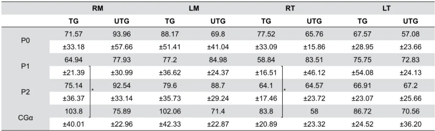
Table 4 presents the results regarding the electromyographic activity of the masseter and
temporalis muscles, in TG and UTG for each study
period. Comparing the groups in P1, the EMG of RM,
RT was lower for TG than the respective CG.
Bite# (maximum
score: 4)
Masticatory type# (maximum score:
10)
Movements of the head+
Altered head posture+
Food escape+
TG UTG TG UTG TG UTG TG UTG TG UTG n (%) n (%) n (%) n (%) n (%) n (%)
P0
Mean±SD 3.61 3.9 4.3 6.20 Presence 4 (31) 2 (20) 5 (38) 8 (80) 0 (0) 1 (10)
±0.87 ±0.31 ±2.56 ±3.3 Absence 9 (69) 8 (80) 8 (62) 2 (20) 13 (100) 9 (90)
Median 4.00 4.00 4.00 6.00
P1
Mean±SD 3.77 4.00 6.30 6.40 Presence 1 (8) 4 (40) 1 (8) 6 (60) 0 (0) 0 (0)
±0.83 ±0.00 ±3.04 ±3.09 Absence 12 (92) 6 (60) 12(92) 4 (40) 13 (100) 10 (100)
Median 4.00 4.00 6.00 6.00
P2
Mean±SD 3.69 3.70 6.92 7.60 Presence 3 (23) 3 (30) 5 (38) 7 (70) 0 (0) 1 (10)
±0.86 ±0.95 ±2.78 ±2.63 Absence 10 (77) 7 (70) 8 (62) 3 (30) 13 (100) 9 (90)
Median 4.00 4.00 8.00 8.00
CG
Mean±SD 3.92 3.90 10.00 9.60 Presence 0 (0) 0 (0) 4 (31) 2 (20) 0 (0) 0 (0)
±0.27 ±0.32 ±0.00 ±1.3 Absence 13 (100) 10 (100) 9 (69) 8 (80) 13 (100) 10 (100)
Median 4.00 4.00 10.00 10.00
*
* *
* *
* *
*
* *
* p≤0.05 statistically signiicant
Legend: P0: before surgery; P1: 3 months after surgery; P2: 6 months after surgery TG: treated group; UTG: untreated group; CG: control group
Table 2- Mean values (±standard deviation) of the OMES-E protocol items according to period of evaluation for the TG, UTG and CG
Tone Mobility
Upper lip Lower lip Tongue Lips Tongue TG UTG TG UTG TG UTG TG UTG TG UTG
n (%) n (%) n (%) n (%) n (%) n (%) n (%) n (%) n (%) n (%)
P0 adequate 7 (53.84) 4 (40.00) 0 (0.00) 1(10.00) 1 (7.69) 1 (10.00) 7 (53.84) 3 (30.00) 4 (30.77) 3 (30.00) alteration 6 (46.15) 6 (60.00) 13 (100) 9 (90.00) 12 (92.31) 9 (90.00) 6 (46.15) 7 (70.00) 9 (69.23) 7 (70.00) P1 adequate 9 (69.23) 6 (60.00) 6 (46.15) 4 (40.00) 6 (46.15) 4 (40.00) 10 (76.92) 3 (30.00) 11 (84.61) 1 (10.00) alteration 4 (30.77) 4 (40.00) 7 (53.84) 6 (60.00) 7 (53.84) 6 (60.00) 3 (23.07) 7 (70.00) 2 (15.38) 9 (90.00) P2
adequate 0 (53.80) 8 (80.00) 8 (61.54) 4 (40.00) 6 (46.15) 5 (50.00) 11 (84.61) 4 (40.00) 11 (84.61) 1 (10.00) alteration 3 (46.15) 2 (20.00) 5 (38.46) 6 (60.00) 7 (53.84) 5 (50.00) 2 (15.38) 6 (60.00) 2 (15.38) 9 (90.00) CG adequate 12 (92.30) 9 (90.00) 11 (84.61) 9 (90.00) 13 (100.00) 9 (90.00) 11 (84.61) 7 (70.00) 8 (61.54) 7 (70.00) alteration 1 (7.69) 1 (10.00) 2 (15.38) 1 (10.00) 0 (0.00) 1 (10.00) 2 (15.38) 3 (30.00) 5 (38.46) 3 (30.00)
* p≤0.05, **p≤0.01 statistically signiicant
Legend: P0: before surgery; P1: 3 months after surgery; P2: 6 months after surgery TG: treated group; UTG: untreated group; CG: control group
Statistical tests used: Comparison between periods and comparison with control: Fisher exact test
Table 3- Frequency of individuals according to muscle tone and mobility in each period of evaluation for the TG, UTG and CG
* *
*
*
* *
* *
**
** ** ** **
** ** **
**
The results concerning duration of the masticatory
act and cycle of each muscle are presented in Table
5. In TG the RM muscle showed lower values in P2
than P0 and P1. The EMG values of RM at P0 were higher than the CG for both groups, whereas for LM
at P0 only UTG showed higher values compared with
their controls. At P1, only TG presented higher values
than CG for RM. The results related to duration of the masticatory cycle. The values for RM in TG at P2 were
signiicantly lower than P0 and P1. The values for RM
and RT at P0 for TG and UTG were higher than CG.
Table 6 contains the values of the TG and UTG on the number of chewing cycles in different periods. At
P2 the TG showed more cycles than in P0. Comparing
the groups before surgery, the UTG showed fewer
masticatory cycles than the CG.
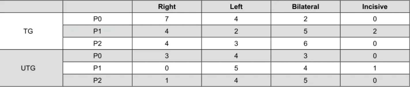
The distribution of individuals according to the
chewing side preference is shown in Figure 4.
Discussion
Besides esthetic and morphological problems, individuals with DFD may present alterations in
stomatognathic functions, particularly in masticatory
muscle activity. Morphological and functional analysis
are important for diagnosis and evaluation of treatment outcomes21,26. Thus, the clinical and
instrumental aspects of masticatory function in
individuals undergoing OGS, as well as the effect of
RM LM RT LT
TG UTG TG UTG TG UTG TG UTG
P0 71.57 93.96 88.17 69.8 77.52 65.76 67.57 57.08
±33.18 ±57.66 ±51.41 ±41.04 ±33.09 ±15.86 ±28.95 ±23.66
P1 64.94 77.93 77.2 84.98 58.84 83.51 75.75 72.83
±21.39 ±30.99 ±36.62 ±24.37 ±16.51 ±46.12 ±54.08 ±24.13
P2 75.14 92.54 79.6 88.7 64.1 64.57 66.91 67.2
±36.37 ±33.14 ±35.73 ±29.24 ±17.46 ±23.72 ±23.07 ±25.66
CGα 103.8 75.89 102.06 71.4 83.8 58 86.72 70.56
±40.01 ±22.96 ±42.33 ±22.87 ±20.89 ±23.32 ±24.52 ±36.20
* *
* p≤0.05 statistically signiicant
P0: before surgery; P1: 3 months after surgery; P2: 6 months after surgery; TG: treated group; UTG: untreated group; CG: control group RM: right masseter; LM: left masseter; RT: right temporalis; LT: left temporalis
αCG was not treated
Statistical tests used: Comparison between periods Anova/Tukey or Friedman/Dunn; Comparison with control: Anova/Dunnet
Table 4- Means and standard deviation of the percentage of muscle activity for the TG, UTG and CG in the evaluation periods
*
Act duration cycle duration
RM LM RT LT RM LM RT LT
TG UTG TG UTG TG UTG TG UTG TG UTG TG UTG TG UTG TG UTG
P0 0.37 0.41 0.34 0.39 0.29 0.31 0.29 0.35 0.84 0.89 0.84 0.89 0.9 1.01 0.86 0.84 ±0.11 ±0.12 ±0.14 ±0.09 ±0.08 ±0.09 ±0.09 ±0.30 ±0.18 ±0.17 ±0.25 ±0.30 ±0.23 ±0.35 ±0.21 ±0.21 P1
0.35 0.32 0.33 0.32 0.29 0.29 0.3 0.3 0.81 0.74 0.8 0.71 0.84 0.79 0.83 0.74 ±0.10 ±0.05 ±0.11 ±0.05 ±0.12 ±0.09 ±0.10 ±0.09 ±0.26 ±0.08 ±0.26 ±0.19 ±0.26 ±0.14 ±0.26 ±0.06 P2 0.29 0.32 0.28 0.3 0.23 0.27 0.25 0.26 0.68 0.77 0.7 0.74 0.72 0.78 0.74 0.78
±0.05 ±0.08 ±0.05 ±0.06 ±0.06 ±0.06 ±0.05 ±0.04 ±0.15 ±0.12 ±0.14 ±0.06 ±0.16 ±0.09 ±0.20 ±0.06
CGα 0.26 0.26 0.26 0.27 0.25 0.24 0.24 0.23 0.74 0.74 0.72 0.74 0.74 0.76 0.74 0.75
±0.05 ±0.04 ±0.04 ±0.06 ±0.04 ±0.04 ±0.04 ±0.04 ±0.09 ±0.09 ±0.09 ±0.09 ±0.08 ±0.12 ±0.07 ±0.13
* *
* * *
*
* * * *
* p≤0.05 statistically signiicant
P0: before surgery; P1: 3 months after surgery; P2: 6 months after surgery; TG: treated group; UTG: untreated group; CG: control group RM: right masseter; LM: left masseter; RT: right temporalis; LT: left temporalis
αCG was not treated
Statistical tests used: Comparison between periods: Anova/Tukey or Friedman/Dunn; Comparison with control: Anova/Dunnet
orofacial myofunctional therapy were veriied.
TG presented increase in maximum scores of OMES-E three and six months after surgery compared
to the preoperative period, indicating improvement
in masticatory function and the effect of OMT. Similar
results were not observed in UTG. Pereira and Bianchini18 (2011) also observed improvement in
masticatory function four months after OGS in patients
with Class II malocclusion submitted to orofacial
myofunctional therapy.
The maximum scores of the TG and UTG differed
from their controls in all periods, showing that in the
TG, although there was improvement six months after
OGS, the values still did not approach the pattern of
control individuals. This inding agreed with Van den
Braber, et al.27 (2006), who observed improvement
in masticatory performance ive years after OGS, but
the function was still impaired when compared with controls.
In the analysis of each item in OMES-E protocol
in relation to the “masticatory type” before surgery, both sub-groups presented alteration in this aspect
compared with the control. A clinical evaluation of
masticatory function in individuals with DFD also found
changes in the mastication type17. In the present study,
six months after surgery, the TG showed signiicant
increase in scores of mastication type, suggesting
improvement in function, and these results were not
observed in UTG. However, comparing the TG and UTG with their counterparts, after surgery, the scores of
the TG were signiicantly lower than the CG; therefore,
despite the improvement, the values did not approach
the control.In relation to the item “bite”, the scores for TG and UTG were similar to their controls at P0.
Moreover, no signiicant differences were observed
after surgery, showing that the DFD did not interfere
with this aspect.
At P0, four individuals of the TG showed alteration
in head movements during chewing, whereas in CG
none was altered, as expected. In UTG two individuals
showed the respective alteration. A direct functional relation between the head and neck posture was
observed during chewing20, and possible changes that
could interfere with it, such as muscles and mandibular
posture, could explain the alteration found in those individuals. At P1 only UTG differed from CG, showing
an improvement in the TG, since only one individual
showed alteration in this aspect. Over time, there was
great variability in this item that could be attributed to individual variation at the moment of evaluation
and also to the subjectivity of the test. Thus, it was
not possible to conirm the effect of OMT for head
movements over the six months after surgery. Only one individual of TG showed alteration in
head posture at P1; nonetheless, recovering was
observed at P2, since the number of individuals with
alteration was similar to P0. Despite this, no signiicant
Number of cycles TG UTG
P0 11.34 10.53
±2.87 ±1.82
P1 12.04 12.63
±2.45 ±1.16
P2 13.79 12.6
±2.44 ±1.24
CGα 12.55 12.6
±1.42 ±1.87
*
*
* p≤0.05 statistically signiicant
P0: before surgery; P1: 3 months after surgery; P2: 6 months after surgery; TG: treated group; UTG: untreated group; CG: control group
RM: right masseter; LM: left masseter; RT: right temporalis; LT: left temporalis
αCG was not treated
Statistical tests used: Comparison between periods: Anova/ Tukey; Comparison with control: Anova/Dunnet
Table 6- Means and standard deviation of the number of cycles for the TG, UTG and CG in the evaluation periods
Right Left Bilateral Incisive
TG
P0 7 4 2 0
P1 4 2 5 2
P2 4 3 6 0
UTG
P0 3 4 3 0
P1 0 5 4 1
P2 1 4 5 0
P0: before surgery; P1: 3 months after surgery; P2: 6 months after surgery TG: treated group; UTG: untreated group
differences were found between periods. It should
be considered that UTG differed from its control at P0 and P2, whereas TG was close to CG with more
individuals without alteration. It has been asserted
that changes in occlusion can inluence the muscular
balance and head position17. Some studies have found
forward head posture, especially in individuals with
Class II malocclusion10. In the UTG there were more
individuals with Class II malocclusion, which may
have contributed to the fact that before surgery more individuals of this group showed alteration in head
posture during chewing.
Food escape was evaluated and it should be
considered that, before surgery, the DFD did not
inluence this aspect, since the values were similar to
the control; no difference was observed after surgery.
The literature shows that many patients experience
paresthesia after orthognathic surgery, mainly at the lips and chin12; thus, food escape could be expected,
although it did not occur. However, the irst evaluation
occurred 3 months after surgery and this period may
sufice for adjustment of this aspect.
The lower lip tone before surgery for TG was
altered in all individuals and in UTG only one individual
presented normality. Similarly to this inding, a study
showed reduced tone of the elevator muscles of the jaw, buccinator muscles and lips in individuals with
DFD1. Three and six months after surgery, in the TG,
there was improvement compared to the preoperative
period and the number of individuals was close to the control, showing improvement in this aspect; the same
was not observed in the UTG. Therefore, there was
effect of therapy in relation to the lower lip tone. In
relation to tone of the tongue, even after surgery, the values were different from the control, showing that
six months were not suficient for tongue adaptation.
After surgery, more individuals of TG presented
adequate lip mobility, but no differences were found between periods, perhaps due to the small number of
subjects. TG presented higher number of individuals
with adequate tongue mobility three and six months
after surgery compared to the preoperative period, and after surgery only the UTG differed from the control.
Therefore, it could suggest that the OMT contributed
to improve muscle mobility. To our knowledge, no
studies could be found that describe this aspect in patients undergoing OGS, evidencing the importance
of the indings and emphasizing that mobility should
be evaluated and treated during OMT.
Regarding EMG of masticatory muscles, the
data for muscle activity were analyzed in different
periods and no signiicant difference was found after OGS. After three months TG presented signiicantly
lower EMG values than CG for the right masseter
and right temporalis. The UTG did not show similar differences. These findings can suggest that the
OMT has little inluence on EMG, probably due to
the evaluation periods after surgery. Thus, the time
needed to obtain improvement of EMG activity after orthognathic surgery can be considered a controversial
issue. Some studies found no difference over a period
of one year13,14, while others showed increase in
EMG activity while chewing, six months22 and three
years23 after surgery compared with the preoperative
period. Moreover, in the present study, even before
surgery the EMG values of the experimental groups
were not different from the controls, probably due to the previous functional adaptation to the abnormal
anatomic structures. The variability of EMG data can
be a contributing factor, despite the care in signal
acquisition, plus the surroundings factors, including muscle length, muscle anatomy, electrode position
and characteristics of contraction ilaments5, which
could inluence the EMG results about the effect of
OMT on EMG data.
The duration of the masticatory act and cycle for
the RM decreased signiicantly in TG over the six
months after OGS, suggesting that the individuals
began to perform chewing cycles with shorter duration, including the occlusal phase. Despite a
possible adaptation to malocclusion in individuals with
DFD, as mentioned above, the abnormalities present
before surgery could be damaging the masticatory
eficiency due to muscle imbalance, increasing cycle
duration to improve mastication. After surgery, the
reestablishment of dentofacial balance added to OMT
may have improved the masticatory eficiency. These indings corroborate the results found by Kobayashi,
et al.16 (2001),who analyzed patients with Class III
malocclusion and found a reduction in the masticatory
rhythm in the postoperative period compared to the preoperative. Conversely, other studies found no
change in this aspect after OGS13,26.
The results conirmed the effect of treatment on
the right masseter muscle. In this context, it can be observed that the side of masticatory preference of TG
was predominantly the right side mainly in P0, which
the right side was predominant at P0, and present at
P1 and P2. The dificulty in maximum intercuspation
in Class II malocclusion associated with mandibular
movement during chewing can determine functional
adaptations, such as unilateral chewing to facilitate the
process18. Thus, the presence of individuals with Class
II malocclusion may have inluenced the unilateral
pattern.
The experimental groups showed significantly
longer cycle duration in RM and RT at P0 than CG. This probably occurred to compensate dental-occlusal and
muscle disorders. According to Engelen, et al.6 (2005),
individuals with impaired masticatory performance
often compensate it by a higher number of chewing cycles, resulting in longer duration of masseter
muscle activity. For act duration, three months after
surgery, only the TG differed from the control for RM,
showing that TG was different from the control. Thus, it is possible to consider that three months were not
enough to detect positive results of OMT. However, six
months after OGS, the groups approached the control
with better results than P1 and P0. During the therapy sessions, masticatory function was exercised using
latex rubber and natural foods in order to promote
balance of this function, relecting an improvement
on the occlusal phase and cycle duration.
The present study did not ind differences in muscle
activity, but the improvement observed in masticatory
duration six months after surgery can suggest that the
effect of treatment remained until this time. The EMG results differ from Ko, et al.15 (2015), who observed
that individuals with Class III malocclusion undergoing
physical therapy after OGC, consisting of active and
passive jaw exercises and dietary instruction, showed greater EMG of the masseter and temporalis muscles
in relation to the untreated group after six weeks.
Nevertheless, after six months no difference between
groups was detected.
An increase was observed in the number of chewing
cycles six months after surgery in the TG, explained
as the result of lower cycle duration, and consequently
more cycles were performed. Corroborating these results, a recent research showed increasing trend of
the total number of chewing cycles after 36 months
of orthodontic-surgical treatment in patients with
Class III malocclusion, determining improvement in the balance of masticatory muscles after surgery19.
Therefore, the OMT brought favorable physiological
changes in the performance of electromyographic
duration with decrease in act and cycle and increase
in the number of chewing cycles after surgery. Furthermore, the clinical results showed that the
orofacial myofunctional therapy could provide
improvement in aspects related to maximum score of
OMES-E, masticatory type, lower lip tone and tongue mobility. It was not possible to prove the enhancement
in all items of the OMES-E protocol, considering that
chewing is a complex physiological function involving
neuromuscular activities12 and individual’s behavior
and attitudes11.
Many studies discuss the results about the
functional characteristics of masticatory muscles in
individuals with DFD undergoing OGS9,15,23 but few
studies have been conducted considering the objective
and subjective chewing aspects18,22. Thus, the present
study contributes to these indings, stressing the
importance of evaluation and myofunctional therapy in cases of OGS. Similar studies should be conducted
with greater number of individuals, and addressing
other orofacial functions.
Conclusion
The effect of treatment was observed in clinical and electromyography aspects. Thus, the importance
of OMT for individuals with DFD undergoing OGS
becomes evident.
Acknowledgments
This research was supported by CAPES – Coordination of Higher Education and Graduate
Training.
References
1- Aléssio CV, Mezzomo CL, Körbes D. The myofunctional treatment in class III patients recommended for orthognathic surgery. Arq Odontol.
2007;43:102-10.
2- Berretin-Felix G, Genaro KF, Trindade IEK, Trindade Júnior AS.
Masticatory function in temporomandibular dysfunction patients: electromyographic evaluation. J Appl Oral Sci. 2005;13:360-5. 3- Castrolorio T, Bracco P, Farina D. Surface electromyography in the assessment of jaw elevator muscles. J Oral Rehabil. 2008;35:638-45.
4- Di Palma E, Gasparini G, Pelo S, Tartaglia GM, Chimenti C. Activities of masticatory muscles in patients after orthognathic surgery. J
Craniomaxillofac Surg. 2009;37(7):417-20.
5- Disselhorst-Klug C, Schmitz-Rode T, Rau G. Surface electromyography
6- Engelen L, Fontijn-Tekamp A, van der Bilt A. The inluence of product and oral characteristics on swallowing. Arch Oral Biol. 2005;50(8):739-46.
7- Felício CM, Ferreira CL. Protocol of orofacial myofunctional evaluation with scores. Int J Pediatr Otorhinolaryngol. 2008;72(3):367-75.
8- Felicio CM, Folha GA, Ferreira CL, Medeiros AP. Expanded protocol of orofacial myofunctional evaluation with scores: validity and reliability.
Int J Pediatr Otorhinolaryngol. 2010;74(11):1230-9.
9- Frongia G, Ramieri G, De Biase C, Bracco P, Piancino MG. Changes in electric activity of masseter and anterior temporalis muscles before
and after orthognathic surgery in skeletal class III patients. Oral Surg Oral Med Oral Pathol Oral Radiol. 2013;116:398-401.
10- Gadotti IC, Berzin F, Biasotto-Gonzalez. Preliminary rapport on head posture and muscle activity in subjects with class I and II. J Oral
Rehabil. 2005;32(11):794-9.
11- Gavião MB, Bilt AV. Salivary secretion and chewing: stimulatory effects from artiicial and natural foods. J Appl Oral Sci. 2004;12(2):159-63.
12- Hanzelka T, Foltan R, Pavlíková G, Horká E, Sedý J. The role of intraoperative positioning of the inferior alveolar nerve on postoperative
paresthesia after bilateral sagittal split osteotomy of the mandible: prospective clinical study. Int J Oral Maxillofac Surg. 2011;40(9):901-6.
13- Iwase M, Sugimori M, Kurachi Y, Nagumo N. Changes in bite force and occlusal contacts in patients treated for mandibular prognathism
by orthognathic surgery. J Oral Maxillofac Surg. 1998;56(7):850-5. 14- Kasai RC, Portella MQ. The phono-audiology treatment for patients
submitted to orthognatic surgery. Rev Dent Press Ortodon Ortop Maxilar 2001;6(2):79-84.
15- Ko EW, Teng TT, Huang CS, Chen YR. The effect of early physiotherapy on the recovery of mandibular function after orthognathic
surgery for class III correction. Part II: electromyographic activity of masticatory muscles. J Craniomaxillofac Surg. 2015;43(1):138-43.
16- Kobayashi T, Honma K, Shingaki S, Nakajima T. Changes in masticatory function after orthognathic treatment in patients with
mandibular prognathism. Br J Oral Maxillofac Surg. 2001;39(4):260-5 17- Pereira AC, Jorge TM, Ribeiro Júnior PD, Berretin-Felix G. Oral
functions characteristics of individuals with Class III malocclusion and different facial types. Rev Dent Press Ortodon Ortop Facial.
2005;10(6):111-9.
18- Pereira JB, Bianchini EM. Functional characterization and temporomandibular disorders before and after orthognathic surgery
and myofunctional treatment of class II dentofacial deformity. Rev CEFAC. 2011;13(6):1086-94.
19- Piancino MG, Frongia G, Dalessandri D, Bracco P, Ramieri G. Reverse cycle chewing before and after orthodontic-surgical correction
in class III patients. Oral Surg Oral Med Oral Pathol Oral Radiol. 2013;115(3):328-31.
20- Shimazaki K, Matsubara N, Hisano M, Soma K. Functional relationships between the masseter and sternocleidomastoid muscle
activities during gum chewing: the effect of experimental muscle fatigue. Angle Orthod. 2006;76(3):452-8.
21- Takeshita N, Ishida M, Watanabe H, Hashimoto T, Daimaruya T, Hasegawa M, et al. Improvement of asymmetric stomatognathic
functions, unilateral crossbite, and facial esthetics in a patient with skeletal Class III malocclusion and mandibular asymmetry, treated with
orthognathic surgery. Am J Orthod Dentofac Orthop. 2013;144(3):441-54.
22- Trawitzki LV, Dantas RO, Mello-Filho FV, Marques W Jr. Effect of treatment of dentofacial deformities on the electromyographic activity
of masticatory muscles. Int J Oral Maxillofac Surg. 2006;35(2):170-3. 23- Trawitsky LV, Dantas RO, Mello-Filho FV, Marques W Jr. Masticatory
muscle function three years after surgical correction of class III dentofacial deformity. Int J Oral Maxillofac Surg. 2010;39(9):853-6.
24- Trench JA, Araújo RP. Dentofacial deformities: orofacial myofunctional characteristics. Rev CEFAC. 2015;17(4):1202-14.
25- Trulsson M, van der Bilt A, Carlsson GE, Gotfredsen K, Larsson P, Müller F, et al. From brain to bridge: masticatory function and dental
implants. J Oral Rehabil. 2012;39:858-77.
26- Ueki K, Marukawa K, Hashiba Y, Nakagawa K, Degerliyurt K,
Yamamoto E. Changes in the duration of the chewing cycle in patients with skeletal class III with and without asymmetry before and after
orthognathic surgery. J Oral Maxillofac Surg. 2009;67(1):67-72. 27- Van den Braber W, van der Bilt A, van der Glas H, Rosenberg T, Koole R. The inluence of mandibular advancement surgery on oral function in retrognathic patients: a 5-year follow-up study. J Oral
Maxillofac Surg. 2006;64(8):1237-40.
28- Youssef RE, Throckmorton GS, Ellis E 3rd, Sinn DP. Comparison