doi: 10.1590/0037-8682-0285-2016
Major Article
Corresponding author: Dr. Thiago José Matos Rocha. e-mail: [email protected]
Received 16 September 2016 Accepted 17 January 2017
Ultrastructural study of morphological changes in
Schistosoma mansoni
after
in vitro
exposure to the
monoterpene rotundifolone
Thiago José Matos-Rocha
[1],[2], Marília Gabriela dos Santos Cavalcanti
[1],[2],
José Maria Barbosa-Filho
[3], Ana Silvia Suassuna Carneiro Lúcio
[3], Dyana Leal Veras
[1],[2],
Márcia Ortiz Mayo Marques
[4], Luiz Carlos Alves
[1],[2]and Fábio André Brayner
[1],[2][1]. Centro de Pesquisas Aggeu Magalhães, Departamento de Parasitologia, Laboratório de Biologia Celular e Molecular, Recife, PE, Brasil. [2]. Universidade Federal de Pernambuco, Laboratório de Imunopatologia Keizo Asami, Recife, PE, Brasil. [3]. Universidade Federal da Paraíba,
João Pessoa, PB, Brasil. [4]. Centro de Pesquisa e Desenvolvimento de Recursos Genéticos Vegetais, Instituto Agronômico de Campinas, Campinas, São Paulo, Brasil.
Abstract
Introduction: Schistosomiasis, a parasitic disease caused by trematode latworms of the genus Schistosoma, affects more than
200 million people worldwide, and its control is dependent on a single drug, praziquantel. Here, we report the in vitro effect of rotundifolone, a monoterpene isolated from Mentha x villosa (Lamiaceae), on Schistosoma mansoni adult worms. Methods: The in vitro effect of rotundifolone on adult Schistosoma mansoni was evaluated by analysis of behavior and mortality and through
a scanning electron microscopic analysis of ultrastructural changes in the tegument of the worms. Results: At concentrations
of 3.54 and 7.09μg/mL-1 rotundifolone, no worm mortality was observed at any of the sampling intervals. A minor reduction
in movement of the tail, suckers, and gynecophoral canal membrane was observed after 96 h of exposure to 7.09μg/mL-1
rotundifolone. At 70.96μg/mL-1, a lack of movement was observed from 72h onwards and all worms were deemed dead; similar
effects were observed at 48h with 177.4μg/mL-1, and at 24h with 354.8μg/mL-1 and 700.96μg/mL-1. Rotundifolone also caused
death of all parasites and separation of coupled pairs into individual males and females after 24h at 354.8μg/mL-1. Conclusions:
The main changes in the tegument induced by the different ROT treatments were: after 24h incubation, bubble lesions spread
over the entire body and loss of tubercles occurred in some regions of the ventral region.
Keywords: Schistosomicidal activity. Schistosoma mansoni. Mentha x villosa. Rotundifolone.
INTRODUCTION
Schistosomiasis is a parasitic disease caused by trematode
latworms of the genus Schistosoma that is common in many tropical countries and affects more than 207 million people
living in conditions of poor sanitation and/or with less developed
social infrastructure1. Chemotherapy is the only immediate
recourse to minimize the prevalence and incidence of this
disease worldwide2. At present, praziquantel (PZQ) is the drug
of choice for the treatment of all forms of schistosomiasis3.
However, dependence on a single drug is a concern because
of the possible development of resistance in some strains4,5. A
second drug, oxamniquine (OXA), acts against Schistosoma mansoni, which is found in Africa, and against Schistosoma haematobium in South America, where it is the only species
of this genus present. OXA is manufactured in Brazil by Pizer6. OXA has a complicated manufacturing process, with
large fermentation tanks required for its biological synthesis, resulting in a higher cost than that required for PZQ synthesis.
Due to this dificulty, PZQ is generally the only drug utilized
at the moment7.
Praziquantel is a pirazyl isoquinoline and is effective against all Schistosoma species infecting humans8; it has been successfully used over the last 20 years as the drug of choice in
most areas where the disease is endemic9-11. Although PZQ has
several advantages, including tolerability, safety, eficacy, and low cost, it does not protect individuals from reinfection and is not active against the immature stages of the worm, such as the
schistosomula, pre-adult and juvenile adult stages12. Therefore,
there is an urgent need to identify new drugs that can combat
S. mansoni.
Investigative methods such as scanning electron microscopy (SEM) have been employed to study the effects of compounds on the tegument of many helminthes13, especially S. mansoni14. In
FIGURE 1 - Chemical structure of rotundifolone. S. mansoni offers a potentially valuable approach15,16. Members
of the plant family Lamiaceae have been widely studied as a source of secondary metabolites with biological activity17,18. Among the species of this family, Mentha x villosa Hudson
has been used in traditional medicine owing to its antiparasitic properties. It is known popularly as hortelã-rasteira, hortelã comum, or hortelã-da-folha-miúda19. A commercial formulation
named Giamebil®, which has as its active compound the dry extract from the leaves and stem of M. x villosa, has been shown to have amebicidal (Entamoeba histolytica) and giardicidal (Giardia lamblia) activities20. Recent studies have also
demonstrated the eficacy of extract from M. x villosa against Trichomonas vaginalis infection21.
Rotundifolone (ROT) is a monoterpene that is present in large amounts in the leaves of M. x villosa. This study had greater impact to ROT, because it represents more than 50% (can reach 70%) of the total essential oil from Mentha x villosa (MVEO) reported previously17. It has the chemical formula
C10H14O2 (Figure 1)and a molecular weight of 166. ROT is an important chemical constituent of the essential oil of many Mentha species (M. rotundifolia, M. suaveolens, M. spicata, M. longifolia, and M. x villosa)21. It has been reported to have
hypotensive and bradycardic22, antimicrobial23, spasmolytic24,
larvicidal25, antitumour effects26and schistosomicidal activity
on adult S. mansoni27.
Considerable efforts are underway to develop novel drugs
for prevention and cure of schistosomiasis. Recent studies by our research group have demonstrated that ROT has in vitro schistosomicidal activity17. However, there are no studies on
the possible ultrastructural changes in adult S. mansoni that are
induced by treatment with ROT.
METHODS
Ethical considerations
All experiments involving the use of experimental animals were performed in accordance with the ethical standards of the Foundation Oswaldo Cruz and were approved by the Animal Experimentation Ethics Committee (No. 06/2010).
Botanical material
Fresh leaves of M. x villosa were collected from the Medicinal Plants Garden of the Pharmaceutical Technology
Laboratory, Federal University of Paraiba (LTF/UFPB) between April and June 2011. The plants were identiied and authenticated by Dr. F. J. Abreu Matos (Laboratory of Natural
Products, Federal University of Ceará) and by Dr. Raymond
Harley of the Royal Botanic Gardens, Kew, England. A voucher specimen was deposited in the Prisco Bezerra Herbarium of the Federal University of Ceará (N. 14996).
Isolation and identiication of rotundifolone
To extract MVEO, 10 kg of leaves were steam-distilled for 8h. The oil obtained (0.1%) was dried over anhydrous sodium
sulfate in the usual manner and stored at 4°C13. We used a gas
chromatograph coupled to a mass spectrometer (Shimadzu
QP-5000) under the following analytical conditions: capillary
column, OV-5 (30m× 0.25mm × 0.25μm); injector (Ohio Valley Specialty Chemical, Inc.), 240°C; detector, 230°C; electron impact, 70 eV; gasdrag, He; low, 1.0mL/min-1; split, 1/20;
program temperature, 60-240°C at 3°C min-1; and solution
injection volume, 1μL (1μL of essential oil per 1mL of ethyl acetate). The compounds were identiied by comparing their
mass spectra using the Gas Chromatography Mass Spectrometry
(GC-MS) database system (Nist 62 lib.) and the Kovats retention index.
The oil was subjected to preparative layer chromatography (Merck silica gel PF254 plates, 40 × 20cm). The plates were eluted three times using hexane as the solvent. Pure ROT
(Figure 1) was thereby obtained from M. x villosa leaves. The
chemical purity of ROT (more than 99.9%) was determined by
high-performance liquid chromatography.
Maintenance and collection of adult Schistosoma mansoni
Schistosoma. mansoni strain BH (Belo Horizonte, Minas
Gerais, Brazil) was used in this study. The parasites were
maintained in Biomphalaria glabrata snails and Swiss strain mice in the laboratory at the Aggeu Magalhães Research
Center of the Oswaldo Cruz Foundation. Female Swiss mice
weighing 20 ± 5g were used as the deinitive host and infected transcutaneously with about 120 cercariae (this is the number
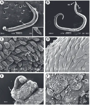
FIGURE 2 - (A-F) Micrographs of adult Schistosoma mansoni worms. (A-B) Micrographs of untreated adult worms. The micrograph of an untreated male shows the gynecophoral canal (gc) and the thinner posterior region (pr). (B) The anterior region of a female Schistosoma mansoni with an oral sucker (os) and ventral sucker (sv). (C) Tubercles (tu) with spines are present on the tegument of a male. (D) Grooves (arrows) are present in the testa (st). (E-F) Micrographs of adult worms treated with PZQ
(0.5μg/mL-1): (E) Worm showing a twisted body shape (arrow)
and extensive destruction of the tegument. (F) Worm showing severe damage to the seed coat, with loss of spicules and extensive ulceration of the tegument (arrows). PZQ: praziquantel.
A B
C D
E F
the tail immersion technique. The animals were kept under
controlled temperature and light conditions and had ad libitum
access to food and water.
At 55 days after infection, adult S. mansoni were recovered from the hepatic portal system of infected mice and left for 2 h
to adapt to the culture medium, washed in RPMI 1640 medium buffered with HEPES (20mM), pH 7.5, supplemented with penicillin (100IU/ml-1), streptomycin (100μg/mL-1) and 10%
fetal bovine serum (Gibco), and placed in petri dishes containing 2mL of sterile culture medium17.
In vitro antischistosomal assay
The in vitro assays of antischistosomal treatments were carried out using ROT dissolved in 100% DMSO. Five
worms were placed into each well and incubated at 37°C
in an atmosphere containing 5% CO2. After 2h to adapt to the culture medium, MVEO and ROT (3.5, 7.0, 71, 177.4,
and 354.8μg/mL-1) were added. As a negative control, adult
worms were incubated in RPMI 1640 and RPMI 1640 + 1.6% DMSO. As a positive control, worms were incubated in 0.5μg/
mL-1 PZQ. The worms were monitored every 24h until 120h
and changes in motor activity were assessed and the mortality
ratewas determined27. The evaluation of the viability of adult
worms was based on the standard procedures for screening schistosomicidal compounds established by the World Health Organization (WHO) Special Program for Research and
Training in Tropical Diseases. In this evaluation system,
(+ + +) indicates normal activity, (+ +) slight loss of movement, with active tail, suckers, and gynecophoral canal membrane, (+) indicates movement of tails and suckers alone, and (−) indicates no movement. The worms were considered to be dead when no movement was identiied after 3 min of observation using an
inverted microscope27.
Scanning electron microscopy
Scanning electron microscopy was used to evaluate
morphological changes in S. mansoni worms after the in vitro
treatments. Worms were incubated for 24h and after their death naturally they were washed in sodium cacodylate buffer (pH = 7.2), ixed in 2.5% glutaraldehyde (pH = 7.4) for 24h, and then ixed in 1% osmium tetroxide for 1h. The samples were
dehydrated through an ethanol series, dried in a critical point
dryer, and then mounted on stubs and coated with gold using a sputter coater. The specimens were examined using a
JEOL-5600LV microscope27.
RESULTS
In vitro effects of rotundifolone on Schistosoma mansoni
adult worms
At ROT concentrations of 3.5 and 7.0μg mL-1, no worm
mortality was observed during the course of the experiment.
A minor loss of movement of the tail, suckers, and gynecophoral
canal membrane was observed after 96h exposure to 7μg/mL-1
ROT. Worms in the negative control groups (RPMI 1640 and
RPMI 1640 + DMSO 1.6%) remained viable throughout the analysis period. In the positive control with PZQ, all parasites
died within 24h. At 71μg mL-1 ROT, mortality and absence
of movement were observed from 72h; similar effects were apparent from 48h exposure to 177.4μg/mL-1 ROT, and from 24h
exposure to 354.8μg mL-1 and 700.96μg mL-1 ROT (Table 1).
Ultrastructural analysis of rotundifolone-induced morphological changes in Schistosoma mansoni
Adult worms in the control groups showed no changes
in behavior up to 120 h and remained vigorous during this
time. Male worms in the control groups had a tegument that was covered with tubercles and tiny projections (spines). The back was long and contained the gynecophoral canal. The area between the oral and ventral suckers did not have any tubercles,
spines, or sensory papillae (Figure 2A and Figure 2B). A large
number of tubercles with typical spines (Figure 2C) as well as sensory papillae (Figure 2D) was observed.
After treatment with PZQ, all worms were dead by 24h of incubation. Under SEM, adult worms treated with 0.5µg/mL-1
showed loss of tubercles and spines and many regions were
ulcerated (Figure 2F).
The main changes in the tegument induced by the different
ROT treatments were: after 24h incubation at 354.8µg/mL-1
ROT, bubble lesions spread over the entire body (Figure 3A) and loss of tubercles occurred in some regions of the ventral region (Figure 3B); after 48h incubation at 177.4μg/mL-1 ROT,
increased rate of death of worms, which showed destruction of
the oral sucker and contraction of the ventral sucker (Figure 3C). The severity of the lesion in the tegument lesion increased after
72h of ROT treatment (71μg/mL-1) ( Figure 3D). The lowest
ROT concentrations (3.54 and 7.09μg/mL-1) did not cause
mortality of adult worms after 120h of exposure; however, changes in the tegument of the worms were observed. At 7.09µg/mL-1 ROT, damage to the tegument was seen at higher
magnifications (Figure 3E) and in the worms treated with
3.54μg/mL-1 there was destruction of some tubercles (Figure 3F).
DISCUSSION
The tegument of S. mansoni is an important structure that is involved in both absorption of nutrients and secretion of some
products; it also has a role in protecting the organism from
the immune response of the host. Therefore, the tegument is a critically important target for antischistosomal drugs28. Electron microscopic analyses, including TEM, have documented damage to the tegument of S. mansoni caused by exposure to synthetic and natural compounds14 for various antischistosomal
prototype compounds29.
Recently, in vitro studies for new active compounds against S. mansoni14-16 showed that ROT, a monoterpene found in
Mentha species, showed promising antischistosomal properties against adult schistosomes17,27.
Matos-Rocha et al.17 determined the composition of MVEO
and evaluated its in vitro effects on the viability of adult S. mansoni. The main constituents of MVEO were identiied
as ROT (70.96%), limonene (8.75%), trans-caryophyllene (1.46%), and β-pinene (0.81%). In viability tests, neither
trans-Groups Incubation period (h) Number of dead worms (%) Changes in motor activity
Control 120 - +++
DMSO 1% 120 - +++
PZQ 24 100.0
-0.7μg mL-1 120 - +++
3.54μg/mL-1 120 - +++
7.09μg/mL-1 96 - ++
70.96μg/mL-1 72 100.0
-177.4μg/mL-1 48 100.0
-354.8μg/mL-1 24 100.0
-700.96μg/mL-1 24 100.0
-TABLE 1
In vitro effect of different concentrations of rotundifolone on mortality of adult Schistosoma mansoni.
Negative control (RPMI 1640; DMSO + RPMI 1640). Positive control with 0.5μg/mL-1 PZQ. Rotundifolone: +++ normal activity; ++ slight loss of movement, with
activity of the tail, suckers, and gynecophoral canal membrane.RPMI 1640:Roswell Park Memorial Institute; DMSO:dimethyl sulfoxide; PZQ: praziquantel.
A B
C D
E F
FIGURE 3 - (A-F) Micrographs of adult Schistosoma mansoni treated with different concentrations of ROT. (A) 24h incubation with 700.96μg/mL-1,
ROT induced disarray in the tegument (arrows); it was not possible to identify tubercles and spikes. (B) Adult Schistosoma mansoni after a 24-h incubation with 354.8μg/mL-1 ROT show minor damage compared to the
highest concentration, and it was possible to identify tubercles (tu); between the tubercles, note sloughing of the tegument (arrow). (C) Anterior region of an adult worm after 48h incubation with 177.4μg/mL-1 ROT. There is clear
damage to the oral sucker oral (so) and ventral sucker (sv). (D) After 72h exposure to 71μg/mL-1 ROT, the worm shows a lattened appearance and
has a twisted tegument (arrow) and supericial ulceration (asterisk). (E) Adult worms after 96h in 7.1μg/mL-1 ROT. Vesicles have formed (arrow).
(F) After 120h of incubation with 3.5μg/mL-1 ROT, no tubercles can be
caryophyllene or β-pinene had anti-schistosomicidal activities.
Rather, the schistosomicidal activity of MVEO appeared to be due to ROT, its major constituent. When analyzed alone, ROT
showed similar activity to MVEO. To date, the possible effects of
ROT on the ultrastructure of S. mansoni has not been examined.
The results of this study show that all tested concentrations, ROT was able to cause damage to the tegument of S. mansoni.
These indings corroborate the report of Lima et al.14 in which they investigated the characteristics of the tegument of adult
worms obtained from mice infected with cercariae of S. mansoni and subjected to treatment with allicin at a concentration of 20mg/mL-1. Lima et al.14 found a variety of structural changes to
the tegument of the worms, including destruction of tubercles, and induction of edema and ulcers. In addition, there were changes to the number and volume of thorns and the tubercles showed a modiied appearance. In the present study, there was a marked difference in the morphology of worms treated with PZQ compared with ROT. Macroscopic examination of adult S. mansoni exposed to PZQ found that they showed muscle contractions that caused them to stay in a retracted or twisted shape. This behavior was not observed in worms treated with MVEO and its constituents.
Mendonça et al.30 compared the in vitro anthelminthic effect
of PZQ against recent S. mansoni isolates by estimating relative
motility, survival indices, and tegumental surface alterations. They found that the damage caused by PZQ on the integument of
adult worms was dose-dependent, and increased with increasing
PZQ concentrations.
The mechanism by which ROT exerts its in vitro effect on S. mansoni is not clear. It has been suggested that, because of the large number of constituents, essential oils may have
no speciic cellular target31. Additionally, as the lipophilic compounds present in essential oils can pass through the cell
wall, integument, and cytoplasmic membrane, they may damage
the structure of the cellular membranes during their passage,
which may cause cellular lysis32,33. Regarding their biological properties, some of the activities of essential oils are associated
with loss of ions and reduction of membrane potential, as well as
collapse of the proton pump and depletion of the ATP pool34,35.
Our analyses here demonstrate the effectiveness of ROT as
a candidate compound with signiicant in vitro effects against
S. mansoni adult worms. This monoterpene was able to cause
morphological alterations in the tegument of adult schistosomes. Overall, these results are important as there is an urgent need to
develop new agents against schistosomes. Because ROT is generally
recognized as safe, this compound may have broad antiparasitic applications. We recommend further analysis to better understand the
in vivo eficacy of this new potential treatment for schistosomiasis.
Conlict of interest
The authors declare that there is no conlict of interest.
Acknowledgments
To Laboratório de Tecnologia Farmacêutica/Universidade Federal da
Paraíba (LTF/UFPB), Laboratório de Imunopatologia Keizo Asami (LIKA/
UFPE) and Núcleo de Plataformas Tecnológicas (NPT) of Aggeu Magalhães
Research Center of the Oswaldo Cruz Foundation for support for the experiments and to CAPES for a scholarship.
REFERENCES
1. Rollinson D, Knopp S, Levitz S, Stothard JR, Tchuenté LA, Garba A, et al. Time to set the agenda for schistosomiasis elimination.
Acta Trop. 2013;128(2):423-40.
2. Hotez PJ, Molyneux DH, Fenwick A, Kumaresan J, Sachs SE, Sachs
JD, et al. Control of neglected tropical diseases. N Engl J Med. 2007;357(10):1018-27.
3. Cioli D, Pica-Mattoccia L. Praziquantel. Parasitol Res.
2003;90(Suppl 1):S3-S9.
4. Stelma FF, Talla L, Sow S, Kongs A, Niang M, Polman K, et al.
Eficacy and side effects of praziquantel in an epidemic focus of Schistosoma mansoni. Am J Trop Med Hyg. 1995;53(2):167-70.
5. Fallon PG, Sturrock RF, Niang AC, Doenhoff MJ. Short report: diminished susceptibility to praziquantel in a Senegal isolate of
Schistosoma mansoni. Am J Trop Med Hyg. 1995;53(1):61-2.
6. Cheetham PSJ, Cabral JMS, Best D, Boross L, Tramper J. Case studies in applied biocatalysis from ideas to products, editors.
Applied Biocatalysis, Harwood Academics Publishers GmbH, 1994. p. 47-108.
7. Araujo N, Mattos AC, Coelho PM, Katz N. Association of
oxamniquine praziquantel and clonazepam in experimental
Schistosomiasis mansoni. Mem Inst Oswaldo Cruz. 2008;103(8):781-5.
8. World Health Organization (WHO). Schistosomiasis: number
of people treated worldwide in 2009. Wkly Epidemiol Rec. 2011;86(9):73-80.
9. Ridi RE, Aboueldahab M, Tallima H, Salah M, Mahana Fawzi NS,
Mohamed SH, et al. In vitro and in vivo activities of arachidonic acid against Schistosoma mansoni and Schistosoma haematobium.
Antimicrob Agents Chemother. 2010;54(8):3383-9.
10. de Moraes J, de Oliveira RN, Costa JP, G Junior AL, de Sousa DP, Freitas RM, et al. Phytol, a diterpene alcohol from chlorophyll, as a drug against neglected tropical disease Schistosomiasis mansoni.
PLoS Negl Trop Dis. 2014;8(1):e2617.
11. Bertão HG, da Silva RA, Padilha RJ, de Azevedo Albuquerque MC, Rádis-Baptista G. Ultrastructural analysis of miltefosine-induced surface membrane damage in adult Schistosoma mansoni BH strain
worms. Parasitol Res. 2012;110(6):2465-73.
12. Holtfreter MC, Loebermanna M, Klammta S, Sombetzkia M, Bodammera P, Riebolda D, Kinzelbachb R, Reisinger EC.
Schistosoma mansoni: schistosomicidal effect of meloquine and
primaquine in vitro. Exp Parasitol. 2011;127(1):270-6.
13. Aires AL, Ximenes EC, Silva RA, Barbosa VX, Góes AJ, Peixoto CA. Ultrastructural analysis of β-lapachone-induced surface membrane damage in male adult Schistosoma mansoni BH strain
worms. Exp Parasitol. 2014;142(1):83-90.
14. Lima CM, Freitas FI, Morais LC, Cavalcanti MG, Silva LF, Padilha RJ. Ultrastructural study on the morphological changes to male
worms of Schistosoma mansoni after in vitro exposure to allicin.
Rev Soc Bras Med Trop. 2011; 44(3):327-30.
15. Silva AP, Silva MP, Oliveira CG, Monteiro DC, Pinto PL, Mendonça RZ. Garcinielliptone FC: antiparasitic activity without cytotoxicity to mammalian cells. Toxicol In Vitro. 2015;29(4):681-7.
16. Dejani NN, Souza LC, Oliveira SR, Neris DM, Rodolpho JM, Correia RO, et al. Immunological and parasitological parameters in
the leaves of Mentha x piperita L. Immunobiology. 2014;219 (8):627-32.
17. Matos-Rocha TJ, Cavalcanti MGS, Barbosa-Filho JM, Lúcio ALSC, Veras DL, Feitosa APS. In vitro evaluation of schistosomicidal activity of essential oil of Mentha x villosa and some of its chemical
constituents in adult worms of Schistosoma mansoni. Planta Med.
2013;79(14):1307-12.
18. Amaral RG, Fonseca CS, Silva TK, Andrade LN, França ME,
Barbosa-Filho JM, et al. Evaluation of the cytotoxic and antitumour effects of the essential oil from Mentha x villosa and its main compound, rotundifolone. J Pharm Pharmacol. 2015;67(8):1100-6. 19. Matos FJA, Machado MIL, Alencar JW, Matos MEO, Craveiro AA.
Plants used in traditional medicine of China and Brazil. Mem. Inst.
Oswaldo Cruz.. 1991;86(suppl 2):13-16.
20. Teles NSB, Fechine FV, Viana FAC, Viana IOL, Nascimento DF,
Leite ALAS. Evaluation of the therapeutic eficacy of Mentha crispa in the treatment of giardiasis. Contemp Clin Trials. 2011;32(6):809-13.
21. Moraes ME, Cunha GH, Bezerra MM, Fechine FV, Pontes AV,
Andrade WS. Eficacy of the Mentha crispa in the treatment of
women with Trichomonas vaginalis infection. Arch Gynecol
Obstet. 2012;286(1):125-30.
22. Guedes DN, Silva DF, Barbosa-Filho JM, Medeiros IA. Calcium
antagonism and the vasorelaxation of the rat aorta induced by
rotundifolone. Braz J Med Biol Res. 2004;37(12 ):1881-7.
23. Arruda TA, Antunes RMP, Catão RMR, Lima EO, Sousa DP, Nunes
XP. Preliminary study of the antimicrobial activity of Mentha x villosa Hudson essential oil, rotundifolone and its analogues. Rev Bras Farmacogn. 2006;16(3):307-11.
24. Lahlou S, Magalhães PJC, Carneiro-Leão RFL, Leal-Cardoso JH.
Involvement of nitric oxide in the mediation of the hypotensive
action of the essential oil of Mentha x villosa in normotensive conscious rats. Planta Med. 2002;68(8):694-99.
25. Lima TC, da Silva TK, Silva FL, Barbosa-Filho JM, Marques MO, Santos RL, et al. Larvicidal activity of Mentha x villosa Hudson essential oil, rotundifolone and derivatives. Chemosphere. 2014;104:37-43.
26. Amaral RG, Fonseca CS, Silva TK, Andrade LN, França ME, Barbosa-Filho JM, et al. Evaluation of the cytotoxic and antitumour effects of the essential oil from Mentha x villosa and its main compound, rotundifolone. J Pharm Pharmacol. 2015,67(8):1100-6. 27. Matos-Rocha TJ, Cavalcanti MG, Veras DL, Feitosa AP, Gonçalves
GG, Portela-Junior NC L, et al. Ultrastructural changes in
Schistosoma mansoni male worms after in vitro incubation with the
essential oil of Menthaxvillosa huds. Rev Inst Med Trop Sao Paulo.
2016;58(1):7. doi.org/10.1590/S1678-9946201658004.
28. Neves BJ, Andrade CH, Cravo PVL. Natural products as leads in schistosome drug discovery. Molecules. 2015;20(2):1872-903.
29. Liang YS, Coles GC, Dai JR, Zhu YC, Doenhoff MJ. Adult worm tegumental damage and egg-granulomas in praziquantel-resistant and –susceptible Schistosoma mansoni treated in vivo. J Helminthol.
2002;76(4):327-33.
30. Mendonça AM, Feitosa AP, Veras DL, Matos-Rocha TJ, Cavalcanti MG, Barbosa CC, et al. The susceptibility of recent isolates of
Schistosoma mansoni to praziquantel. Rev Inst Med Trop São Paulo. 2016;58:7. doi.org/10.1590/S1678-994620165800.
31. Moraes J De, Nascimento C, Lopes PO, Nakano E, Yamaguchi LF, Kato MJ. Schistosoma mansoni: in vitro schistosomicidal activity of piplartine. Exp Parasitol. 2011;127(2):357-64.
32. Bakkali F, Averbeck S, Averbeck D, Idaomar M. Biological effects
of essential oils-a review. Food Chem Toxicol. 2008;46(2):446-75.
33. Eissa MM, El-Azzouni MZ, Amer EI, Baddour NM. Miltefosine, a promising novel agent for schistosomiasis mansoni. Int J Parasitol.
2011;41(2):235-42.
34. Esperandim VR, da Silva Ferreira D, Sousa Rezende KC, Magalhães LG, Medeiros Souza J, Pauletti PM, et al. In vitro antiparasitic activity and chemical composition of the essential oil obtained from the fruits of Piper cubeba. Planta Med. 2013;79(17):1653-5. 35. Godinho LS, Aleixo de Carvalho LS, Barbosa de Castro CC, Dias
MM, Pinto PFS, Crotti AEM, et al.Anthelmintic activity of crude
extract and essential oil of Tanacetum vulgare (Asteraceae) against