Cop
yright
© ABE&M t
odos os dir
eit
os r
eser
vados
.
Arq Bras Endocrinol Metab. 2011;55/5 314
original article
Correspondence to: Francisco Bandeira Divisão de Endocrinologia, Hospital Agamenon Magalhães, Universidade de Pernambuco, Estrada do Arraial, 2723 52021-380− Recife-PE, Brazil [email protected]
Received on Nov/12/2010 Accepted on May/25/2011
1 Division of Endocrinology
and Diabetes, Hospital Agamenon Magalhães, Brazilian Ministry of Health (MS/ SUS), Universidade de Pernambuco (UPE), Medical School, Recife, PE, Brazil
Normocalcemic primary
hyperparathyroidism in
clinical practice: an indolent
condition or a silent threat?
Hiperparatireoidismo primário normocalcêmico na prática clínica: condição indolente ou ameaça silenciosa?
Thyciara Fontenele Marques1, Renata Vasconcelos1, Erik Diniz1, Daniela Rêgo1, Luiz Griz1, Francisco Bandeira1
ABSTRACT
Objective: To describe the characteristics of normocalcemic primary hyperparathyroidism (NPHPT) in patients seen for osteoporosis evaluation.Patients and methods: We examined the records of 156 women who came to the hospital to be screened for osteoporosis. Measu-rements of total calcium, PTH, 25-hydroxy vitamin D, and β-C-telopeptide were recorded. Bone mineral density and T-scores were evaluated by densitometry of the lumbar spine, femoral neck and distal one-third of the radius. The latter was only measured in patients with primary hyperparathyroidism. Nephrolithiasis and bone fractures were documented by a review of the medical records.Results: We identified 14 patients with NPHPT, accounting for 8.9% of the po-pulation studied. In the medical records, the occurrence of kidney stones was reported in 28.6% of the patients with NPHPT, in contrast with only 0.7% of the noncarriers. Regarding the presen-ce of general fractures, 21.4% of the patients with NPHPT were affected versus 16.2% of
noncar-riers. Conclusion: Data from our study suggest that NPHPT has a diverse phenotypic presen-tation, implying that this may not be an “indolent” disease. Arq Bras Endocrinol Metab. 2011;55(5):314-7
Keywords
Normocalcemic hyperparathyroidism; osteoporosis; fractures; kidney stones
RESUMO
Objetivo: Avaliar as características do hiperparatireoidismo primário normocalcêmico (HPTPN) em pacientes atendidos para avaliação de osteoporose. Pacientes e métodos: Foi realizada análi-se de um banco de dados de 156 mulheres que procuraram atendimento para avaliação de oste-oporose. Todas apresentavam dosagem de cálcio sérico, PTH, 25-hidroxi-vitamina D e C-telopep-tídeo. A densidade mineral óssea e escore-T foram avaliados por meio de densitometria óssea de coluna lombar, colo do fêmur e rádio distal, este último apenas em pacientes com hiperpara-tireoidismo renal primário. Nefrolitíase e fraturas ósseas foram documentadas pela revisão dos prontuários. Resultados: Foram identificadas 14 pacientes com HPTPN, correspondendo a 8,9% da população estudada. Nos registros médicos, o relato da existência de litíase renal ocorreu em 28,6% dos portadores de HPTN em contraste com apenas 0,7% nas mulheres não portadoras, com um p < 0,001. Conclusão: Os dados do estudo sugerem que HPTPN tem uma apresentação fenotípica variada, podendo não ser uma patologia “indolente”. Arq Bras Endocrinol Metab. 2011;55(5):314-7
Descritores
Hiperparatireoidismo normocalcêmico; osteoporose; fraturas; cálculo renal
INTRODUCTION
N
ormocalcemic primary hyperparathyroidism(NPHPT), which is characterized by persistently
Cop
yright
© ABE&M t
odos os dir
eit
os r
eser
vados
.
315 Arq Bras Endocrinol Metab. 2011;55/5
Normocalcemic primary hyperparathyroidism in clinical practice
irst used in 1960 by Wills, who described a group of patients with characteristics different from those with classic primary hyperparathyroidism (1). In this situ-ation, a thorough search for causes of secondary hy-perparathyroidism, in particular 25-hydroxy vitamin D deiciency, is imperative (2).
Studies on the evaluation of this speciic popu-lation are scarce, especially since there is no routine measurement of PTH. As a result, there are few data on the impact of this condition on the health of indi-viduals. This has been the great challenge in the area of bone metabolism.
Could normocalcemic primary hyperparathyroi-dism be an indolent disease or could it have an im-pact similar to classic primary hyperparathyroidism on bone metabolism and with renal involvement? It is extremely important to conduct studies on this pa-thological condition in order to achieve a more pre-cise epidemiological characterization and deinition of the clinical signiicance of this disorder.
PATIENTS AND METHODS
Selection of patients and study design
A survey was conducted using the database from the Center for Endocrinology and Bone Metabolism of Pernambuco, Recife, Brazil. From this database, the records of 179 women who came to the hospital to be screened for osteoporosis were analyzed.
The following cases were excluded from the analysis: patients taking bisphosphonates, diuretics, anticonvulsants and lithium; as well as those suffe-ring from kidney failure with GFR < 40 mL/min, using the formula Modiication of Diet in Renal Di-sease Study (MDRD); 25-hydroxy vitamin D < 30 ng/mL; metabolic bone diseases (e.g., classic prima-ry hyperparathyroidism); gastroenterological disea-ses associated with malabsorption; liver disease and those whose records were incomplete (3).
One hundred and ifty-six patients were eligible for the study, and the following measurements had been recorded in all cases: at least two samples of serum total calcium (normal range: 8.5 to 10.4 mg/dL), serum PTH (normal range between 10 and 65 pg/mL), 25-hydroxy vitamin D (25OHD, normal > 30 ng/ mL) and C-telopeptide (normal: 50-450 pg/mL). Bone mineral density (BMD) and T-scores were evaluated by the DXA technique (Lunar
Corpora-tion Madison, Wisconsin, USA) in the lumbar spine (L1-L4), femoral neck and distal one-third of the ra-dius. The latter measurement was only recorded in patients with primary hyperparathyroidism. Correc-tion of serum calcium in relaCorrec-tion to albumin levels was performed using the formula: corrected calcium = calcium recorded + (4 − serum albumin) x 0.8.
Analyses of serum albumin, calcium and creatinine were measured by dry chemistry in VITROS 250/950 (Johnson & Johnson®). PTH levels were measured by means of chemiluminescence in Immulite 200, which shows intra- and inter-assay coeficients of variation of 4.2% to 5.7% and 6.3% to 6.8%, respectively. Serum
β-CTX levels were measured by electrochemilumines-cence (Elecsys systems, Roche diagnostics, Mannheim, Germany). The measurement interval was 10-6,000 pg/mL, with the lowest detection threshold equal to 10 pg/mL, and intra- and inter-assay coeficients of variation of 10% and 12%, respectively. Concentration of 25OHD was determined by competitive chemilumi-nescent immunoassay (DiaSorin-Liaison), which has a coeficient of variation of 10%, and the following refe-rence values: normal, 30 to 60 ng/mL; borderline, 20 to 30 ng/mL; insuficient, 10 to 20 ng/mL; poor, less than 10 ng/mL.
Nephrolithiasis and bone fractures were docu-mented based on a review of the medical records. The retrospective review of medical records for the purposes of this study was approved by the board of directors and Ethics Committee of the hospital.
Statistical analysis
Analysis of absolute data and percentage distributions was done by mean of descriptive statistics, and pre-sented as mean and standard deviations. Fisher’s exact test was used when the conditions were not right for chi-square test, and Student’s t-test was employed for equal or unequal variances (inferential statistics). Le-vene F test was used to verify the equality of variances.
The level of signiicance adopted for the statistical tests was 5.0%. Data were entered on an Excel sprea-dsheet, and statistical calculations were done in SPSS 15.0 (Statistical Package for Social Sciences).
RESULTS
Cop
yright
© ABE&M t
odos os dir
eit
os r
eser
vados
.
316 Arq Bras Endocrinol Metab. 2011;55/5
Normocalcemic primary hyperparathyroidism in clinical practice
Bone Metabolism reference center to be screened for osteoporosis. Fourteen patients with normocalcemic hyperparathyroidism were identiied, representing 8.9% of the study population. Baseline characteristics of all patients are shown in Table 1.
with normocalcemic hyperparathyroidism, mean bone mineral density in the distal one-third of the ra-dius was 0.580 g/cm2 (± 0.110 g/cm2), and mean T-score was -1.97.
In the medical records, kidney stones were repor-ted in 28.6% of the women with NPHPT, in contrast to only 0.7% in the noncarriers (p < 0.001). Regar-ding the presence of general fractures, 21.4% of the patients with NPHPT were affected versus 16.2% in the noncarriers (p = 0.705). Fractures were observed in three women with NPHPT, one in the lumbar spine, one in the humerus, and the other in the wrist.
DISCUSSION
The present study demonstrated a prevalence of 8.9% of normocalcemic hyperparathyroidism in a sample of women referred to a reference center for osteoporosis screening. There are very few data in the literature re-garding this condition. In a population-based study in Sweden, prevalence of normocalcemic hyperparathyroi-dism among postmenopausal women was 0.6% (4).
In fact, NPHPT clinical condition has not been well characterized yet, and it remains unclear whe-ther it could be merely an incipient and indolent form of classic hyperparathyroidism (5,6).
It has been suggested that normocalcaemia does not accurately relect the biologically active free frac-tion of calcium in these patients, making it essential to assay ionized calcium levels. Subsequently, it was shown that even when this laboratory technique was used, cases of normocalcemia persisted (7,8).
Rao and cols. proposed a biphasic progression towards the hypercalcemic form of the disease, with an early, subclinical phase with normal serum cal-cium levels, during which some features of the di-sease might be detected, but with no progression (9). A study by Bilezikian and cols., corroborated this inding, and reported that the analysis of bone mineral density in trabecular and cortical areas, and biochemical tests showed the disease to be stable for over a decade (10,11).
After this initial phase, the disease is said to pro-gress to its classic form, with a rise in serum calcium levels, and greater abundance of clinical indings. It is important to emphasize that this disease model proposed by Rao and cols. is not a deinitive one. Further studies are needed to substantiate this hy-pothesis (12).
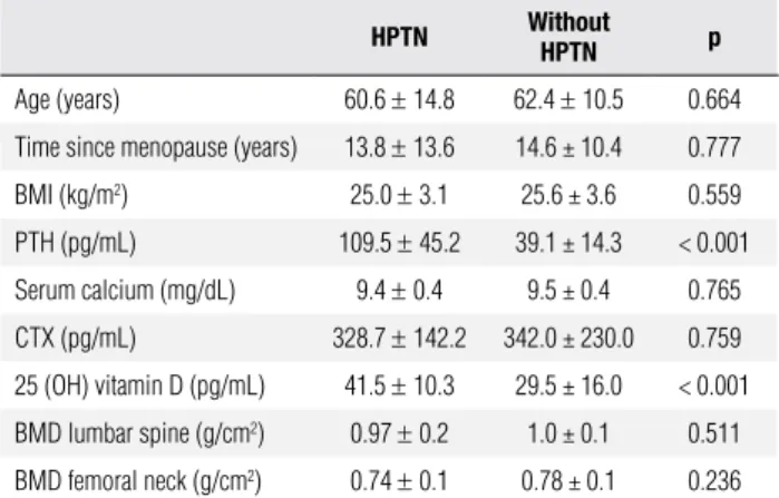
Table 1. Baseline characteristics and biochemical data of patients with and
without NPHPT
HPTN Without HPTN p
Age (years) 60.6 ± 14.8 62.4 ± 10.5 0.664
Time since menopause (years) 13.8 ± 13.6 14.6 ± 10.4 0.777
BMI (kg/m2) 25.0 ± 3.1 25.6 ± 3.6 0.559
PTH (pg/mL) 109.5 ± 45.2 39.1 ± 14.3 < 0.001
Serum calcium (mg/dL) 9.4 ± 0.4 9.5 ± 0.4 0.765
CTX (pg/mL) 328.7 ± 142.2 342.0 ± 230.0 0.759
25 (OH) vitamin D (pg/mL) 41.5 ± 10.3 29.5 ± 16.0 < 0.001
BMD lumbar spine (g/cm2) 0.97 ± 0.2 1.0 ± 0.1 0.511
BMD femoral neck (g/cm2) 0.74 ± 0.1 0.78 ± 0.1 0.236
In the overall analysis, mean age was 62.2 years. Only one patient was of childbearing age, the others were menopausal for an average of 14.5 years. Mean BMI was 25.5 kg/m2 and there were no statisti-cal differences between those with normostatisti-calcemic hyperparathyroidism and the other patients. Overall mean serum calcium, corrected for albumin, was 9.4 mg/dL in the group with normocalcemic hyperpara-thyroidism, a value similar to that of the patients wi-thout the disorder, which was 9.5 md/dL. In the as-sessment of 25-hydroxy vitamin D status, excluding the group with normocalcemic hyperparathyroidism − in which 25OHD deicit was an exclusion criterion − 26.1% of the patients had levels below 20 ng/mL, and 62% had levels below 30 ng/mL.
The analysis of serum PTH showed mean values of 39.1 pg/mL and 109.5 pg/mL in the groups without and with normocalcemic hyperparathyroi-dism, respectively (p = 0.001). Serum levels of C-telopeptide were similar in both groups.
Cop
yright
© ABE&M t
odos os dir
eit
os r
eser
vados
.
317 Arq Bras Endocrinol Metab. 2011;55/5
A more recent study, conducted by Maruani and cols., in 2003, demonstrated that a signiicant pro-portion of patients with normocalcemic hyperpara-thyroidism have lower kidney and bone sensitivity to the biological effect of PTH. The explanation for this lower sensitivity needs further investigation (13).
With the increasing medical concern on bone he-alth, some specialists in bone metabolism routinely perform PTH measurement when there is a decrease in bone mineral density, making it possible to iden-tify patients with normocalcemic hyperparathyroi-dism. The main issue now is whether normocalcemia is a guarantee of the indolent nature of this clinical condition.
Kidney stones were found in 28.6% of the women with normocalcemic hyperparathyroidism. It is there-fore possible that NPHPT has a phenotype similar to that of the classic form of the disease, which leads us to believe that this clinical condition may cause some adverse health outcomes, deserving greater attention.
In the study by Lowe and cols., involving 37 pa-tients with NPHPT, 11% had fractures and 2.5% had kidney stones, which is less than we found in our stu-dy. They found osteoporosis in 57% of the patients versus 35.7% of our patients. They also demonstra-ted that the sites preferentially affecdemonstra-ted by osteopo-rosis were the femoral neck and lumbar spine, unlike the results of our study, in which the most affected site was the distal one-third of the radius (3).
In conclusion, our data suggest that NPHPT has a diverse phenotypic presentation, which may resem-ble, in some way, the hypercalcemic form of prima-ry hyperparathyroidism. As this was a retrospective study based on medical records, further studies are needed to support this conclusion.
Disclosure: no potential conlict of interest relevant to this article was reported.
REFERENCES
1. Silverberg SJ, Bilezikian JP. Primary hyperparathyroidism. Endo-crinology. 2001;1075-93.
2. Silverberg SJ, Lewiecki EM, Mosekilde L, Peacock M, Rubin MR. Presentation of asymptomatic primary hyperparathyroidism: proceedings of the third international workshop. J Clin Endocri-nol Metab. 2009;94(2):351-65.
3. Lowe H, McMahon DJ, Rubin MR, Bilezikian JP, Silverberg SJ. Normocalcemic primary hyperparathyroidism: further charac-terization of a new clinical phenotype. J Clin Endocrinol Metab. 2007;92:3001-5.
4. Lundgren E, Rastad J, Thurfjell E, Akerstrom G, Ljunghall S. Po-pulation based screening for primary hyperparathyroidism with serum calcium and parathyroid hormone values in menopausal women. Surgery. 1997;121:287-94.
5. Bilezikian JP, Potts JT. Asymptomatic primary hyperparathyroi-dism: new issues and new questions bridging the past with the future. J Bone Miner Res. 2002;17(suppl 2):N57-67.
6. Glendenning P, Gutteridge DH, Retallack RW, Stuckey BG, Kermo-de DG, Kent GN. High prevalence of normal total calcium and in-tact PTH in 60 patients with proven primary hyperparathyroidism. Aust N Z J Med. 1998;2:173-8.
7. Forster J, Monchik JM, Martin HF. A comparative study of serum ultrafiltrable, ionized, and total calcium in the diagnosis of prima-ry hyperparathyroidism in patients with intermittent or no eleva-tion in total calcium. Surgery. 1988;04:1137-42.
8. Muldowney FP, Freaney R, McMullin JP, Towers RP, Spillane A, O’Connor P, et al. Serum ionized calcium and parathyroid hormo-ne in renal stohormo-ne disease. Q J Med. 1976;45:75-86.
9. Rao DS, Wilson RJ, Kleerekoper M, Parfitt AM. Lack of biochemi-cal progression or continuation of accelerated bone loss in mild asymptomatic primary hyperparathyroidism. J Clin Endocrinol Metab. 1988;67:1294-8.
10. Parfitt AM, Rao DS, Kleerekoper M. Asymptomatic primary hyper-parathyroidism discovered by multichannel biochemical scree-ning: clinical course and considerations bearing on the need for surgical intervention. J Bone Miner Res. 1991;6(Suppl 2):S97--S101; discussion S121-4.
11. Silverberg SJ, Shane E, Jacobs TP, Siris E, Bilezikian JP. A 10-year prospective study of primary hyperparathyroidism with or wi-thout parathyroid surgery. N Engl J Med. 1999;341:1249-55. 12. Silverberg SJ, Bilezikian JP. “Incipient” primary
hyperparathyroi-dism: a “forme fruste” of an old disease. J Clin Endocrinol Metab. 2003;88(11):5348-52.
13. Maruani G, Hertig A, Paillard M, Houillier P. Normocalcemic pri-mary hyperparathyroidism: evidence for a generalized target-tis-sue resistance to parathyroid hormone. J Clin Endocrinol Metab. 2003;88(10):4641-8.