Authors
Samara Rodrigues Moreira Eloi1
José Luiz Nishiura1
Ita Pfeferman Heilberg1
1Polycystic Kidney
Outpa-tient Clinics of the Federal University of São Paulo (Universidade Federal de São Paulo – UNIFESP)
Submitted: 4/19/2010 Accepted: 9/21/2010
Corresponding author: Profa. Dra. Ita Pfeferman Heilberg
Rua Botucatu, 740. Vila Cle-mentino – São Paulo – São Paulo - Brasil
CEP: 04023-900
Phone: 55 (11) 5904-1699 Fax.: 55 (11) 5904-1684 E-mail: ipheilberg@nefro. epm.br
Financial support: CAPES - Coordenação de Aperfeiçoamento de Pesso-al de Nível Superior.
This study was carried out at the Federal University of São Paulo.
We declare no conflict of interest.
A
BSTRACTIntroduction: Pain is a common symptom in patients with autosomal dominant polycys-tic kidney disease (ADPKD), affecting around 60% of cases. Objective: Translate a pain questionnaire developed and valida-ted for ADPKD in USA into Portuguese and to perform its cultural adaptation and apply it. Method: The cultural adaptation perfor-med by a panel of experts resulted in small changes consisting of words substitution by synonyms or deletion of terms not com-monly used in our culture in 12 out of the 46 questions posed, to solve patients diffi-culties in understanding the questionnaire. Results: There has been equivalence betwe-en the adapted form of the instrumbetwe-ent with the back-translation. The final form of the questionnaire applied in 97 patients with ADPKD (64F/33M, 35 ± 12 years) showed that 65 (67%) had isolated or associated pain in multiple locations , more often at lumbar region (77%), followed by abdomi-nal (66%), headache (15%) and chest (4%). The questionnaire revealed that after family history, pain was the second factor contri-buting to the diagnosis of ADPKD in this population (55% and 22% of cases, respec-tively). Discussion: Clinical and laboratory data from medical records showed that pa-tients referring pain had renal volume and size of the largest cyst significantly higher than those without pain. Conclusion: We conclude that the use of a specific pain ques-tionnaire for ADPKD population provided a better characterization of this symptom, as well as its relationship with the associated complications that commonly occur in this setting.
Keywords: polycystic kidney diseases, pain measurement, low back pain.
[J Bras Nefrol 2010;32(4): 386-399]©Elsevier Editora Ltda.
Translation, cultural adaptation and aplication of a pain
questionnaire for patients with polycystic kidney disease
I
NTRODUCTIONAutosomal dominant polycystic kidney disease (ADPKD), characterized by the progressive development of bilateral renal cysts, has an incidence ranging from 1:400 to 1:1000 live births. It accounts for up to 10% of the patients with end-stage renal disease (ESRD) undergoing renal repla-cement therapy (dialysis or transplanta-tion).1,2 In ADPKD, cysts may be found in
other organs, such as the spleen, pancreas, and, most commonly, the liver.2,3 In
addi-tion, other extra-renal manifestations, su-ch as abdominal wall hernias, diverticu-litis, valvular abnormalities, and cerebral aneurysms, can be found.4,5 Among the
renal manifestations of ADPKD, the follo-wing are the most common: arterial hyper-tension; urinary infection; nephrolithiasis; low back pain; hematuria; increased kid-ney volume; and progressive loss of renal function.6 Arterial hypertension is an
ear-ly and frequent complication of ADPKD. It affects 60% of the patients, even before renal function impairment, probably due to stimulation of the renin-angiotensin--aldosterone system caused by the growth of the cysts.7 Hematuria can result from
the rupture of cysts8 or from the presence
of kidney stones that are very frequent in ADPKD due to anatomical and/or meta-bolic abnormalities.9,10,11 Urinary tract
in-fection (UTI) and inin-fection of the cysts are also frequent in ADPKD.12 End-stage
re-nal disease, which occurs in almost 50% of the patients with ADPKD until the sixth decade of life,5 results from the
re-duction in the renal parenchyma replaced by the cysts, and from vascular sclerosis and interstitial fibrosis.1,2 The diagnosis
innumerous conditions associated. Ultrasonography is the most used imaging test for the diagnosis of ADPKD, and has a sensitivity close to 100% for indi-viduals over the age of 30 years suspected of having ADPKD;13 for younger individuals, nuclear magnetic
resonance is the method of choice.14
Although ADPKD usually remains asymptomatic for many years, pain is a common symptom, and can even affect 60% of the adult population with that di-sease.15 Bajwa et al16 have reported the following most
frequent locations of pain in that population: low ba-ck; abdomen; chest; lower limbs; and head. The low back and/or abdominal pain in patients with ADPKD is multifactorial. It can occur acutely due to cyst ruptu-re or infection, kidney stone elimination, and UTI, or can be chronic resulting from increased kidney or liver volume, due to cyst expansion,6 or even from
diverti-culitis. As the cysts are associated with excessive an-giogenesis, polycystic kidneys are specially susceptible to traumas, which can lead to hemorrhage or bleeding in the retroperitoneal space, usually accompanied by intense pain.17 Intracystic hemorrhages occur usually
in 90% of ADPKD patients and are characterized by the presence of hyperdense cysts on imaging tests.18
Some patients do not associate the pain with the cysts, and, thus, the episodes of cystic hemorrhage can pass undiagnosed when gross hematuria is absent.18 The
headache of ADPKD patients can be related to the presence of cerebral aneurysms, but the frequency of headache does not differ from that of the general po-pulation.19 Thus, that diversity of factors turns pain
into a challenging diagnosis in such patients. The pain reported by ADPKD patients is usually treated with analgesics, but, when the pain is severe, special thera-pies, such as transcutaneous stimulation, use of local anesthetics, or even open or laparoscopic surgery, may be required.20,21
The high percentage of ADPKD patients repor-ting pain emphasizes the importance of a specific pain questionnaire that better characterizes that sympto-matology, providing better assessment to determine more adequate therapeutic measures for each patient. The cost and complexity involved in the elaboration of a questionnaire can be minimized by the use of a translated questionnaire already validated in other countries.22 However, transcultural adaptation of such
questionnaires is required for their use in each coun-try.23,24 The present study aimed at performing the
Portuguese translation and transcultural adaptation of a pain questionnaire specific for ADPKD patients, de-veloped and validated by Bajwa et al16 in 2004, and at
its application in a sample of ADPKD patients.
P
ATIENTS ANDMETHODSThe pain questionnaire developed and validated by the group of Steinman for the North-American po-pulation with ADPKD16 has 46 questions. It
inclu-ded since how and when the diagnosis of ADPKD was made to pain characteristics, such as location, frequency, intensity, and associated pathologies. The initial translation of the pain questionnaire in-to Portuguese was performed by a nephrologist and revised by a multidisciplinary team (two physicians, one nurse, two biologists, and two nutritionists), ai-ming at assessing the clarity and understanding of the questions. The differences found or expressions consi-dered difficult to be understood by the lay population were replaced with a more simple language, in such a way not to change the conceptual translation of the question. Eventually, the version considered definitive underwent backtranslation by an independent trans-lator, aiming at comparing the Portuguese translated and the English original versions.
SELECTION OFPATIENTS
One hundred adult patients, followed up at the Polycystic Kidney Outpatient Clinics of the Discipline of Nephrology of the Federal University of São Paulo (UNIFESP) were selected. The diagnosis of ADPKD was made based on the presence of a family history of the disease (an affected parent) and data obtained with kidney ultrasonography, meeting the criteria proposed by Pei13 for each age bracket. Data regarding kidney
volume, number of cysts, and size of the largest cyst were obtained from the ultrasound reports on the me-dical records. Kidney volume was determined by use of the formula of the modified ellipsoid = 4/3 π x (ante-roposterior diameter/4 + width/4)2 x length/2,25 and, in
the present study, it was considered as the sum of the volumes of both kidneys.26
records. Chronic kidney disease (CKD) stage 1 was de-fined as kidney damage caused by the presence of cysts on imaging tests, and CrCl ≥ 90 mL/min/1.73m2. Stage
2 was defined as the presence of kidney damage and CrCl between 60 and 89 mL/min/1.73m2; and stage 3
was defined as CrCl below 60 mL/min/1.73m2.27
APPLICATIONOFTHEQUESTIONNAIRE
The questionnaire was applied to 97 patients out of the 100 previously selected, considered as having a good capacity of understanding. They had no pain at the time of questionnaire application to reduce the possibility of influencing their responses. Three pa-tients refused to or could not participate in the study because they could not understand the questions pro-posed. The questionnaire was individually applied at a private room, with no interference of a third party, for a mean time of 15 minutes. All patients included in the study were informed about the research and provided written informed consent. The study was ap-proved by the Committee on Ethics of the UNIFESP.
Pain intensity was measured by use of the visual analogue scale (VAS),28 present in the original
ques-tionnaire16 and used in clinical practice and other
stu-dies. That scale comprises a score ranging from zero to ten, where zero corresponds to lack of pain and ten corresponds to maximum pain intensity.16
STATISTICALANALYSIS
The numerical variables were expressed as medians (minimum value – maximum value), and the Mann-Whitney test was used to compare the groups. The categorical variables were expressed as numbers and
percentages, and the Chi-square test and Fisher exact test, when indicated, were used for comparisons. Non-parametric tests were used due to lack of nor-mal distribution of the numerical variables between both groups. The significance level adopted for the statistical tests was 5% (p value < 0.05), and the sof-tware used for the analyses was the SAS System for Windows (Statistical Analysis system), version 8.02.
R
ESULTSTRANSLATIONANDTRANSCULTURALADAPTATIONOFTHE
QUESTIONNAIRE
The initial translation from English to Portuguese was assessed by the multidisciplinary team. Some expres-sions originally used required changes to be better un-derstood by the patients. The first change was in the title of the questionnaire adopted in the English origi-nal version: from “The questionnaire employed in the PKD population – headache and pain in polycystic kidney disease project” to “Questionnaire for patients with polycystic kidney disease”. As shown in Table 1, only some words present in 12 of the 46 questions re-quired changes for adaptation to local culture. Those changes consisted basically in replacing some words or expressions by synonyms, or in including between parentheses terms to which no equivalent was found in Portuguese, aiming at enhancing understanding by the patients. For example, as shown in Table 1, in item D3, in the description of the type of chest pain, the term “undefined” was added between parentheses to the expression “aching”, to exclude all other adjec-tives for defining that pain (stabbing, dull pressure, cramping, intermittent, and continuous). That change
Table 1 MODIFICATIONSOFTHEDESCRIPTORSOFTHEORIGINALQUESTIONNAIRESUGGESTEDBYTHEMULTIDISCIPLINARYTEAM
Item of the questionnaire/ words Initial translation (Version 1) Modification (Version 2)
10/ Has your pain ever been associated with blood in your urine?
Alguma vez esta dor “associou-se” com sangue na urina?
Alguma vez esta dor foi
acompanhada de sangue na urina?
A4/... Ice massage ... “Massagem fria” ... “Gelo”
A5/... Abdominal fullness? ... “Sensação de plenitude abdominal”? ... “sensação de estômago cheio”?
D3/ Dull press Pressão “incômoda” Pressão “desagradável”
D3/Type of pain ...Aching Tipo de dor ... “dolorida” Tipo de dor: ... “dolorida” (indefinida)
D3/Type of pain…Intermittent “Intermitente” “Intermitente” (que vai e volta)
E3/ Front of the head Região “frontal” “Frente da cabeça”
E3/ Back of the head Região “occipital” “Atrás da cabeça”
E4/ …Throbbing ... “Pulsátil” ... “Latejante”
6 a/... “Aura” ... “Aura” ... “Sensação diferente”
6 a/... Nausea ... “Náusea” ... “Enjoo”
was performed to not jeopardize that item in the ba-cktranslation, and also to better express missing ter-ms or those that made no sense in Portuguese. In the same question, we added the expression “that comes and goes” to the term “intermittent”, and replaced the adjective in the expression “dull pressure” by “un-pleasant” to improve understanding. Backtranslation performed by an independent translator showed no conceptual difference when compared to the text of the English original questionnaire. When assessing the equivalence between the backtranslation and the English original version of the questionnaire, it was evident that only a few items had been altered, because the translation suppressed and/or modified certain words. Those changes were aimed at pro-viding semantic equivalence (equivalence between words) and idiomatic equivalence (items that nee-ded to be replaced). For example, expressions of the English original questionnaire, such as “do you ex-perience abdominal pain?” translated and adapted to Portuguese as “você tem dor abdominal?” were backtranslated as “do you have abdominal pain?”. In other words, the term “experience” disappeared from the backtranslation because it is not part of an usual expression in Portuguese, being suppressed already in the phase of translation and adaptation. The final version of the questionnaire later applied to patients is found in the Appendix.
APPLICATIONOFTHEQUESTIONNAIRE
Ninety-seven adult patients with ADPKD (64 F/33 M; 35 ± 12 years) were assessed by use of a pain ques-tionnaire, 57% being Caucasians, 18% afro descen-dents, 21% mixed heritage, and 2% Asians. Figure 1 shows the percentage of patients with a family history of ADPKD and signs and/or symptoms contributing to the diagnosis of ADPKD. The presence of a family history of ADPKD was the most contributing factor
for the diagnosis, and was observed in 55% of the patients. Other elements leading to the diagnosis of ADPKD were: pain (22%); assessment of hyperten-sion or hematuria (18%); UTI (6%); and incidental findings on periodic tests or medical consultations not related to nephrology (9%). The sum of all factors reported by patients exceeds 100% (110%), because some patients looked for medical assessment due to the presence of some symptoms and/or because they also had a family history of ADPKD, although they did not relate the symptom reported to ADPKD prior to the diagnosis.
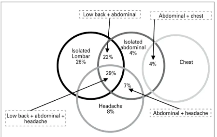
According to the answers to the pain questionnai-re, pain at any location was present in 67% (65/97) of the patients. Low back pain was more frequent when considered in isolation (26%), followed by heada-che (8%), and abdominal (4%) or heada-chest pains (4%). However, patients most commonly reported pain in its associated form, as shown in Figure 2, which hi-ghlights the greater frequency of the association be-tween low back and/or abdominal pains (52% of the reports of pain) and the additional association with headache (36% of the reports). Based on the patients’ report in the present series, the following percenta-ge distribution of pain was observed: low back pain, 77%; abdominal pain, 66%; headache, 15%; and chest pain, 4%.
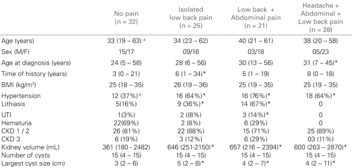
Table 2 shows the clinical and ultrasonographic characteristics of the patients who reported isola-ted low back pain, the association of low back and abdominal pains, and the association of low back and abdominal pains with headache. Patients wi-th isolated low back pain had a longer history as compared with the group with no pain. The kidney volume of patients with isolated low back pain, or with the association of abdominal pain and/or hea-dache was significantly greater than that of patients with no pain. A higher percentage of patients with
Figure 1. Family history, symptoms and/or signs leading to the diagnosis of ADPKD.
55% 60%
50%
40%
30%
20%
10%
0%
13%
Family history
9% 9% 9%
6%
Low back pain
Abdominal pain
Hematuria Incidental findings
UTI
% of patients
Figure 2. Location of the pain (% of patients).
Isolated Lombar 26%
Isolated abdominal
4%
Headache 8%
Chest 29%
7% 4% 22%
Low back + abdominal + headache
hypertension and lithiasis was observed in all groups with pain. However, even excluding hypertensive pa-tients from the analysis, the kidney volume of nor-motensive patients with pain (n = 26) was also sig-nificantly greater than that of normotensive patients with no pain (n = 20), 430 mL vs 315 mL, respecti-vely (p < 0.036) (data not shown in table). When pa-tients with lithiasis are excluded, the kidney volume of ADPKD patients without lithiasis but with pain (n = 41) is significantly greater than that of patients without pain (n = 27), 591 mL vs 335 mL, respec-tively (p < 0.001). As the total number of patients reporting isolated abdominal pain (n = 4) or isolated headache (n = 8) was small, those patients were not included in the statistical analysis. A greater number of patients with UTI was observed in the group of low back pain associated with abdominal pain, and a BMI significantly greater was observed in patients with isolated abdominal pain. No statistically sig-nificant difference was observed in age, number of cysts, and loss of renal function between the groups. The female sex predominated in the total group of pain, especially in that of low back pain.
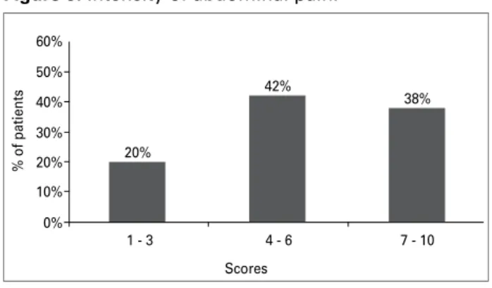
Figure 3 shows that 46% of the patients with low back pain reported its intensity as moderate (scores 4 - 6). The frequency of low back pain was once a week in 39% of the patients (Figure 4). Among pa-tients with abdominal pain, 42% reported its inten-sity as moderate (scores 4 - 6) (Figure 5), and 37% as less frequently than once a month (Figure 6).
Patients complaining of headache described their pain as throbbing (44%), pressure (28%), pounding (8%), exploding (5%), and stabbing (3%), occur-ring once a week in 36% of the cases.
Table 2 CLINICALANDULTRASONOGRAPHICCHARACTERISTICS
No pain (n = 32)
Isolated low back pain
(n = 25)
Low back + Abdominal pain
(n = 21)
Headache + Abdominal + Low back pain
(n = 28)
Age (years) 33 (19 – 63) a 34 (23 – 62) 40 (21 – 61) 38 (20 – 58)
Sex (M/F) 15/17 09/16 03/18 05/23
Age at diagnosis (years) 24 (5 – 58) 28 (6 – 56) 30 (13 – 56) 31 (7 – 45)*
Time of history (years) 3 (0 – 21) 6 (1 – 34)* 5 (1 – 19) 8 (0 – 18)
BMI (kg/m2) 25 (18 – 35) 26 (19 – 36) 25 (19 – 35) 25 (19 – 35)
Hypertension Lithiasis
12 (37%) b
5(16%) 16 (64%)* 9 (36%)* 16 (76%)* 14 (67%)* 18 (64%)* 0 UTI Hematuria CKD 1 / 2 CKD 3
Kidney volume (mL) Number of cysts Largest cyst size (cm)
1(3%) 22(69%) 26 (81%) 6 (19%) 361 (180 - 2482)
15 (4 – 15) 3 (2 – 6)
2 ((8%) 2 (8%) 22 (88%)
3 (12%) 646 (251-2150)*
15 (4 – 15) 5 (2 – 8)*
3 (14%)* 6 (29%) 15 (71%)
6 (29%) 657 (216 – 2394)*
15 (4 – 15) 4 (2 – 7)*
0 0 25 (89%) 03 (11%) 600 (263 – 2870)*
15 (4 – 15) 4 (2 – 11)*
a Median (minimum – maximum); b number (percentage) of patients; *p < 0.05 vs no pain.
Figure 3. Intensity of low back pain.
60% 50% 40% 30% 20% 10% 0% 20% 35% 46%
1 - 3 4 - 6 7 - 10
% of patients
Scores
Figure 4. Frequency of low back pain.
% of patients
50% 40% 30% 20% 10% 0%
Always once a day
once a week
once a month
D
ISCUSSIONResearchers interested in better assessing and charac-terizing specific conditions in the health area, but who do not have instruments built in their countries and languages, can choose to create a new instrument or adapt questionnaires already validated in another lan-guage. Factors such as time and cost for elaborating a questionnaire lead most researchers to choose trans-lation and transcultural adaptation of questionnaires already existing in other languages.22,24,29 Guillemin et al30 have standardized the process of translation
and transcultural adaptation into five steps: transla-tion; review of the translation by a multidisciplinary committee; translation into the original language (ba-cktranslation); pre-test to assess cultural equivalence; and assessment of the need to attribute weight to the scores. However, the simplification of that method has already been suggested by Da Mota-Falcão et al,31
who have reported that even simplified questionnai-res still satisfy the objectives proposed.
Pain has been reported as a very frequent symptom of ADPKD patients, affecting up to 60% of them15,32
and jeopardizing their quality of life.33 Because that
symptom involves a series of sensorial qualities and subjective aspects,22,32 it requires a careful
investi-gation by a multiprofessional team. Duarte et al23
have emphasized the importance of using proper instruments to assess the situations and experiences
in the health area, so that reliable information is ob-tained. Despite the existence of some pain questio-nnaires translated and validated in Brazil,22,34 none
of them has the necessary and/or adequate specifi-cations to assess pain in ADPKD. Thus, this study aimed at performing a translation and transcultural adaptation of an English pain questionnaire, already validated to be used in ADPKD patients.16 That
va-lidated pain questionnaire is currently being used in the HALT PKD study.35 A simplified version of the
method developed by Guillemin30 was adopted, since
the questionnaire to be used in our study approa-ched simple questions, which did not require weight attributions, using only scores of pain intensity of the visual analogue scale (VAS).16,28
The modification of the title of the pain question-naire adopted in this study to “Questionquestion-naire for pa-tients with polycystic kidney disease”, suppressing the subtitle of the English original version containing the terms “pain” and “headache”, and the simplification of the terminology aimed at neither inducing patients to report non-existing pains nor overestimating the value of existing pains. In addition, in our opinion, our decision not to include patients with pain on the occasion of completing the questionnaire contributes to legitimate the results, a preoccupation not expres-sed by the authors of the English original version, but which we consider important.
The comparison of the initial translation and that suggested by the multidisciplinary team provided equivalence and reconciliation between most items. In the phase of transcultural adaptation, the use of more simple expressions or synonyms, in addition to the suppression of words in the backtranslation, has alre-ady occurred in other studies of translation and trans-cultural adaptation performed in Brazil,22,24 without
jeopardizing the final result of the study. Differently from the English original questionnaire that was self--applied,16 in our study, the questionnaire was
ap-plied during the pre-consultation, so that the greatest amount of information could be obtained from the patient, in addition to preventing and minimizing the number of non-responders usually found with self--applied questionnaires. However, the questionnaire proved to be of rapid and easy application, and can be self-applied in the future.
By applying the questionnaire in our ADPKD sample, 67% (65/97) of the patients assessed could be identified as having some type of pain, a preva-lence similar to that reported in the literature, which is around 60%.6,15,22,32 The use of that
questionnai-re has also evidenced that the pquestionnai-resence of a family
Figure 5. Intensity of abdominal pain.
60%
50%
40%
30%
20%
10%
0%
20%
38% 42%
1 - 3 4 - 6 7 - 10
% of patients
Scores
Figure 6. Frequency of abdominal pain.
% of patients
50%
40%
30%
20%
10%
0%
7% 7%
22% 26%
37%
Always once a day
once a week
once a month
history of ADPKD was the finding that most frequen-tly led to the diagnosis of ADPKD (55% of the ca-ses). However, low back pain was the symptom most frequently reported by the patients (13%), and, when added to abdominal pain, it reached 22% of the pa-tients, confirming that pain can contribute to the diagnosis of ADPKD.16 In addition, it is worth noting
that of the patients who sought a specialist due to a positive family history, 21% reported pain before the diagnosis, but had not related it to polycystic kidney disease, in accordance with that reported by Bajwa et al.16 Low back and abdominal pains were the most
frequent ones, followed by headache and chest pain, as reported by Bajwa et al.16 and, in Brazil, by Romão et al.36 The present study evidenced that those pains
(low back, abdominal, and headache) were reported in isolation or, more frequently, in association, which is also in accordance with the study by Bajwa et al,16
in which 70% of the patients had more than one type of pain, and in only 18% the pain had a single loca-tion. The frequencies of low back (once a week) and abdominal (less than once a month) pains found in our study differed from those of the North-American population, which characterized those pains as conti-nuous. The intensity of the low back/abdominal pains was similar to that in the literature.16 The reasons
why the patients reported a lower recurrence of the episodes of low back/abdominal pain in the present series are not clear, but that finding may be attribu-ted to a shorter time interval between the diagnosis of ADPKD and the application of the questionnaire, which was six years, while in the study by Bajwa et al16 that interval was around 16 years. In addition, as
the English original questionnaire was self-applied, it may have caused an overestimation of the frequency of pains that can or cannot be related to polycystic kidney disease.
In the present study, the female sex predominated among patients reporting pain in general. In the study by Bajwa et al,16 the number of women in the total
sample was also greater. However, because of the cha-racteristics of the present instrument, the greater re-port of pain in women could represent a bias of pains originating from the female genital tract not related to ADPKD.
Regarding the clinical manifestations and labora-tory alterations and their associations with the pre-sence of pain, the present sample showed a greater percentage of hypertensive patients with associated nephrolithiasis in the groups of patients with pain, especially low back and/or abdominal pain. Although UTI episodes were slightly more frequent in the total
group with pain, the number of confirmations with urine cultures on medical records was small, thus, hindering the relevance of that finding. Surprisingly, microscopic hematuria did not differ between the groups with and without pain. As hematuria is usu-ally related to the rupture of cysts,1,37 the urine
sedi-ment exams reported on the medical records, required on a routine basis and not during an episode of pain, may explain the lack of a temporal relation between hematuria and the episodes of pain in our study. As low back pain could be related to excessive weight, we also assessed the relation between the presence of pain and BMI. That association, however, has not be-en detected.
Although the median age of patients with isolated or associated low back pain and that of patients with no pain did not differ, the median age at the diag-nosis of ADPKD tended to be greater in the groups with low back and abdominal pains (associated or not with headache), despite the lack of statistical sig-nificance. The estimated time of history (from diag-nosis to application of the questionnaire) was longer in patients with pain, but statistical significance was reached only by those with isolated low back pain. Still, in accordance with data reported by Bajwa et al,16 the pain in the present sample appeared early in
the course of ADPKD, when approximately 80% of the patients still had normal renal function. Thus, no association was observed between pain and alteration in renal function, emphasizing that pain is not a late manifestation in the course of ADPKD, as already re-ported by other authors.16
The progressive increase in kidney volume do-es not associate only with the deterioration of renal function in ADPKD,26,38 but also with the presence
of hypertension39 and nephrolithiasis, as previously
reported by Nishiura et al 10 at our service, and
In conclusion, the results of this study emphasize the importance of the use of a specific instrument to assess pain in patients with ADPKD. The pain ques-tionnaire revealed that pain, in addition to being a very frequent and early symptom in the course of ADPKD, can help in the early diagnosis of that disea-se. The use of that tool has allowed a better characte-rization of pain location, intensity, and frequency, in addition to the assessment of the association of pain
with other clinical, laboratory, and imaging manifes-tations in ADPKD. A greater frequency of low back and/or abdominal pains and its significant association with the increase in kidney volume has been observed. Thus, we believe that the use of a specific pain ques-tionnaire for assessing ADPKD patients will draw attention to the pain complaints of that population, contributing to the follow-up and control of their as-sociated complications.
DATE OF COMPLETION _____/_____/____
Name :
___________________________________________________________________________
Address:
___________________________________________________________________________
Age: _____ Sex: M [ ] F [ ] Birth date: __/__ /__
Race: Black [ ] Caucasian [ ] Asian [ ] Other [ ]
1. Year you were first aware that you had polycystic kidney disease (PKD): __________
2. Age at diagnosis: ______
3. What led to the diagnosis? (please explain)
• Finding during evaluation of another problem [ ]
• Routine evaluation done because of family history of polycystic kidneys [ ]
• Symptoms led to a physician ordering exams [ ]
• Abdominal pain [ ]
• Low back pain [ ]
• Blood in the urine [ ]
• Other [ ] Please, explain:
________________________________________________________________________
4. Do you know of any blood relatives who have polycystic kidneys: Y [ ] N [ ]
5. If yes, please list with relationship and age of relative at the time of diagnosis of polycystic kidneys:
Age Relationship
Age at time of test Results reported to you
Abdominal ultrasound Intravenous pyelogram Abdominal CT scan Cerebral CT scan Abdominal MRI Cerebral MRI
6. Which diagnostic tests have you had for polycystic kidneys? Please, list the results:
7. Did you experience abdominal or low back pain before the diagnosis of polycystic kidneys? Y [ ] N [ ]
8. How was that pain treated before the diagnosis of polycystic kidneys?
9. Since the diagnosis of polycystic kidneys, have you experienced persistent or chronic pain in the following locations?
Mark with an X, Yes or No:
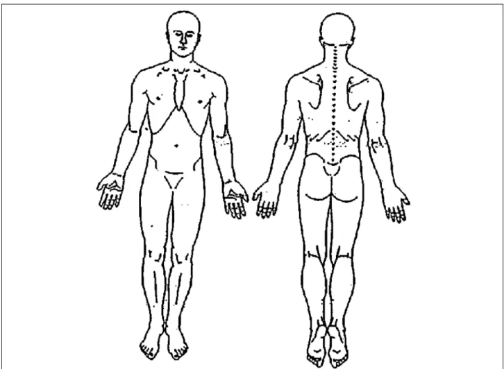
Head – Y [ ] N [ ] Chest - Y [ ] N [ ] Back – Y [ ] N [ ] Abdomen Y [ ] N [ ] Legs – Y [ ] N [ ]
10. Did you ever think that those pains were related to polycystic kidneys? Y [ ] N [ ]
11. Has that pain ever been associated with blood in your urine? Y [ ] No [ ]
Section A – Abdominal pain
1. Do you experience abdominal pain? Y [ ] N [ ]
If your answer is yes, please mark the location on Chart 1 (front and back) on the last page and complete this section. If your answer is no, please skip to Section B.
2. How often? [ ] always
[ ] at least once a day [ ] about once a week [ ] about once a month [ ] less than once a month
3. Can you rate this pain on average by placing an X on the following scale:
(no pain) 0 1 2 3 4 5 6 7 8 9 10 (worst pain possible)
4. Have you had any of the following treatments for this pain? Mark with an X, Yes [ ] or No [ ]
a) surgery: Y [ ] N [ ] b) analgesics Y [ ] N [ ]
If yes, please name:_______________________________ c) ice Y [ ] N [ ]
d) heat Y [ ] No [ ]
5. Do you experience stomach fullness? Y [ ] No [ ]
6. Do you feel full after eating a small amount of food? Y [ ] N [ ] 7. Is your appetite poor? Y [ ] No [ ]
8. Is your appetite poor because of stomach fullness? Y [ ] N [ ]
9. Is your appetite poor because of nausea? Y [ ] N [ ]
Section B – Back pain
1. Do you experience low back pain? Y [ ] N [ ]
If your answer is yes, please mark the location of pain on Chart 1 attached and complete this section. If your answer is no, please skip to Section C.
2. How often? [ ] always
[ ] at least once a day [ ] about once a week [ ] about once a month [ ] less than once a month
Section C – Back pain shooting to the hips or legs
1. Do you experience back pain shooting down to your hips or legs? Y [ ] N [ ]
If yes, please mark the location on Chart 1 attached and complete this section. If no, skip to Section D.
2. How often? [ ] always
[ ] at least once a day [ ] about once a week [ ] about once a month [ ] less than once a month
3. Can you rate this pain on average by placing an X on the following scale:
(no pain) 0 1 2 3 4 5 6 7 8 9 10 (worst pain possible)
4. Have you had any of the following treatments for this pain? a) surgery Y [ ] N [ ]
b) analgesics Y [ ] N [ ]
If yes, please name: _________________________ c) ice Y [ ] N [ ]
d) heat Y [ ] N [ ]
Section D – Chest pain
1. Do you experience chest pain? Yes [ ] No [ ]
If your answer is yes, please mark the location of pain on Chart 1 attached and complete this section. If your answer is no, skip to Section E.
2. How often? [ ] always
[ ] at least once a day [ ] about once a week [ ] about once a month [ ] less than once a month
3. Can you rate this pain on average by placing an X on the following scale:
(no pain) 0 1 2 3 4 5 6 7 8 9 10 (worst pain possible) Type of pain:
[ ] stabbing [ ] dull pressure [ ] cramping
[ ] aching (undefined)
[ ] intermittent (that comes and goes) [ ] continuous
4. Have you had any of the following treatments for this pain? a) surgery Y [ ] N [ ]
b) analgesics Y [ ] N [ ] If yes, please name:____________________________ c) ice Y [ ] N [ ]
d) heat Y [ ] N [ ]
e) others (please specify):________________________________________________
Section E - Headache
1. Do you experience chronic headache? Y [ ] N [ ]
2. How often? a) daily b) once a week c) 5 – 10 per month d) once a month e) rarely
4. My headache feels like: a) throbbing
b) pounding c) pressure d) stabbing e) exploding
f) other (please, specify):______________________________________________________________________
5. Have you ever experienced a very strong headache? Y [ ] No [ ]
If the answer is yes, how was it treated? Please, describe:__________________________________________
6. Is your headache:
a) preceded by an “aura” (different sensation): Y [ ] N [ ] b) associated with nausea: Y [ ] N [ ]
c) associated with vomiting: Y [ ] N [ ]
7. Have you ever had a CT or MRI of your head? Y [ ] N [ ]
If yes, result: _______________________________________________________________________________
8. Do you suffer from migraine? Y [ ] N [ ]
Who diagnosed the migraine? ________________________________________________________________
9. Timing – at what time of the day? a) morning
b) afternoon c) evening d) night
10. Have you or a family member ever been diagnosed with a brain hemorrhage? Y [ ] N [ ]
If yes, please explain: _________________________________________________________________________
Section F – List antiallergic medications that you take: ____________________________________________
Section G - List medications for high blood pressure that you take: _________________________________
Section H - List other current medications: ______________________________________________________
Section I – List any other treatment for polycystic kidneys: _________________________________________
Section J – Do you suffer from any other medical problem? If yes, please list: _________________________
Figure 1. Questionnaire for patients with polycystic kidneys.
R
EFERENCES1. Torres VE, Harris PC. Autosomal dominant polycys-tic kidney disease: the last 3 years. Kidney Int 2009; 76:146-168.
2. Chang MY, Ong ACM. Autosomal dominant polycys-tic kidney disease: recent advances in pathogenesis and treatment. Nephron Physiol 2008; 108:1-7.
3. Garcia-Gonzalez MA, Menezes LF, Piontek KB et al. Genetic interaction studies link autosomal dominant and recessive polycystic kidney disease in a common pathway. Hum Mol Genet 2007; 16:1940-50.
4. Torres VE, Harris PC. Mechanisms of disease: autoso-mal dominant and recessive polycystic kidney diseases. Nat Clin Prac Nephrol 2006; 2:40-54.
5. Masoumi A, Reed-Gitomer B, Kelleher C, Bekheirnia MR, Schier RW. Developments in the management of autosomal dominant polycystic kidney disease. Ther Clin Risk Manag 2008; 4:393-407.
6. Grantham JJ, Chapman AB,Torres VE. Volume pro-gression in autosomal dominant polycystic kidney di-sease: the major factor determining clinical outcomes. Clin J Am Soc Nephrol 2006; 1:148-57.
7. Gabow PA, Chapman AB, Johnson AM et al. Renal structure and hypertension in autosomal dominant polycystic kidney disease. Kidney Int 1990; 38:1177-80.
8. Torres VE, Harris PC, Pirson Y. Autosomal dominant polycystic kidney disease. Lancet 2007; 369:1287-301. 9. Torres VE, Wilson DM, Hattery RR, Segura JW. Renal
stone in autosomal dominant polycystic kidney disease. Am J Kidney Dis 1993; 22:513-9.
10. Nishiura JL, Neves RFCA, Eloi SRM, Cintra SMLF, Ajzen SA, Heilberg IP.Evaluation of nephrolithiasis in autosomal dominant polycystic kidney disease patients. Clin J Am Soc Nephrol 2009; 4:838-44.
11. Grampsas SA, Chandhoke PS, Fan J et al. Anatomic and metabolic risk factors for nephrolithiasis in pa-tients with autosomal dominant polycystic kidney dise-ase. Am J Kidney Dis 2000; 36:53-7.
12. Sallée M, Rafat C, Zahar JR et al. Cyst infections in patients with Autosomal Dominant Polycystic Kidney Disease. Clin J Am Soc Nephrol 2009; 4:1183-9. 13. Pei Y, Obaji J, Dupuis A et al. Unified criteria for
ultra-sonographic diagnosis of ADPKD. J Am Soc Nephrol 2009; 20:205-12.
14. Torres VE, King BF, Chapman AB et al. Consortium for radiologic imaging studies of polycystic kidney disease (CRISP); Magnetic resonance measurements of renal blood flow and disease progression in autosomal domi-nant polycystic kidney disease. Clin J Am Soc Nephrol 2007; 2:112-20.
15. Steinman TI. Pain management in polycystic kidney di-sease. Am J Kidney Dis 2000; 35:770-2.
16. Bajwa ZH, Gupta S, Warfield CA, Steinman TI. Pain patterns in patients with polycystic kidney disease. Kidney Int 2004; 66:1561-9.
17. Bello-Reuss E, Holubec K, Rajaraman S. Angiogenesis in autosomal dominant polycystic kidney disease. Kidney Int 2001; 60:37-45.
18. Levine E, Grantham JJ. Calcified renal stone and cyst calcifications in autosomal dominant polycystic kidney disease: clinical and CT study in 84 patients. AJR Am J Roentgenol 1992; 159:77-81.
19. Bajwa ZH, Gupta S, Warfield CA, Steinman TI. Pain management in polycystic kidney disease. Kidney Int 2001; 60:1631-44.
20. Desai PS, Castle EP, Daley SM, Humphreys MR, Andrews PE. Bilateral laparoscopic nephrectomy for significantly enlarged polycystic kidneys: a technique to optimize outcome in the largest of specimens. British Journal Urology Int 2008; 101:1019-23.
21. Lee DI, Andreoni CR, Rehman J et al. Laparoscopic cyst decortication in autosomal dominant polycystic kidney disease: impact on pain, hypertension, and renal function. J Endourol 2003; 6:345-54.
22. Varoli FK, Pedrazzi V. Adapted version of the McGill pain questionnaire to brazilian portuguese. Braz Dent J 2006; 17:328-35.
23. Duarte PS, Miyzaki MCOS, Ciconelli RM, Sesso R. Tradução e adaptação cultural do instrumento de qua-lidade de vida para pacientes renais crônicos. Rev Assoc Med Bras 2003; 49:375-81.
24. Fonseca ESM, Camargo ALM, Castro RA et al. Validação do questionário de qualidade de vida (Kings Health Questionnaire) em mulheres brasileiras com in-continência urinária. Rev Bras Ginecol Obstet 2005; 27 (5): 235-242.
25. Schrier RW, McFann KK, Johnson AM. Epidemiological study of kidney survival in autosomal dominant polycystic kidney disease. Kidney Int 2003; 63:678-85. 26. Grantham JJ, Torres VE, Chapman AB et al. Volume
progression in polycystic kidney disease. N Engl J Med 2006; 354:2122-30.
27. Levey AS, Eckardt K, Tsukamoto Y et al. Definition and classification of chronic kidney disease: a position statement from kidney disease: improving global outco-mes (KDIGO). Kidney Int 2005; 67:2089-100.
28. Lund I, Lundeberg T, Sandeberg L, Budh CN, Kowalski J, Svensson E. Lack of interchangeability between visu-al anvisu-alogue and verbvisu-al rating pain scvisu-ales: a cross sectio-nal description of pain etiology groups. BMC Medical Research Methodology 2005; 5:1-9.
29. Pereira GIN, Costa CDS, Geoczer L. Tradução e vali-dação para a língua portuguesa (Brasil) de instrumen-tos específicos para avaliação de qualidade de vida na doença do refluxo gastresofágico. Arq Gastroenterol 2007; 44:168-77.
30. Guillemin F, Bombardier C, Beaton D. Cross-Cultural adaptation of health related quality of life measu-res: literature review and proposed guidelines. J Clin Epidemiol 1993; 46:1417-32.
31. Da Mota-Falcão D, Ciconelli RM, Ferraz MB.
Translation and cultural adaptation of quality of li-fe questionnaires: an evaluation of methodology. J Rheumatol 2003; 30:379-85.
32. Heiwe S, Bjuke M. “An evil heritage”: interview study of pain and autosomal polycystic kidney disease. Pain Management Nursing 2009; 10:134-41.
33. Rizk D, Jurkovitz C, Veledar E et al. Quality of life in autosomal dominant polycystic kidney disease pa-tients not yet on dialysis. Clin J Am Soc Nephrol 2009; 4:560-6.
34. Calil AM, Pimenta CAM. Intensidade da dor e adequa-ção de analgesia. Rev Latino-Am Enfermagem 2005; 13:692-9.
35. Chapman AB, Torres VE, Perrone RD et al. The Halt polycystic kidney disease trials: design and implementa-tion. Clin J Am Soc Nephrol 2010; 5:102-9.
36. Romão EA, Moyses NM, Teixeira SR, Muglia VF, Neto OMV, Dantas MI. Renal and extrarenal manifestations of autosomal dominant polycystic kidney disease. Braz J Med Biol Res 2006; 39:533-8.
37. Grantham JJ. Autosomal dominant polycystic kidney disease. N Engl J Med 2008; 359:1477-85.
38. Bae KT, Grantham JJ. Imaging for the prognosis of au-tosomal dominant polycystic kidney disease. Nat Rev Nephrol 2010; 6:96-106.