Cop
yright
© ABE&M t
odos os dir
eit
os r
eser
vados
.
Cop
yright
© ABE&M t
odos os dir
eit
os r
eser
vados
.
Effects of endocrine disruptors
in the development of the
female reproductive tract
Efeitos dos desreguladores endócrinos no desenvolvimento do trato reprodutivo feminino
Elaine Maria Frade Costa1, Poli Mara Spritzer2, Alexandre Hohl3, Tânia A. S. S. Bachega4
ABSTRACT
Environmental agencies have identiied a growing number of environmental contaminants that have endocrine disrupting activity, and these can become a major public health problem. It is suggested that endocrine disruptors could account for the higher-than-expected increase in the prevalence of some non-communicable diseases, such as obesity, diabetes, thyroid diseases, and some cancers. Several endocrine Disrupting Chemicals (EDCs), such as pesticides, bisphenol A, phthalates, dioxins, and phytoestrogens, can interact with the female reproductive system and lead to endocrine disrup-tion. Initially, it was assumed that EDCs exert their effects by binding to hormone receptors and trans-cription factors, but it is currently known that they may also alter the expression of enzymes involved in the synthesis or catabolism of steroids. Biomonitoring studies have identiied these compounds in adults, children, pregnant women, and fetuses. Among the diseases of the female reproductive tract associated with EDCs exposure are the following: precocious puberty, polycystic ovary syndrome, and premature ovarian failure. The different populations of the world are exposed to a great number of chemicals through different routes of infection; despite the various available studies, there is still much doubt regarding the additive effect of a mixture of EDCs with similar mechanisms of action. Arq Bras Endocrinol Metab. 2014;58(2):153-61
Keywords
Endocrine disruptors; female development; precocious puberty; polycystic ovary syndrome; premature ovarian failure
RESUMO
As diversas agências de controle ambiental têm identiicado um crescente número de contaminantes ambientais que apresentam atividade de desregulador endócrino e estes poderão se tornar um dos maiores problemas de saúde pública. Sugere-se que os Desreguladores Endócrinos (EDCs) poderiam justiicar o aumento na prevalência de algumas doenças não transmissíveis acima do esperado, como, por exemplo, obesidade, diabetes, doenças tireoidianas e alguns tipos de cânceres. Vários EDCs, como pesticidas, bisfenol A, ftalatos, dioxinas e itoestrógenos, podem interagir com o siste-ma reprodutivo feminino e levar à desregulação endócrina. Inicialmente, supunha-se que os EDCs exercessem seus efeitos através da ligação com receptores hormonais e fatores de transcrição, mas, atualmente, sabe-se que também podem alterar a expressão de enzimas envolvidas na síntese ou no catabolismo dos esteroides. Estudos de biomonitoramento têm identiicado esses compostos em adultos, crianças, gestantes e em fetos. Entre as patologias do trato reprodutor feminino associadas à exposição aos EDCs, destacam-se: puberdade precoce, síndrome dos ovários policísticos e falência ovariana prematura. As diversas populações estão expostas a um grande número de substâncias químicas, através de diferentes vias de contaminação. Apesar dos diferentes estudos disponíveis, ainda permanece uma grande dúvida sobre o efeito aditivo de uma mistura de EDCs com similar mecanismo de ação. Arq Bras Endocrinol Metab. 2014;58(2):153-61
Descritores
Desreguladores endócrinos; desenvolvimento feminino; puberdade precoce; síndrome dos ovários policísticos; falência ovariana prematura
1 Unidade de Endocrinologia do
Desenvolvimento, Laboratório de Hormônios e Genética Molecular LIM42, Disciplina de Endocrinologia e Metabologia, Hospital das Clínicas, Faculdade de Medicina da Universidade de São Paulo (HC-FMUSP), São Paulo, SP, Brazil
2 Divisão de Endocrinologia,
Unidade de Ginecologia Endócrina, Hospital de Clínicas de Porto Alegre, Departamento de Fisiologia, Laboratório de Endocrinologia Molecular, Universidade Federal do Rio Grande do Sul (UFRGS), Porto Alegre, RS, Brazil
3 Serviço de Endocrinologia
e Metabologia do Hospital Universitário, Universidade Federal de Santa Catarina (UFSC), Florianópolis, SC, Brazil
4 Unidade de Adrenal, Laboratório
de Hormônios e Genética Molecular LIM42, Disciplina de Endocrinologia e Metabologia, HC-FMUSP, São Paulo, SP, Brazil
Correspondence to: Tânia A. S. S. Bachega Laboratório de Hormônios e Genética Molecular/LIM42 Hospital das Clínicas, Faculdade de Medicina, Universidade de São Paulo Av. Dr. Enéas de Carvalho Aguiar, 155, 2º andar, Bloco 6 05403-900 – São Paulo, SP, Brazil [email protected]
Cop
yright
© ABE&M t
odos os dir
eit
os r
eser
vados
.
INTRODUCTION
I
n an era focused on sustainability, environment preservation is an international concern, since large amounts of chemicals have been released to the envi-ronment, especially after the expansion of the industrial revolution. These chemicals are known as Emerging Organic Contaminants (EOCs), and many of them can alter the normal balance of endocrine systems. Because of this, this latter group of chemicals has been termed Endocrine Disrupting Chemicals (EDCs) (1).A number of environmental agencies worldwide have identiied a growing number of environmental contami-nants that have endocrine disrupting activity. Currently, these data have been one of the greatest public health concerns. It has been suggested that EDCs could ex-plain the higher-than-expected increasing prevalence of some diseases in growing populations, such as diabetes, infertility, thyroid diseases, and some cancers (2). Con-sidering the increasing prevalence of obesity, it has been assumed that the presence of chemicals in the environ-ment could predispose to its developenviron-ment in addition to the classic etiologic factors, such as increasing consump-tion of high caloric foods, and a sedentary lifestyle. All this evidence can help us better understand the mecha-nisms by which EDCs can alter hormonal control of the metabolism and how they contribute to the ontogeny of diseases, which is of great importance for the health of populations and for environmental management.
EDCs can originate naturally from plants or ani-mals; however, artiicial chemical compounds have cur-rently been the main concern all over the world. EDCs potentially interfere with the production, secretion, metabolism, transport or peripheral action of endog-enous hormones by means of their binding to hormone receptors (2). After binding to the receptors, the EDCs can trigger two types of response: mimicking hormonal action, which is called an agonistic effect, or leading to a lack of response and preventing the binding of the natural hormone, which is called antagonistic effect.
Initially, it was thought that EDCs worked only by binding to hormone receptors; however, currently it is known that EDCs may act by recruiting of coacti-vators or correpressors in enzymatic pathways, altering hormone synthesis, metabolism pathways, modifying plasmatic clearance, or acting directly on gene expres-sion by means of epigenetic modiications without changing the nucleotide sequences (1,3). The main ex-ample of epigenetic modiication is the methylation of cytosine residues in the promoter regulatory regions,
which means the takeover of a methyl group at the 5th
carbon of a cytosine followed by a guanine chain, and resulting in aberrant gene silencing. This is the caus-ative mechanism of many diseases, such as tumor de-velopment (3). A classic example of epigenetic effect secondary to EDCs was demonstrated after exposure of pregnant women to diethylstilbestrol (DES), used to prevent miscarriages. It was observed that female fe-tuses exposed in the irst trimester of pregnancy had a higher incidence of infertility in adult life, and higher incidence of vaginal clear cell carcinoma. The highest incidence of reproductive disorders was observed in the second generation of these women exposed to DES in utero, suggesting that epigenetic changes can be trans-mitted to subsequent generations (4).
Based on the data above, the United State Environ-mental Protection Agency (EPA) proposed a broader deinition for endocrine disruptors, which also consi-ders the wide diversity of mechanisms involved in endo-crine dysfunctions. The agency describes an endoendo-crine disrupting chemical as an exogenous substance, or mix-ture that alters one or more functions of the endocrine system, as well as its structure, causing adverse effects on both the exposed organism and its offspring (1).
For many years, toxicologists believed that the EDCs toxic effects were directly proportional to the ingested dose (monotonic curve); however, several studies refute this concept. It has been suggested that, similar to the hormones, EDCs have a biphasic, U-shaped, or invert-ed-U dose response curve (i.e., dose-response curve is not monotonic). Hence, very low doses could have sig-niicant effects on cell proliferation or development (3).
In addition, exposure to EDCs during the develop-mental period is an important factor. Although expo-sure of adult populations may cause damage to health, exposure of fetuses and infants is even more relevant, since the susceptibility to the adverse effects of EDCs is much higher during these periods. These harmful effects during developmental periods may occur with exposure to much lower doses than those considered harmful to adults (3). The reasons for this increased sensitivity could be explained by the lack of protective mechanisms generally present in adults, such as mechanism of DNA repair, presence of eficient detoxifying enzymes, liver maturity enabling serum clearance of chemical com-pounds, and the protection of the blood-brain barrier.
Cop
yright
© ABE&M t
odos os dir
eit
os r
eser
vados
.
for example, diseases of reproductive tract and meta-bolic diseases. Beyond the fetal period, other periods of increased sensitivity to the effects of EDCs include childhood and adolescence (5).
MAJOR CHEMICAL COMPOUNDS WITH
ENDOCRINE DISRUPTING ACTIVITY
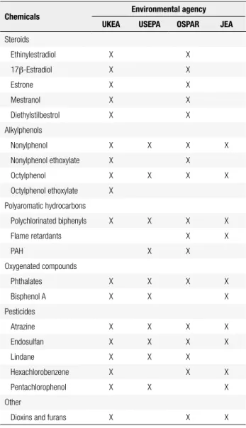
A wide variety of chemical compounds with endo-crine disruption activity has been recognized by environ-mental agencies around the world, including pesticides, pollutants and substances used in plastic manufacturing (Table 1). EDCs can be classiied according to their use, (e.g., pesticides), or to its structural property, (e.g., dioxins, steroids, and polyaromatic hydrocarbons). All
of them are widely found in the environment and may even be transported over long distances. In addition, a number of EDCs can be stored for several years in the fatty tissue of humans and animals, while others are quickly eliminated from the body. However, they can still cause serious effects, if exposure occurs during critical periods of development (3).
Below, some of the chemicals that have endocrine disrupting activity and are emphasized in the scientiic literature will be discussed in detail.
Plastics
Due to their low cost of production, variety and dura-bility, plastics have an important place in modern life. Their worldwide production exceeded 300 million tons in 2012 (3). Despite their importance, there are many controversies about the possible adverse effects of plas-tic components on human health, or even plasplas-tic addi-tive compounds, with particular emphasis on phthalates and bisphenol A.
Bisphenol A (BPA) is one of the chemical compounds of highest production worldwide: more than 6 billion pounds are produced annually (6). BPA is a monomer used in the production of polycarbonate plastics and ep-oxy resin covering the inside surface of food cans. After polymerization, some BPA molecules remain unbound and can migrate and contaminate foods and drinks. BPA can also be used as a dental sealant, and as an additive in other plastics, such as PVC and halogenated derivatives, which are widely used as lame retardants.
Human BPA exposure is widespread; it has been identiied in about 95% of urine samples of the Ameri-can population. BPA has also been identiied in the se-rum of pregnant women, in the amniotic luid and in umbilical cord blood at birth, indicating human expo-sure during one of the windows of increased suscepti-bility, the prenatal period (3,6). It is estimated that the use of BPA by the ingestion of foods packaged in cans that are coated internally with epoxy resin, is around 6.6 mg/person/day (6).
Among the BPA endocrine disrupting effects, the most recognized are on thyroid hormones and its es-trogenic and adipogenic activities; consequently, it has been debatable that there are possible adverse effects of BPA exposure on human health. Several studies ind an association between serum and urinary BPA concen-trations with increased prevalence of hypothyroidism, infertility, cardiovascular disease, and diabetes (6).
Table 1. Some of chemicals worldwide recognized as Endocrine Disrupting
Chemicals
Chemicals Environmental agency
UKEA USEPA OSPAR JEA
Steroids
Ethinylestradiol X X
17β-Estradiol X X
Estrone X X
Mestranol X X
Diethylstilbestrol X X
Alkylphenols
Nonylphenol X X X X
Nonylphenol ethoxylate X X
Octylphenol X X X X
Octylphenol ethoxylate X
Polyaromatic hydrocarbons
Polychlorinated biphenyls X X X X
Flame retardants X X
PAH X X
Oxygenated compounds
Phthalates X X X X
Bisphenol A X X X
Pesticides
Atrazine X X X X
Endosulfan X X X X
Lindane X X X
Hexachlorobenzene X X X
Pentachlorophenol X X X
Other
Dioxins and furans X X X
Cop
yright
© ABE&M t
odos os dir
eit
os r
eser
vados
.
Phthalates are plasticizers used as softeners, because they determine lexibility, suppleness, and elasticity of rigid polymers, such as PVC. They have been produced in large quantities since the 1930s, and are also found in most industrial paints, solvents, toys, personal and medical care products, such as cosmetics and blood transfusion bags. The main routes of human contami-nation are food and skin absorption; exposure during the early stages of development can occur by transpla-cental route and during breastfeeding (6).
Among all phthalates, the most worrisome is DEHP, Di (2-ethylhexylphthalate), whose annual pro-duction exceeds 2 million tons. Studies with low doses of DEHP in mice identiied toxicity to the reproductive system and increased proliferation and differentiation of adipocytes, which predisposes to visceral obesity (7). In humans, epidemiological studies suggest a correla-tion between phthalate concentracorrela-tions in cord blood and lower gestational age at delivery (6).
Dioxins
Dioxins comprise a group of organochlorine com-pounds including polychlorinated dibenzodioxins (PCDDs), polychlorinated dibenzofurans (PCDFs), and polychlorinated biphenyls (PCBs). The PCDDs and PCDFs are not produced commercially, but they are present in compound impurities from the produc-tion of other chemicals, such as PCBs. These are used as insulators, lame-retardants, lubricants, and machine and transformer luids. Dioxins are also formed during combustion processes, such as waste incineration and in smelting and reining of metals (8).
Another group of dioxins are polybrominated di-phenyl ethers (PBDE), which are used primarily as lame-retardants. Whereas these compounds are added to some products, rather than chemically bound, they easily release material and still contaminate the environ-ment. PBDEs also have bioaccumulation properties, and studies with human biological materials found that their concentrations have risen, especially in children, making it essential to carry out studies to assess the en-docrine toxicity (6).
Dioxins are fat-soluble and easily contaminate the food chain by accumulating in adipose tissue. The main source of human exposure to dioxins and PCBs are foods of animal origin (9). In addition, dioxins are not readily metabolized and excreted and have a long half-life, about 8 years in case of TCDD (2,3,7,8-tetrachlo-rodibenzo-p-dioxin).
Several studies have evaluated the effects of human exposure to dioxins. In Vietnam War, veterans exposed to Agent Orange revealed increased prevalence of sev-eral types of cancers, degenerative diseases of the cen-tral nervous system, thyroid diseases, and disorders of sexual development. Exposure to dioxins leads to del-eterious effects on reproductive functions by both anti-estrogenic and anti-androgenic activities in animals, and carcinogenic activity was also demonstrated (3).
Organotins
Organotins, persistent organic pollutants, are being widely used in agriculture as fungicides, rodent repel-lents, and ship hull paints; they are also used as stabili-zers for plastics, such as PVC.
Human exposure to organotins occurs mainly by food sources: contaminated ish, oysters, or water. Contamination by chloride tributyltin (TBT) may be associated with effects on male and female reproductive tracts and, recently, its association with adipogenic ef-fects has also been discussed (10).
Pesticides
Human exposure to pesticides is ubiquitous, and recent an UK study reported that there are approximately 127 pesticides that have endocrine disrupting activity. Among the classes of pesticides that stand out are organophos-phates, carbamates, and organochlorines. The latter is very persistent in the environment. Although dichlorodi-phenyltrichloroethane (DDT) has been banned in most developed countries, human contamination is still identi-ied in samples from adipose tissue and breast milk (10).
Atrazine is the most commonly detected pesticide contaminant of ground water and surface water. It is also an endocrine disruptor that, among other effects, alters reproductive tissues when animals are exposed during development (11).
Although EDCs may interfere with the endocrine system as a whole, many of the effects in humans are due to changes in estrogen signaling, one of the most conserved pathways in the evolution of the species. An-ti-androgenic and anti-thyroid activities are well-known and can determine changes in the reproductive system, sexual differentiation and puberty, when exposure oc-curred in early embryonic development (12).
Cop
yright
© ABE&M t
odos os dir
eit
os r
eser
vados
.
Pubertal development
Over the past century, the average age of menarche has diminished in all the socioeconomic and ethnic groups, from 16 or 17 years to less than 13. Regarding telarche, there is even stronger evidence that its onset is also oc-curring earlier (13). Besides the possibility of psycho-logical disturbances, precocious puberty is associated with a greater prevalence of various co-morbidities, among of which are: insulin resistance, metabolic syn-drome, breast and reproductive system cancers (14).
It is widely known that nutritional factors inluence the onset of puberty; however, in spite of the global epidemic of obesity, only increased body mass index does not justify this inding. Also, an increase in the fre-quency of cases of central idiopathic precocious puberty has been observed. For these data, besides the known inluence of genetic and nutritional factors that trigger puberty, environmental effects are also discussed. From this discussion, the inluence of EDCs is a noteworthy inding. These compounds act by binding to the estro-gen receptor or by post-receptor signaling. Additionally, EDCs may also act in the brain by stimulating hypotha-lamic neurons, thereby releasing kisspeptin and promot-ing the maturation of the hypothalamus, causpromot-ing earlier onset of puberty, or even precocious puberty (15).
The consumption of soy milk formulas, due to the presence of phytoestrogens, has been associated with pre-cocious telarche (13). Some EDCs, such as DES, poly-brominated biphenyls, DDT, DDE, and phthalates have also been associated with increased prevalence of preco-cious puberty in humans. Interestingly, exposure during the prenatal period may change developmental program-ming, and be associated with pubertal development af-terwards without the need of a second exposure (16).
Reproductive tract
The differentiation of germ cells in the ovaries begins in the irst trimester of intrauterine life; later, between the second and third quarter, they form primordial fol-licles, and afterwards, the follicles enter a latency period that can last from 15 to 50 years old in women. The oocytes are non-degenerate cells with a longer life in the human body, and consequently they are exposed to the effects of the environment during the entire period of their existence.
The correct formation of ovarian follicles in the fetus depends on a balance between systemic concentrations of estrogen, inhibin and activin. Therefore, estrogen
exposure during the critical period of formation of folli-cles can change follicular dynamics. A classic example of the effects of EDCs on the ovaries is the identiication of multioocyte follicles (MOF) in alligators inhabiting a lake in California; these animals were exposed during the early phases of the development to estrogenic com-pounds (17). This inding shows that maintenance of homeostasis of local and systemic hormones during fol-licle formation is required for normal development and subsequent maintenance of the quality of the oocyte. However, the mechanism by which endocrine disrup-tors alter folliculogenesis and have an impact on ovarian function in adult women remains unknown.
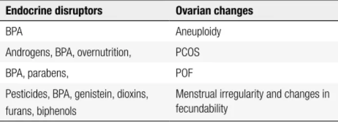
Assuming the hypothesis that EDCs contribute to the development of ovarian disease and thus reduce conception rates, two questions must be considered: what are the ovarian structures that would be target of EDCs? How do these changes are manifested? The re-productive disorders that may be related to these issues are: aneuploidy, polycystic ovary syndrome, primary ovarian failure, and changes in the menstrual cycle and in fecundability.
Aneuploidy
Several years ago, it was observed that mice that lived in damaged cages made of plastic with polycarbonates had a high incidence of meiotic changes in oocytes, and this observation led to the investigation of the estro-genic effects of the plasticizer bisphenol A (BPA) on oocytes (18). The authors concluded that BPA came from the water consumed by the animals. When they were maintained in intact cages and received water with BPA, similar changes in oocytes were also found, and some of these meiotic changes have resulted in aneu-ploidies (Table 2).
Table 2. Ovarian changes related to exposure to Endocrine Disrupting
Chemicals
Endocrine disruptors Ovarian changes
BPA Aneuploidy
Androgens, BPA, overnutrition, PCOS
BPA, parabens, POF
Pesticides, BPA, genistein, dioxins, furans, biphenols
Menstrual irregularity and changes in fecundability
BPA: bisphenol A; PCOS: polycystic ovary syndrome; POF: primary ovarian failure.
Cop
yright
© ABE&M t
odos os dir
eit
os r
eser
vados
.
cause similar meiotic changes in adult animals. How-ever, changes also occur with exposure to signiicantly higher doses of BPA, during the fetal period of oogen-esis. It is suggested that these effects may be due to the interference of BPA on estrogen receptor β (ERb) (19). Primary ovarian failure
Primary ovarian failure (POF) occurs in about 1% of the female population under 40 years of age, leading to reproductive disorders, early symptoms of menopause and other comorbidities (20). There are three possible mechanisms involved in the development of POF, in-cluding acceleration of apoptosis, blockage of follicle maturation and premature activation of the follicle. Several causes have been studied and may have some association with EDCs: (a) chromosomal abnormalities or genetic mutations, mainly involving the X chromo-some, such as the premutation in FMR1, (b) metabolic disorders such as galactosemia and 17-hydroxylase de-iciency, (c) virus infection, such as HIV, (d) iatrogenic causes: radiotherapy or chemotherapy, (e) autoimmune causes, (f) other factors: smoking, toxins, and other en-vironmental factors. The cause of most cases of POF is not identiied, and it is believed to be multifactorial (21).
Many EDCs lead to multioocyte follicles (MOF), a potential precursor of premature ovarian failure, with the process being mediated by ERb agonist actions (22). After the administration of BPA to pregnant mice at doses of 0.1-1,000 μg/kg between the 9th and 16th
day of pregnancy, the appearance of ovarian cysts was observed in adulthood, which were signiicantly more numerous in the group that received 1 μg/kg BPA. Moreover, cystic adenomas were observed in the groups receiving higher doses, but not in the controls; these data support the hypothesis that BPA causes adverse effects on the reproductive system, if exposure occurs during critical periods of differentiation (23). The do-ses used in this study are relevant for human exposure, since the estimated daily dose intake of BPA in the in-fant’s formula is around 1 to 13 mg/kg. The increased expression of genes involved in ovarian meiosis can ex-plain the effects of BPA on female germ cells (24).
Another example of EDC that can affect folliculo-genesis are parabens, which stimulate mRNA expres-sion of anti-Mullerian hormone, and thus may inhibit the early stage of folliculogenesis in ovaries of newborn rats. In addition, parabens may regulate steroidogenesis
by inhibiting follicular Foxl2, the main transcriptional repressor (25).
The compound 4-vinylcyclohexene diepoxide (VCD) is considered an occupational chemical ovo-toxic (26). Repeated doses of VCD may accelerate the apoptotic process of atresia and selectively destroy primordial and primary follicles in rats and mice (27). Therefore, women exposed to VCD are considered at risk of POF.
Methoxychlor (MXC) is an organochlorine pesti-cide used as a substitute for DDT. MXC is an estro-genic compound that was demonstrated to have lower binding afinity for the estrogen receptor (28). The major MXC metabolites, HPTE [2, 2-bis-(p-hydroxy-phenyl)-1, 1, 1- trichloroethane] and mono-OH MXC, have estrogenic, anti-estrogenic, or anti-androgenic ac-tions (29). Studies have demonstrated that adult mice and rats that were exposed to MXC showed inhibition of growth and implantation of embryo and ovarian at-rophy due to inhibition of folliculogenesis, leading to reduced atretic follicles and ovulation (30).
Diethylstilbestrol is a synthetic non-steroidal estro-gen that was prescribed to pregnant women at doses of 5-150 mg/day to prevent abortions between the years 1940-1970. Many reproductive abnormalities, both cardiovascular and immunological, were reported in male and female offspring of women treated with DES, and were validated in animal models (31). Recent stud-ies have shown that neonatal exposure to 3 μg/kg DES induces MOF (22).
Menstrual irregularity and fecundability
Interference in the hormonal regulation of the men-strual cycle results in irregular or long cycles that will reduce fecundity (ability to conceive in a menstrual cycle). Studies in humans suggest that fetal and neona-tal exposure to EDCs can change menstrual cycles due to hormonal interference (32). Exposure to organo-chlorine pesticides may make menstrual cycles shorter (33). Moreover, women who are exposed to non-or-ganochlorine pesticides have 60 to 100% higher risk of developing long cycles or absence of menstrual cycles (34). Animal studies support the indings in humans;
Cop
yright
© ABE&M t
odos os dir
eit
os r
eser
vados
.
to a change in hypothalamic control of LH secretion and ovulation (36).
A recent Brazilian study quantiied organochlorine compounds in infertile women and compared them to a control group of fertile women. Exposure to organo-chlorine compounds was most prevalent in infertile women, conirming the data from literature, indicating that dichlorodiphenyl dichloroethylene (pp’DDE) may negatively affect female fertility (37).
Polycystic ovary syndrome
Polycystic ovary syndrome (PCOS) is a prevalent endo-crine disorder in women and, in its classical presenta-tion, is characterized by chronic anovulation and hyper-androgenism. More recently, some milder phenotypes have been recognized, in which either anovulation or hyperandrogenism may be absent. Frequently, these women present a characteristic polycystic ovarian ap-pearance at ultrasound and high prevalence of obesity, insulin resistance, and metabolic comorbidities, mainly in its classical phenotype.
While the pathogenesis of PCOS is still not well established, evidence suggests that both genetic and environmental factors may contribute to the clinical de-velopment of the disease. Among these, BPA, an endo-crine disruptor with well-known estrogen-like actions, has been implicated in its development (38).
In fact, studies in animal models have shown that BPA acts in different ways to disturb reproductive func-tions, including both estrogen and androgen pathways. BPA in vitro increases testosterone synthesis in rat ovarian theca-intersticial cells (39), and in male rats, it seems to compete with endogenous androgens for binding on sex hormone-binding globulin (SHBG), in-creasing serum free androgen levels. BPA also has been demonstrated to decrease hepatic androgen-related hy-droxylases, inhibiting testosterone degradation, with an expected increase in hormone levels (40). Moreover, neonatal exposure to BPA could result in PCOS de-velopment (41). Additionally, BPA may disturb insulin action and glucose metabolism contributing to insulin resistance in intact animals (42).
In humans, studies have reported higher BPA circu-lating levels in anovulatory women (38) and in PCOS patients (43), in comparison with controls. The mecha-nisms behind this association between BPA and PCOS are not clear but may be, at least in part, linked to an-drogen secretion, as suggested by data on animal stud-ies. In this sense, BPA was found to be positively
corre-lated with androgen levels in PCOS women, suggesting an additive effect of BPA in an individual genetically susceptible to an ovarian enzymatic defect (43).
In turn, dietary products have been emerging, as other environmental factors associated with the devel-opment of PCOS in susceptible individuals. Advanced glycated end-products (AGEs) are reactive derivatives of non-enzymatic glucose-protein reactions. AGEs are endogenously produced by aging, hyperglycemia, and oxidative stress, or exogenously ingested mainly from high-temperature processed, protein-rich foods (44). AGEs promote oxidative stress and insulin resistance in peripheral tissues by activating protein kinase C (45).
Serum AGEs levels are increased in women with PCOS, and these have been positively correlated with serum androgen levels (46). AGEs seem to contribute to the elevated risk of diabetes and cardiovascular dis-ease in the general population (45). Moreover, insulin-resistant PCOS women have increased levels of both serum AGEs and their respective receptor (RAGE), localized in theca and granulosa cells (46), suggesting a putative direct action of AGEs on ovarian function.
Interestingly, women with classic PCOS phenotype have higher serum AGEs levels than ovulatory PCOS women, indicating that AGEs are associated with the phenotypical severity (46). AGEs are related to the number of oocytes and pregnancy rates in PCOS wo-men (47).
It is important to underline that the environmental source of AGEs can be reduced by diet modiications that lead to a decrease on oxidative stress markers as well as serum testosterone levels (48).
CONCLUSIONS
Cop
yright
© ABE&M t
odos os dir
eit
os r
eser
vados
.
exposure and adult reproductive dysfunction. Finally, it is a matter of concern that the endocrine disrupting potential of the majority of chemicals in production, including those that are found in common consumer products, has not been assessed systematically in regard to their effects on reproduction.
Disclosure: no potential conlict of interest relevant to this article was reported.
REFERENCES
1. Bachega TASS, Verreschi IT, Frade EMC, D’Abronzo FH, Lazaretti-Castro M. The environmental endocrine disruptors must receive the attention of Brazilian endocrinologists. Arq Bras Endocrinol Metab. 2011;55(2):175-6.
2. Decherf S, Demeneix BA. The obesogen hypothesis: a shift of focus from the periphery to the hypothalamus. J Toxicol Environ Health B Crit Rev. 2011;14(5-7):423-48.
3. Schug TT, Janesick A, Blumberg B, Heindel JJ. Endocrine disrupting chemicals and disease susceptibility. J Steroid Biochem Mol Biol. 2011;127(3-5):204-15.
4. Christensen BC, Marsit CJ. Epigenomics in environmental health. Front Genet. 2011;2:84.
5. Diamanti-Kandarakis E, Bourguignon JP, Giudice LC, Hauser R, Prins GS, Soto AM. Endocrine-disrupting chemicals: an Endocrine Society scientiic statement. Endocr Rev. 2009;30(4):293-342. 6. Zoeller RT. Endocrine disruptors: do family lines carry an
epigenetic record of previous generations’ exposures? Endocrinology. 2006;147(12):5513-4.
7. Hao C, Cheng X, Xia H, Ma X. The endocrine disruptor mono-(2-ethylhexyl) phthalate promotes adipocyte differentiation and induces obesity in mice. Biosci Rep. 2012;32(6): 619-29.
8. Chang JW, Chen HL, Su HJ, Liao PC, Guo HR, Lee CC. Dioxin exposure and insulin resistance in Taiwanese living near a highly contaminated area. Epidemiology. 2010;21(1):56-61.
9. Liem AK, Fürst P, Rappe C. Exposure of populations to dioxins and related compounds. Food Addit Contam. 2000;17(4):241-59. 10. Casals-Casas C, Desvergne B. Endocrine disruptors: from
endocrine to metabolic disruption. Annu Rev Physiol. 2011;73:135-62.
11. Hayes TB, Anderson LL, Beasley VR, de Solla SR, Iguchi T, Ingraham H, et al. Demasculinization and feminization of male gonads by atrazine: consistent effects across vertebrate classes. J Steroid Biochem Mol Biol. 2011;127(1-2):64-73.
12. Bigsby R, Chapin RE, Daston GP, Davis BJ, Gorski J, Gray LE, et al. Evaluating the effects of endocrine disruptors on endocrine function during development. Environ Health Perspect. 1999;107(4):613-8.
13. Fortes EM, Malerba MI, Luchini PD, Sugawara EK, Sumodjo L, Ribeiro Neto LM, et al. [High intake of phytoestrogens and precocious thelarche: case report with a possible correlation]. Arq Bras Endocrinol Metab. 2007;51(3):500-3.
14. Buttke DE, Sircar K, Martin C, et al. Exposures to endocrine-disrupting chemicals and age of menarche in adolescent girls in NHANES (2003-2008). Environ Health Perspect. 2012;120:1613-8. 15. Patisaul HB. Effects of environmental endocrine disruptors and
phytoestrogens on the kisspeptin system. Adv Exp Med Biol. 2013;784:455-79.
16. Mouritsen A, Aksglaede L, Sørensen K, Mogensen SS, Leffers H, Main KM, et al. Hypothesis: exposure to endocrine-disrupting
chemicals may interfere with timing of puberty. Int J Androl. 2010;33:346-59.
17. Guillette LJ Jr, Gross TS, Masson GR, Matter JM, Percival HF, Woodward AR. Developmental abnormalities of the gonad and abnormal sex hormone concentrations in juvenile alligators from contaminated and control lakes in Florida. Environ Health Perspect. 1994;102(8):680-8.
18. Hunt PA, Koehler KE, Susiarjo M, Hodges CA, Ilagan A, Voigt RC, et al. Bisphenol a exposure causes meiotic aneuploidy in the female mouse. Curr Biol. 2003;13(7):546-53.
19. Eichenlaub-Ritter U, Vogt E, Cukurcam S, Sun F, Pacchierotti F, Parry J. Exposure of mouse oocytes to bisphenol A causes meiotic arrest but not aneuploidy. Mutat Res. 2008;651(1-2):82-92. 20. Nelson LM. Clinical practice. Primary ovarian insuficiency. N
Engl J Med. 2009;360(6):606-14.
21. Cooper AR, Baker VL, Sterling EW, Ryan ME, Woodruff TK, Nelson LM. The time is now for a new approach to primary ovarian insuficiency. Fertil Steril. 2011;95(6):1890-7.
22. Kirigaya A, Kim H, Hayashi S, Chambon P, Watanabe H, Lguchi T, et al. Involvement of estrogen receptor beta in the induction of polyovular follicles in mouse ovaries exposed neonatally to diethylstilbestrol. Zoolog Sci. 2009;26(10):704-12.
23. Newbold RR, Jefferson WN, Padilla-Banks E. Prenatal exposure to bisphenol A at environmentally relevant doses adversely affects the murine female reproductive tract later in life. Environ Health Perspect. 2009;117(6):879-85.
24. Brieño-Enríquez MA, Reig-Viader R, Cabero L, Toran N, Martínez F, Roig I, et al. Gene expression is altered after bisphenol A exposure in human fetal oocytes in vitro. Mol Hum Reprod. 2012;18(4):171-83.
25. Ahn HJ, An BS, Jung EM, Yang H, Choi KC, Jeung EB. Parabens inhibit the early phase of folliculogenesis and steroidogenesis in the ovaries of neonatal rats. Mol Reprod Dev. 2012;79(9):626-36. 26. Hoyer PB, Devine PJ, Hu X, Thompson KE, Sipes IG. Ovarian
toxicity of 4-vinylcyclohexene diepoxide: a mechanistic model. Toxicol Pathol. 2001;29(1):91-9.
27. Kappeler CJ, Hoyer PB. 4-vinylcyclohexene diepoxide: a model chemical for ovotoxicity. Syst Biol Reprod Med. 2012;58(1):57-62. 28. Cummings AM. Methoxychlor as a model for environmental
estrogens. Crit Rev Toxicol. 1997;27(4):367-79.
29. Gaido KW, Maness SC, McDonnell DP, Dehal SS, Kupfer D, Safe S. Interaction of methoxychlor and related compounds with estrogen receptor alpha and beta, and androgen receptor: structure-activity studies. Mol Pharmacol. 2000;58(4):852-8. 30. Martinez EM, Swartz WJ. Effects of methoxychlor on the
reproductive system of the adult female mouse. Gross and histologic observations. Reprod Toxicol. 1991;5(2):139-47. 31. Newbold RR. Lessons learned from perinatal exposure to
diethylstilbestrol. Toxicol Appl Pharmacol. 2004;199(2):142-50. 32. Chao HR, Wang SL, Lin LY, Lee WJ, Papke O. Placental transfer
of polychlorinated dibenzo-p-dioxins, dibenzofurans, and biphenyls in Taiwanese mothers in relation to menstrual cycle characteristics. Food Chem Toxicol. 2007;45(2):259-65.
33. Axmon A, Rylander L, Strömberg U, Hagmar L. Altered menstrual cycles in women with a high dietary intake of persistent organochlorine compounds. Chemosphere. 2004;56(8):813-9. 34. Farr SL, Cooper GS, Cai J, Savitz DA, Sandler DP. Pesticide use and
menstrual cycle characteristics among premenopausal women in the Agricultural Health Study. Am J Epidemiol. 2004;160(12):1194-204.
Cop
yright
© ABE&M t
odos os dir
eit
os r
eser
vados
.
36. Rubin BS, Lenkowski JR, Schaeberle CM, Vandenberg LN, Ronsheim PM, Soto AM. Evidence of altered brain sexual differentiation in mice exposed perinatally to low, environmentally relevant levels of bisphenol A. Endocrinology. 2006;147(8):3681-91. 37. Bastos AM, de Souza Mdo C, de Almeida Filho GL, Krauss
TM, Pavesi T, da Silva LE. Organochlorine compound levels in fertile and infertile women from Rio de Janeiro, Brazil. Arq Bras Endocrinol Metabol. 2013;57(5):346-53.
38. Takeuchi T, Tsutsumi O, Ikezuki Y, Takai Y, Taketani Y. Positive relationship between androgen and the endocrine disruptor, bisphenol A, in normal women and women with ovarian dysfunction. Endocr J. 2004;51:165-9.
39. Zhou W, Liu J, Liao L, Han S, Liu J. Effect of bisphenol A on steroid hormone production in rat ovarian theca-interstitial and granulosa cells. Mol Cell Endocrinol. 2008;283:12-8.
40. Hanioka N, Jinno H, Nishimura T, Ando M. Suppression of male-speciic cytochrome P450 isoforms by bisphenol A in rat liver. Arch Toxicol. 1998;72:387-94.
41. Fernández M, Bourguignon N, Lux-Lantos V, Libertun C. Neonatal exposure to bisphenol A and reproductive and endocrine alterations resembling the polycystic ovarian syndrome in adult rats. Environ Health Perspect. 2010;118:1217-22.
42. Alonso-Magdalena P, Morimoto S, Ripoll C, Fuentes E, Nadal A. The estrogenic effect of bisphenol A disrupts pancreatic beta-cell function in vivo and induces insulin resistance. Environ Health Perspect. 2006;114:106-12.
43. Kandaraki E, Chatzigeorgiou A, Livadas S, Palioura E, Economou F, Koutsilieris M, et al. Endocrine disruptors and polycystic ovary syndrome (PCOS): elevated serum levels of bisphenol A in women with PCOS. J Clin Endocrinol Metab. 2011;96:E480-4. 44. Pasquali R, Stener-Victorin E, Yildiz BO, Duleba AJ, Hoeger
K, Mason H, et al. PCOS Forum: research in polycystic ovary syndrome today and tomorrow. Clin Endocrinol (Oxf). 2011;74:424-33.
45. Uribarri J, Tuttle KR. Advanced glycation end products and nephrotoxicity of high-protein diets. Clin J Am Soc Nephrol. 2006;1:1293-9.
46. Diamanti-Kandarakis E, Katsikis I, Piperi C, Kandaraki E, Piouka A, Papavassiliou AG, et al. Increased serum advanced glycation end-products is a distinct inding in lean women with polycystic ovary syndrome (PCOS). Clin Endocrinol (Oxf). 2008;69:634-41. 47. Jinno M, Takeuchi M, Watanabe A, Teruya K, Hirohama J,
Eguchi N, et al. Advanced glycation end-products accumulation compromises embryonic development and achievement of pregnancy by assisted reproductive technology. Hum Reprod. 2011;26:604-10.
48. Diamanti-Kandarakis E, Piouka A, Livadas S, Piperi C, Katsikis I, Papavassiliou AG, et al. Anti-mullerian hormone is associated with advanced glycosylated end products in lean women with polycystic ovary syndrome. Eur J Endocrinol. 2009;160:847-53. 49. Costa EMF, et al. Desreguladores Endócrinos. In: Proendócrino.