Case 10646
Fibrothecoma of the ovary
Elisa Melo Abreu, Teresa Margarida Cunha
Genital (Female) Imaging
Section:
2015, Jan. 2
Published:
70 year(s), female
Patient:
Authors' Institution
Department of Radiology, Instituto Português de Oncologia de Lisboa Francisco Gentil, Lisbon, Portugal
Clinical History
A 68-year-old woman was referred to our hospital to charaterize a pelvic mass, an incidental finding on a routine ultrasound. A computed tomography (CT) and a magnetic resonance imaging (MRI) were performed. She was asymptomatic and laboratorial analysis was unremarkable.
Imaging Findings
Contrast enhanced CT (CECT) shows a well-defined, predominantly solid and heterogeneous pelvic mass, with mild contrast enhancement (Fig. 1).
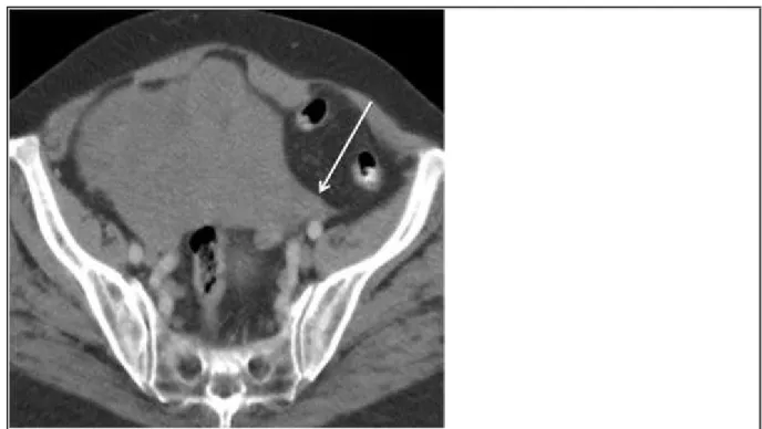
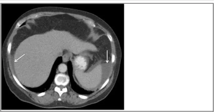
Following the left ovarian vein to the level of the suspensory ligament, as it approaches the mass, has shown "the ovarian suspensory ligament sign", confirming the left ovarian origin (Fig. 1a). There is no attachment between the lesion and uterus (Fig. 1c), and subphrenic ascites coexists (Figs. 1d).
After paramagnetic contrast administration it has only mild, diffuse and slight heterogeneous enhancement (less than myometrium) (Figs. 2e-2f).
Discussion
Ovarian fibrothecomas are benign tumors arising from the stromal component of the ovary [1-3]. According to the World Health Organization classification they represent a subgroup of the granulosa-theca cell tumors, belonging to the thecoma-fibroma group.
Histologically, fibrothecomas differ slightly from fibromas, demonstrating theca-like cells [1, 4]. These tumors usually are asymptomatic. They are often an unexpected finding on imaging performed for other reason. Symptoms related to pleural effusion or peritoneal fluid (Meigs' syndrome) may occur [1, 3].
In all cases, it is fundamental to differentiate fibrothecoma from a malignant tumor and imaging plays an important role.
On ultrasound, fibrothecoma appears as a primarily hypoechoic ovarian mass with posterior wall attenuation [2]. Most tumors are solid, with smooth contours, but some of them demonstrate cystic areas [1, 2]. Color Doppler findings are variable, although most of fibrothecomas show minimal to moderate vascularization.
A fibrothecoma with atypical appearance may be mistaken for a malignancy, particularly if
associated with high color content on Doppler evaluation, with ascites or raised CA 125 levels, and further evaluation is needed [2-4].
On sectional imaging, knowledge of the morphologic features and ligamentous attachments of the ovaries (particularly the suspensory ligament), the relationship of the ovary to the ureter, and the course of the ovarian vein and artery is fundamental not only for confident identification of the laterality of the lesion, but also to differentiate ovarian from non-ovarian masses [3, 5]. On CECT fibrothecomas are described as well-defined, solid, homogeneous or slightly heterogeneous lesions, with delayed contrast enhancement [2, 5].
On MRI, because of their high content in collagen, fibrothecomas have low-signal intensity on T1WI and very low-signal on T2WI, characteristics that display a relatively specific appearance [3-4].
Therefore, after analyse the relationship of the mass with ovaries and other anatomic pelvic structures, if an ovarian origin is proven and the lesion demonstrates the described signal intensity, a fibrothecoma is likely.
Nevertheless, when the diagnosis of a fibrothecoma is likely, it is important to know that the prognosis is extremely good. They are almost always benign and elective surgical ressection is sufficient. Even in cases associated with ascites (Meigs' syndrome), fluid collections usually regress following surgical removal of the tumor [3, 4].
Knowledge of the anatomic relationships and of the key features of ovarian tumors, particularly of fibrothecomas, will provide the criteria for making a specific diagnosis or substantially narrowing the differential diagnosis, preventing unnecessary therapies.
Differential Diagnosis List
Fibroma of the ovary, Pedunculated leiomyoma, Broad ligament leiomyoma
Figures
Figure 1 CECT images
Axial CECT image shows the left fan-shaped suspensory ligament (white arrow) joining to a
large left ovarian mass, a finding known as "ovarian suspensory ligament sign".
© Cunha TM, Department of Radiology, IPOLFG, Lisbon, Portugal
Area of Interest: Genital / Reproductive system female;
Imaging Technique: CT;
Coronal CECT image shows the left ovarian large mass, predominantly solid and
heterogeneous, with small areas of fluid-attenuation. Note that the mass shows mild
enhancement.
© Cunha TM, Department of Radiology, IPOLFG, Lisbon, Portugal
Area of Interest: Abdomen;
Imaging Technique: Absorptiometry / Bone densiometry;
Procedure: Abscess delineation;
Special Focus: Abscess;
Area of Interest: Genital / Reproductive system female;
Imaging Technique: CT;
Procedure: Diagnostic procedure;
Special Focus: Neoplasia;
Axial CECT image demonstrates the coexistence of ascites in the upper abdomen,
particularly in peri-hepatic and peri-splenic locations (white arrows).
© Cunha TM, Department of Radiology, IPOLFG, Lisbon, Portugal
Area of Interest: Genital / Reproductive system female;
Imaging Technique: CT;
Procedure: Diagnostic procedure;
Special Focus: Neoplasia;
Figure 2 Pelvic MRI
Axial T1WI shows a hypointense left ovarian mass with smooth contours.
© Cunha TM, Department of Radiology, IPOLFG, Lisbon, Portugal
Area of Interest: Genital / Reproductive system female;
Imaging Technique: MR;
Axial T2WI shows a heterogeneous, predominantly hypointense left ovarian mass. The
remnant high signal intensity areas are consistent with cystic areas.
© Cunha TM, Department of Radiology, IPOLFG, Lisbon, Portugal
Area of Interest: Genital / Reproductive system female;
Imaging Technique: MR;
Procedure: Diagnostic procedure;
Special Focus: Neoplasia;
Sagittal T2WI shows a heterogeneous, predominantly hypointense left ovarian mass, that
extends until the level of L4 vertebra. Note the small amount of peritoneal fluid seen in the
pelvic cavity.
© Cunha TM, Department of Radiology, IPOLFG, Lisbon, Portugal
Area of Interest: Genital / Reproductive system female;
Imaging Technique: CT;
Coronal T2WI shows a heterogeneous, predominantly hypointense left ovarian mass. The
hyperintense components are consistent with cystic areas, that are isointense to peritoneal
fluid seen in the pelvic cavity.
© Cunha TM, Department of Radiology, IPOLFG, Lisbon, Portugal
Area of Interest: Genital / Reproductive system female;
Imaging Technique: MR;
Procedure: Diagnostic procedure;
Special Focus: Neoplasia;
Axial fat-supressed gadolinium-enhanced T1WI shows mild enhancement of the left ovarian
heterogeneous mass.
© Cunha TM, Department of Radiology, IPOLFG, Lisbon, Portugal
Area of Interest: Genital / Reproductive system female;
Imaging Technique: MR;
Sagittal fat-supressed gadolinium-enhanced T1WI shows mild enhancement of the left
ovarian mass (less than myometrium). Note there is no attachment between the lesion and
the uterine fundus.
© Cunha TM, Department of Radiology, IPOLFG, Lisbon, Portugal
Area of Interest: Genital / Reproductive system female;
Imaging Technique: MR;
Procedure: Diagnostic procedure;
Special Focus: Neoplasia;
MeSH
[A05.360.576.497]
Ovary
The reproductive organ (GONADS) in female animals. In vertebrates, the ovary contains two functional parts: the OVARIAN FOLLICLE for the production of female germ cells
(OOGENESIS); and the endocrine cells (GRANULOSA CELLS, THECA CELLS, and LUTEAL CELLS) for the production of ESTROGENS and PROGESTERONE.
[C13.371.056.630.705]
Ovarian Neoplasms
Tumors or cancer of the OVARY. These neoplasms can be benign or malignant. They are classified according to the tissue of origin, such as the surface epithelium, the stromal endocrine cells, and the totipotent germ cells.
References
[2] Jung SE, Lee JM, Rha SE, Byun JY, Jung JI, Hahn ST (2002) CT and MR Imaging of ovarian tumors with emphasis on differential diagnosis Radiographics 22(6):1305-25
[3] Troiano RN, Lazzarini KM, Scout LM, Lange RC, Flynn SD, McCanthy S (1997) Fibroma and fibrothecoma of the ovary: MR imaging findings Radiology 204(3):795-8
[4] Shinagara AB, Meylaerts LJ, Laury AR, Mortele KJ (2012) MRI features of ovarian fibroma and fibrothecoma with histopathologic correlation AJR 198:296-303
[5] Saksouk FA, Johnson SC (2004) Recognition of ovaries and ovarian origin of pelvic masses Radiographics 136-146
Citation
Elisa Melo Abreu, Teresa Margarida Cunha (2015, Jan. 2)
Fibrothecoma of the ovary {Online}