Rev Odontol UNESP. 2014 Jan-Feb; 43(1): 1-7 © 2014 - ISSN 1807-2577
ORIGINAL ARTICLE
Doi: http://dx.doi.org/10.1590/S1807-25772014000100001
Inluence of the addition of chlorhexidine diacetate on bond
strength of a high-viscosity glass ionomer cement
to sound and artiicial caries-afected dentin
Inluência da adição do diacetato de clorexidina na resistência de união de um cimento de ionômero de
vidro de alta viscosidade à dentina sadia e afetada por cárie artiicial
Ana Carolina de Oliveira BECCI
a, Luana Mafra MARTI
a, Angela Cristina Cilense ZUANON
a,
Fernanda Lourenção BRIGHENTI
a, Denise Madalena Palomari SPOLIDÓRIO
a,
Elisa Maria Aparecida GIRO
a,
aFaculdade de Odontologia, UNESP - Univ Estadual Paulista, Araraquara, SP, Brasil
Resumo
Introdução: A adição da clorexidina (CLX) ao cimento de ionômero de vidro (CIV) visa melhorar a sua propriedade antibacteriana, podendo contudo interferir na adesão à dentina. Objetivo: Avaliar a influência da adição de diacetato de CLX em diferentes concentrações a um CIV de alta viscosidade, na sua adesão à dentina sadia e afetada por cárie artificial. Material e método: Foram utilizados 80 terceiros molares, que tiveram a superfície de dentina exposta na face oclusal. Metade dos dentes foram mantidos hígidos e a outra metade foi submetida à indução artificial de cárie. A CLX foi misturada ao pó do CIV nas concentrações de 0,5%, 1% e 2%. O CIV sem CLX foi usado como controle. Em cada superfície dentinária foi confeccionado um espécime com 1 mm de diâmetro e 1 mm de altura. Estes foram mantidos a 37 °C com 100% de umidade por 24 horas, e, submetidos ao teste de microcisalhamento. Os resultados foram analisados pelos testes de Kruskal-Wallis e Mann Whitney (α=0,05). Resultado: Não houve diferença
estatística entre os valores de resistência de união para dentina hígida e afetada (p>0,05). Para as duas condições do substrato, os grupos CIV, CIV+CLX 0,5% e CIV+CLX 1% apresentaram resistência de união estatisticamente semelhante (p>0,05), e superior ao CIV+CLX 2% (p≤0,025). Houve predominância de fraturas mistas e coesivas do material para todos os grupos. Conclusão: A adição de CLX nas concentrações de 0,5% e 1% não influenciou negativamente na resistência de união de um CIV de alta viscosidade à dentina sadia e afetada por cárie.
Descritores: Cimentos de ionômeros de vidro; clorexidina; propriedades físicas; dentina.
Abstract
Introduction: The aim of adding chlorhexidine (CHX) to glass ionomer cements (GIC) is to improve their antibacterial property, but it may interfere with their bond to dentin. Objective: To evaluate the influence of adding chlorhexidine diacetate at different concentrations to a high-viscosity GIC on its bond to sound and artificial caries-affected dentin. Material and method: Eighty human third molars were used, on which an area of dentin was exposed on the occlusal surface. Half of the specimens were kept sound and the other half were subjected to artificially induced caries. CHX was mixed with GIC powder at 0.5%, 1% and 2% (w/w). GIC without CHX was used as control. On each dentin surface a specimen measuring 1 mm in diameter and 1 mm high was made. The samples were kept at 37 °C and 100% humidity for 24 hours and subject to microshear testing. The results were analyzed using Kruskal-Wallis and Mann Whitney tests (α=0.05). Result: There was no significant difference between bond
strength of sound and caries-affected dentin (p>0.05). For both substrate conditions, groups GIC, GIC+0.5% CHX and GIC+1% CHX showed statistically similar bond strength (p>0.05), and higher than that of GIC+2% CHX (p≤0.025). Cohesive and mixed failures were predominant in all groups. Conclusion: The addition of 0.5% and 1% chlorhexidine did not result in negative changes in the bond strength of GIC to caries-affected and sound dentin.
Descriptors: Glass ionomer cements; chlorhexidine; physical properties; dentin.
INTRODUCTION
he minimal intervention technique for the treatment of caries lesions has been widely used over the last few decades. In this technique, the recommendation is to remove the layer of infected, more disorganized dentin, and preserve the afected dentin that has the potential to be remineralized1,2.
contribute to the remineralization process3,4. However, the low mechanical strength of the conventional formulations of this material5 is a limiting factor for its use in cavities subjected to high masticatory stresses, such as extensive Class I and Class II restorations. his has led to the development of a new category of GICs with enhanced physical properties.
hese GICs present greater wettability of the powder by the liquid component than the conventional GICs, which has resulted in easier and faster manipulation and greater viscosity of the product6. As a disadvantage, they present lower cumulative luoride ion release7-9. Although the efects of this lower luoride ion release on the inhibition of residual caries lesions are not yet known, various researchers10-14 have proposed the association of anti-septic agents with GICs, in order to improve their antibacterial properties.
Chlorhexidine (CHX) has been used in association with GICs, particularly in the forms of digluconate10,13 and diacetate12,13,15. Nevertheless, their antibacterial efect is concentration-dependent11, and at high concentrations, CHX may interfere in the physical and mechanical properties of GIC10,11.
In order to attain the clinical success of a restorative technique, it is important to point out that the modiied material must present adequate physical properties, and its property of bonding to dental structures must not be altered, since the anticariogenic efect depends on a combination of antibacterial agent release and retention time of the material in the cavity11. As studies of the bond strength of GIC associated with chlorhexidine are scarce12 and there are no studies testing this property in caries-afected dentin, the aim of this study was to evaluate the inluence of the addition of diferent concentrations of CHX diacetate, on the bond strength of a high viscosity GIC to sound and caries-afected dentin.
MATERIAL AND METHOD
1.
Selection and Preparation of Teeth
Eighty sound extracted human third molars were obtained from the Tooth Bank of the Araraquara School of Dentistry – UNESP, with approval from the Research Ethics Committee (Report 68.388, of August 07, 2012). Ater the removal of tissue remainders, prophylaxis and washing, only teeth without anatomical and structural defects were selected. hese were stored in a 0.1% thymol solution, under refrigeration (4 °C) up to the time of being used.he teeth were randomly divided into
eight groups (n=10) according to the condition of the substrate (caries-afected dentin and sound dentin) and concentration of chlorhexidine diacetate added to the material (0%, 0.5%, 1% and 2%). he material used was a high viscosity GIC (Ketac Molar Easymix - 3M-ESPE Dental Products, St. Paul, MN, USA) (Table 1).
1.1. Obtaining the dentin surface
he teeth were cut in the transverse direction in the occlusal third of the crown, for the purpose of producing a lat surface in dentin. he cut was made with a metallographic cutter (ISOMET 1000, Buehler Ltd., Lake Bluf, IL, EUA) equipped with a diamond disc (No.11-4254, Buehler LTD., Lake Bluf IL, USA), at a speed of 300 rpm and force of 200 gf, under constant cooling. he surfaces were inspected under a stereomicroscopy (Model SZX7, Olympus, São Paulo, Brazil) at 40x magniication to prove the absence enamel remainders on the dentin surface.
1.2. Artiicial induction of caries lesions
In half of the selected teeth (n=40) one of the roots of each tooth was perforated and transixed with orthodontic wire to enable the teeth to be suspended. he teeth were sealed with two layers of acid resistant enamel, leaving only the dentin surface exposed, and were then sterilized with ethylene oxide. Ater this, the teeth were suspended in a cariogenic solution (BHI broth supplemented with 2% sucrose, 1% glucose and 0.5% yeast extract; 25 mL/tooth) and inoculated with 105 CFU/mL of Streptoccocus mutans ATCC 25175 (Tropical Culture Collection – André Toselo Foundation). he set was incubated under microaerophilic conditions at 37 °C for 14 days, with changes of cariogenic solution every 48 hours, without the inoculation of new microorganisms. Ater the incubation period, the bioilm was removed with gauze and the sealing material with the aid of scalpel blades. he teeth were abundantly washed in deionized water and the dentin surface was found to be darkened and sotened when it was touched with an exploratory probe without using pressure16.
1.3. Carious tissue removal and obtainment of dies
In the teeth with artiicially induced caries lesions, the infected dentin was manually removed with 320 grain silicon carbide abrasive papers, until hardened dentin, resistant to the touch of a sharp exploratory probe, without using pressure, was obtained (afected dentin). In an attempt to obtain a similar dentin depth among the caries-afected teeth and the teeth that were maintained sound, the latter were also worn with the same type of abrasive paper.
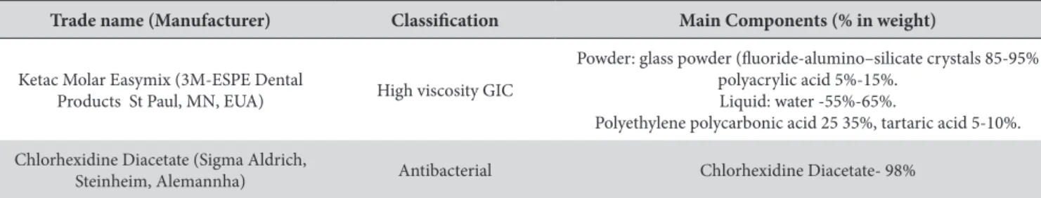
Table 1. Trade name, manufacturer, classiication and main components of the materials used in the study
Trade name (Manufacturer) Classiication Main Components (% in weight)
Ketac Molar Easymix (3M-ESPE Dental
Products St Paul, MN, EUA) High viscosity GIC
Powder: glass powder (luoride-alumino–silicate crystals 85-95% polyacrylic acid 5%-15%.
Liquid: water -55%-65%.
Polyethylene polycarbonic acid 25 35%, tartaric acid 5-10%.
Chlorhexidine Diacetate (Sigma Aldrich,
Ater this, the teeth were washed in an ultrasonic bath, embedded in self-polymerizing acrylic resin, using a cylindrical PVC tube as matrix, with an external diameter of 20 mm by 18 mm high, so that the dentin surface would be centralized and parallel to the base of the tube. Ater complete polymerization of the acrylic resin, all the teeth were carefully abraded manually with 320 grit silicon carbide abrasive papers lubricated with water, for 15 seconds, in order to produce a standardized smear layer16. All the procedures were carried out by one single, previously trained operator.
2.
Test Specimen Fabrication
On the dentin surface of each tooth a test specimen was constructed. Initially, the bonding area was delimited using double-faced acid resistant adhesive tape (3M do Brasil, Sumaré, SP, Brazil) with a perforation (1.0 mm in diameter) made with a rubber mat perforator (model Ainsworth, Wilcos do Brasil Indústria e Comércio Ltda, Petrópolis, RJ, Brazil). Ater this, the dentin surfaces were etched with polyacrylic acid for 10 seconds and then washed with a jet of water-air for 10 seconds and dried with cotton wool balls. A transparent cylindrical silicone matrix 1 mm high with an oriice 1 mm in diameter obtained from a disposable urethral probe (Embramed, São Paulo, SP, Brazil) was placed so that its internal diameter would coincide with the perforation in the adhesive tape, and this was used as a matrix for fabricating the test specimens. he material was manipulated manually at a controlled room temperature (24 ± 1 °C), in accordance with the Manufacturer’s recommendations (3M – ESPE Dental Products, St. Paul, MN,USA) and was inserted into the matrix with the aid of a Centrix syringe (DFL, Indústria e Comércio S.A, Jacarepaguá, RJ, Brazil). he test specimens were protected with vaseline on their top portion and the set (die + microtube + test specimen) was stored at 37 °C with 100% humidity for 24 hours. Ater this the matrix were removed with the aid of a scalpel blade no.15 (Embramed, Jurubatuba, SP, Brazil). he test specimens were observed under a stereomicroscopy at 40x magniication to certify the absence of defects at the bond interface.
2.1. Bond Strength Determination by means of the
microshear test
he mechanical microshear test was performed in a mechanical testing machine (DL-Digital Line, EMIC, Paraná, Brazil), previously adjusted for tensile forces. To perform the test, a metal wire 0.2 mm in diameter was used simultaneously
lassoing the test specimen as closely as possible to the material/ dentin bond and prolongation of the load cell. he movements of traction were made at a speed of 0.5 mm/min. he tests were started by means of a speciic computerized program (Tesc-Test Script, EMIC Equipamentos de Ensaio Ltda, São José dos Pinhais, Paraná, Brazil) and proceeded until fracture. he maximum stress values in MegaPascal withstood by the dentin/material bond were recorded.
3.
Fracture Pattern Analysis
he fracture pattern of each specimen was evaluated by a single trained examiner, with the aid of light microscope (Model SZX7 Olympus, São Paulo, Brazil) at a magniication that would allow adequate analysis (approximately 40x). he fractures were classiied as adhesive (failure between the substrate and restorative material), cohesive in dentin or in material (failure in dentin or in material, respectively) and mixed (combination of adhesive and cohesive failures). he operator was blind to the group to which each test specimen belonged.
4.
Analysis of the Results
he bond strength data (in MPa) were evaluated as regards the normality and homogeneity of variance. As these conditions were not met, the non parametric Kruskall-Wallis test was applied, complemented with the Mann-Whitney test. he level of signiicance adopted for decision making was 5%. Fracture pattern analysis was done in a descriptive manner.
RESULT
he bond strength values considering the variables chlorhexidine diacetate concentration and condition of the substrate are presented in Table 2. In afected dentin, two specimens (one from Group GIC and the other from Group GIC+2%CHX) fractured during removal of the matrix and were excluded from the analysis.
Condition of the substrate (sound and caries-afected dentin) had no inluence on the immediate bond strength values (p>0.05). For both conditions, the addition of chlorhexidine diacetate to GIC in the concentrations of 0.5% and 1% showed statistically similar bond strength results to those of the control group without the addition of chlorhexidine (GIC) (p>0.05), and all the groups presented higher bond strength than the group
Table 2. Bond Strength (MPa) of Ketac Molar Easymix to dentin considering concentration of chlorhexidine diacetate added and condition of the dental substrate
Substrate (Dentin)
Material
(n=10)
GIC GIC+CHX 0.5% GIC+CHX 1% GIC+CHX 2%
Sound 4.55 (3.30-5.77) Aa 4.70 (2.27-6.28) Aa 4.62(3.01-6.68) Aa 2.42(1.79-3.23) Ab
Afected 3.25 (2.36-3.94) Aa 3.96 (1.98-5.89) Aa 3.62(3.30-3.74) Aa 2.15(1.44-2.50) Ab
in which CHX diacetate at the concentration of 2% was added (p≤0.025), (Table 2).
he distribution of the fracture types for each study group may be observed in Figure 1. he cohesive in material and mixed fractures were predominant for all groups, irrespective of the condition of the substrate. No cohesive failure was observed in dentin.
In addition, it may be observed that when CHX diacetate was added to GIC, there was an increase in the percentage of the adhesive failures in caries-afected dentin, and this was proportional to the increase in concentration. For sound dentin, the increase in the percentage of adhesive failures occurred only when 2% CHX diacetate was used.
DISCUSSION
Bonding is a phenomenon by which two surfaces remain attached by means of chemical or chemical-physical interactions. One of the main characteristics of GICs is the bond to dental structures without any pre-treatment of the surface14. However, this property may be afected by the conditions of the substrate17-19.
Previous studies have shown that the values of shear bond strength of GICs to sound dentin are low, generally between 1 and 3 MPa and they rarely exceed 5 MPa20-22. hese values tend to be higher in microtensile tests due to the diferences in stress distribution and reduction in bonding area when compared with shear tests23,24. In the present study, the option taken was to use the microshear test, with the purpose of obtaining specimens with a
reduced area, without the need for a great deal of manipulation of the specimens, as occurs during their obtainment for the microtensile test. In spite of this, the bond strength medians were lower than 5 MPa.
Low bond strength values may have occurred because before the mechanical test, the plastic matrix used for fabrication of the test specimens was removed with the aid of a scalpel blade, and the pressure exerted for cutting may have been transferred to the cylinder of material, forming cracks in it, and also cause some level of stress at the bond interface, thus interfering in the real bond strength25. Maintaining the plastic matrix would be an alternative, however, it is resilient and capable of absorbing stress during the test. Andrade et al.25 (2012) evaluating the bond strength of a resin composite to dentin, with two adhesive systems, veriied no signiicant diference between the groups either with maintaining or removing the plastic matrix. Higher bond strength values were obtained by the authors when the resin composite was pre-polymerized in the matrix and it was removed before the bond to dentin was performed. As the application of this methodology is not possible when it concerns the GIC/dentin bond, which is chemically determined during the setting reaction of the material, the option was taken to remove the plastic matrix before performing the microshear test, which is the method most frequently used in the literature.
he inluence of the addition of CHX diacetate in diferent concentrations on the bond strength of a high viscosity GIC to sound and caries-afected dentin was evaluated in this study. he results showed that the condition of the substrate did not signiicantly inluence the immediate bond strength of the material, irrespective of whether or not CHX was added.
Up to now, few studies have evaluated the bond strength of GIC to carious dentin. As was demonstrated in the present study, Way et al.26 (1996) using resin-modiied GIC, and Palma-Dibb et al.27 (2003) using conventional and resin-modiied GICs, also observed no signiicant diference in the bond strength to caries-afected and sound dentin. On the other hand, Choi et al.23 (2006) using a conventional GIC and another resin-modiied GIC veriied a signiicantly lower microtensile bond strength to caries-afected dentin. Although the bond mechanism of GICs to dental structure has not yet been well elucidated, these authors considered that the loss of calcium ions in carious dentin is unfavorable to bonding, because in the irst moment the chemical bond of GICs to mineralized dental tissues involves the chelation of the carboxylic groups of polyalkenoic acid with the calcium of hydroxyapatite28. Moreover, the afected-dentin surface is more porous and capable of retaining traces of lactic acid involved in the caries lesion22,29. he GICs rapidly react with this acids30 and this may be the reason why the bond of some cements to sound and caries-afected dentin is diferent.
In the present study, as in the study of Palma-Dibb et al.27 (2003)the sotened dentin was removed until a surface well resistant to touch (afected dentin) was obtained. Ater the teeth were embedded in acrylic resin, this dentin was again abraded with 320 grit silicon carbide abrasive papers for 15 seconds to produce a standardized smear layer on all the test specimens16.
his procedure may have contributed to obtaining a dentin surface with characteristics close those of sound dentin. herefore, it is considered that the loss of calcium ions in this dentin was not enough to determine a signiicant reduction in bond strength with GIC when compared with that obtained to sound dentin. When comparing the bond strength of GIC to sound dentin and dentin ater the process of erosion, Cruz et al.31 (2012) also observed no statistically signiicant diferences. Although dentin that has gone through the process of erosion would be highly demineralized, with a low calcium and hydroxyapatite content, this condition did not afect the bond of the material, but it may be related to the alterations in the fracture patterns31.
High viscosity glass ionomer cements, such as Ketac Molar Easymix were especially developed for use in the Atraumatic Restorative Treatment technique. hese materials present high performance physical properties6, however, they have demonstrated a lower level of luoride release in comparison with the conventional GICs8,9. Since the efect of this lower level of luoride release on residual bacteria has not yet been well elucidated, the association of these GICs with antimicrobial agents such as chlorhexidine, ciproloxacin, metronidazol and minocycline10-14 has been suggested in order to increase their antibacterial properties. In this study, CHX in the form of diacetate was chosen because of being more stable than CHX digluconate, and because it is presented in the form of power that would be more easily added to the GIC powder15. Türkün et al.15 (2008) using CHX digluconate and diacetate in the concentrations of 0.5%, 1.25% and 2.5% veriied that 1.25% CHX diacetate presented better antibacterial properties without altering the mechanical properties of GIC. Mixed with GIC in the concentrations of 0.5% and 1% in the present study, CHX diacetate presented similar bond strength to dentin results to those obtained with GIC without any association, and higher than those presented for the concentration of 2%. hese results are compatible with those obtained in the study of Takahashi et al.12 (2006), in which statistically signiicant diference in the bond strength of a GIC with the addition of various concentrations of CHX diacetate were found only in the groups with 2% and 3% CHX. hese authors demonstrated that the incorporation of 1% CHX diacetate was excellent for increasing the antibacterial activity, without afecting the mechanical properties and bonding capacity. Other studies have also veriied an increase in antibacterial activity of GIC when both CHX diacetate and CHX digluconate were added in concentrations as from 0.5%10,11,13. Nevertheless, the physical and mechanical properties of GIC were compromised by the addition of CHX in concentrations higher than 2%.
he addition of CHX in high concentrations appears to interfere in luoride release. According to Hoszek, Ericson11 (2008), GICs with additions of CHX in concentrations of 3% release up to 30% less luoride in 60 days than GIC without
CHX. A possible explanation for this is that the molecules of CHX may interact with those of the luoride ions, resulting in the precipitation of salts with low solubility, leading to a lower level of its release11. his factor, associated with the cationic properties of CHX salts, which interfere in the reaction of polyacrylic acid with the glass particles12, may harm the mechanical properties of the material when high concentrations of CHX are added. It is therefore suggested that the mechanical properties of the material depend on the concentration of CHX added to it, and that concentrations equal to or higher than 2% may cause a signiicant reduction in the bond strength of GIC to sound and caries-afected dentin.
1.
Fracture Pattern Analysis
In this study, cohesive in material and mixed fractures were predominant. To Cruz et al.31 (2012), fracture patterns are related to the properties of all components of the bonded joint: material, bonding interface and substrate, in addition to the mechanics of the test assembly. Higher prevalence of cohesive in material fractures has also been observed in other studies10,23,27 and has been related to the low tensile strength and low cohesive strength of GICs, as well as to the low values of bond strength. his in the majority of instances does not represent the real bond strength of the material to dentin23.
In the present study, although no statistically signiicant diference was detected between the median of the bond strength values of GIC to sound dentin (4.55 MPa) and to caries-afected dentin (3.25 MPa), the latter was shown to be numerically lower. When the bond to afected dentin was established, there was an increase in the percentage of adhesive fractures proportional to the increase in concentration of CHX diacetate added to the GIC, whereas for sound dentin the increase in the percentage of adhesive fractures occurred only for 2% CHX. his may be explained by a lower degree of bond of the modiied material to dentin, particularly when it is afected by caries.
Although low bond strength values have been observed for both sound dentin and caries-afected dentin when high viscosity GIC modiied by the addition of CHX diacetate, or not, was used, these results cannot yet be considered conclusive, and further studies are necessary, using other tests, including determination of the real advantage in the reduction of microorganisms determined by this modiication of the material.
CONCLUSION
REFERENCES
1. Mount GJ, Ngo H. Minimal intervention: a new concept for operative dentistry. Quintessence Int. 2000 Sep; 31(8):527-33. PMid:11203973. 2. Peters MC, McLean ME. Minimally invasive operative care. II. Contemporary techniques and materials: an overview. J Adhes Dent. 2001
Spring; 3(1):17-31.; 3: 17-31
3. Moshaverinia A, Chee WW, Brantley WA, Schricker SR. Surface properties and bond strength measurements of N-vinylcaprolactam (NVC) containing glass-ionomer cements. J Prosthet Dent. 2011 Mar; 105(3):185-93. http://dx.doi.org/10.1016/S0022-3913(11)60027-9 4. ten Cate JM, van Duinen RN. Hypermineralization of dentinal lesions adjacent to glass-ionomer cement restorations. J Dent Res. 1995
Jun; 74(6):1266-71. PMid:7629335. http://dx.doi.org/10.1177/00220345950740060501
5. Xie D, Brantley WA, Culbertson BM, Wang G. Mechanical properties and microstructures of glass-ionomer cements. Dent Mat. 2000 Mar;16(2):129-38. http://dx.doi.org/10.1016/S0109-5641(99)00093-7
6. Peez R, Frank S. he physical-mechanical performance of the new Ketac Molar Easymix compared to commercially available glass ionomer restoratives. J Dent. 2006 Sep; 34(8):582-7. PMid:16581174. http://dx.doi.org/10.1016/j.jdent.2004.12.009
7. Frencken JE, Makoni F. A treatment technique for tooth decay in deprived communities. World Health.1994; 1: 15-7.
8. Gao W, Smales R J, Gale M S. Fluoride release/uptake from newer glass ionomer cements used with the ART approach. Am J Dent. 2000 Aug; 13(4):201-4. PMid:11763931.
9. Smales RJ, Yip HK. he atraumatic restorative treatment (ART) approach for the management of dental caries. Quintessence Int. 2002 Jun; 33(6):427-32. PMid:12073723.
10. Jedrychowski JR, Caputo AA, Kerper S. Antibacterial and mechanical properties of restorative materials combined with chlorhexidines. J Oral Rehabil. 1983 Sep; 10(5):373-81. PMid:6355413. http://dx.doi.org/10.1111/j.1365-2842.1983.tb00133.x
11. Hoszek A, Ericson D. In vitro luoride release and the antibacterial efect of glass ionomers containing chlorhexidine gluconate. Oper Dent. 2008 Nov-Dec;33(6):696-701. PMid:19051864. http://dx.doi.org/10.2341/08-20
12. Takahashi Y, Imazato S, Kaneshiro AV, Ebisu S, Frencken JE, Tay FR. Antibacterial efects and physical properties of glass-ionomer cements containing chlorhexidine for the ART approach. Dent Mat. 2006 22(7):647-52. PMid:16226806. http://dx.doi.org/10.1016/j. dental.2005.08.003
13. Tüzüner T, Kuşgöz A, Er K, Taşdemir T, Buruk K, Kemer B. Antibacterial activity and physical properties of conventional glass-ionomer cements containing chlorhexidine diacetate/cetrimide mixtures. J Esthet Restor Dent. 2011 Feb; 23(1):46-55. PMid:21323839. http:// dx.doi.org/10.1111/j.1708-8240.2010.00385.x
14. Yesilyurt C, Er K, Tasdemir T, Buruk K, Celik D. Antibacterial activity and physical properties of glass-ionomer cements containing antibiotics. Oper Dent. 2009 Jan-Feb; 34(1):18-23. PMid:19192833. http://dx.doi.org/10.2341/08-30
15. Türkün LS, Türkün M, Ertugrul F, Ates M, Brugger S. Long-term antibacterial efects and physical properties of a chlorhexidine containing glass ionomer cement. J Esthet Restor Dent. 2008; 20(1):29-44. PMid:18237338. http://dx.doi.org/10.1111/j.1708-8240.2008.00146.x 16. Ricci HA, Schefel DLS, Santos FJ, Jafelicci-Junior M, Hebling J. Inluência da clorexidina na capacidade de umectablidade da dentina
hígida e afetada por cárie por um sistema adesivo. ROBRAC: Rev Odontol Brasil Central. 2011; 20 (53): 119-124.
17. Glasspole EA, Erickson RL,Davidson CL. Efect of surface treatments on bond strength of glass ionomers to enamel. Dent Mater. 2002 Sep; 18(6):454-62. http://dx.doi.org/10.1016/S0109-5641(01)00068-9
18. Gordan VV. Efect of conditioning times on resin-modiied glass-ionomer bonding. Am J Dent. 2000 Feb; 13(1):13-6. PMid:11763896. 19. Kobayashi CA, Fujishima A, Miyasaki T, Kimura Y, Matsumoto K, Osada T , et al. Efect of Nd:YAG laser irradiation on shear bond
strength of glass-ionomer luting cement to dentin surface. Int J Prothodont. 2003 Sep-Oct; 16(5):493-8. PMid:14651233.
20. Berry EA, Powers JM. Bond strength of glass ionomers to coronal and radicular dentin. Oper Dent. 1994 Jul-Aug;19(4):122-6. PMid:9028230.
21. Cattani-Lorente MA, Godin C, Meyer JM. Early strength of glass ionomer cements. Dent Mater. 1993 Jan; 9(1):57-62. http://dx.doi. org/10.1016/0109-5641(93)90107-2
22. Czarnecka B, Deregowska-Nosowicz P, Limanowska-Shaw H, Nicholson JW. Shear bond strengths of glass-ionomer cements to sound and to prepared carious dentine. J Mater Sci: Mater Med. 2007 May; 18(5):845-9. PMid:17203413. http://dx.doi.org/10.1007/s10856-006-0085-y 23. Choi K, Oshida Y, Platt JA, Cochran MA, Matis BA, Yi K. Microtensile bond strength of glass ionomer cements to artiicially created carious
dentin. Oper Dent. 2006 Sep-Oct;31(5):590-7. PMid:17024948. http://dx.doi.org/10.2341/05-108
24. Garcia FCP, Terada RSS, Carvalho RM. Testes mecânicos para a avaliação laboratorial da união resina /dentina. Rev Fac Odontol Bauru. 2002; 10(3): 118
25. Andrade AM, Garcia E, Moura SK, Reis A, Loguercio A, Silva LM, et al. Do the microshear test variables afect the bond strength values? Int J Dent. 2012. doi: 10.1155/2012/618960. http://dx.doi.org/10.1155/2012/618960
26. Way JL, Caputo AA, Jedrychowski JR. Bond strength of light-cured glass ionomers to carious primary dentin. J Dent Child. 1996 Jul-Aug; 63(4):261-4. PMid:8893978.
28. Yoshida Y, Van Meerbeek B, Nakayama Y, Snauwaert J, Hellemans L, Lambrechts P, et al. Evidence of chemical bonding at biomaterial-hard tissue interfaces. J Dent Res. 2000 Feb; 79(2):709-14. PMid:10728971. http://dx.doi.org/10.1177/00220345000790020301
29. Sano H. Relationship between caries detector staining and structural characteristics of carious dentin. J. Stomatol. Soc. Jpn. 1987 Mar; 54(1):241-70. http://dx.doi.org/10.5357/koubyou.54.241
30. Nicholson JW, Aggarwal A, Czarnecka B, Limanowska-Shaw H. he rate of change of pH of lactic acid exposed to glass-ionomer dental cements. Biomaterials. 2000 Oct; 21(19):1989-93. http://dx.doi.org/10.1016/S0142-9612(00)00085-5
31. Cruz JB, Lenzi TL, Tedesco TK, Guglielmi Cde A, Raggio DP. Eroded dentin does not jeopardize the bond strength of adhesive restorative materials. Braz Oral Res. 2012 Jul-Aug; 26(4):306-12. PMid:22714927. http://dx.doi.org/10.1590/S1806-83242012005000009
CONFLICTS OF INTERESTS
he authors declare no conlicts of interest.
CORRESPONDING AUTHOR
Elisa Maria Aparecida Giro
Departamento de Clínica Infantil, Faculdade de Odontologia de Araraquara, UNESP – Univ Estadual Paulista, Rua Humaitá, 1680, 14801-903 Araraquara - SP, Brasil
e-mail: [email protected]