○ ○ ○ ○ ○ ○ ○ ○ ○ ○ ○ ○ABSTRACT○ ○ ○ ○ ○ ○ ○ ○ ○ ○ ○ ○ ○ ○ ○ ○ ○ ○ ○ ○ ○ ○ ○ ○ ○ ○
INTRODUCTION
It is widely accepted that automation has afforded high precision and accuracy for plate-let counting in normal individuals.1-5 How-ever, automated counting is still very contro-versial in the case of samples from thrombo-cytopenic or other patients in which other small particles could generate electrical or optical signals that are similar to platelets, such as debris and red cell fragments.4,6-11 Most counters nowadays employ the principle of electrical impedance or optical signals for counting the platelets in peripheral blood, using the particle volume for counting them.12 On the other hand, the presence of large plate-lets beyond the upper threshold may lead to underestimation of the platelet counts.13-15 The use of multiple light scatter parameters rather than impedance alone has improved the abil-ity to discriminate platelets.6
Prophylactic platelet transfusions have been successfully employed in hematological patients under chemotherapy when the plate-let levels drop to lower than 20,000/µL. Nev-ertheless, in an attempt to lower the risks in platelet transfusions in bone marrow trans-plants, as well as reducing the cost, there is a great tendency to use 10,000/µL,16-21 or even 5,000/µL as advocated by Gmür et al.,22 as the threshold for prophylactic or therapeutic platelet transfusions. Thus, higher precision and accuracy in platelet counting is required.6 In fact, the Consensus Conference on Plate-let Transfusion Therapy of the National Insti-tute of Health,23 reported that there was a lack of reproducibility and a variability in platelet counts at low levels. This fact is a great
prob-lem in recommending a standard threshold for platelet transfusion in thrombocytopenic patients.
Manual platelet counting in the Neubauer chamber, by means of a phase-contrast mi-croscope,24,25 has been recommended as the reference method for assessing the platelet number by the International Committee for Standardization in Hematology (ICSH -1984).26 Quite recently, the International Council for Standardization in Hematology and the International Society for Laboratory Hematology27 have recommended the use of labeled platelets in a fluorescence-flow cytom-eter, together with a semiautomated, single-channel aperture-impedance counter as the reference method for platelet counting, but few centers are able to afford this.
This investigation was thus carried out with the objective of studying the accuracy and precision of automated instruments and comparing these with the recommended manual method (ICSH 1984) for low plate-let counts. Different instruments based on different technical characteristics, such as re-fraction index and platelet size, were used.
○ ○ ○ ○ ○ ○ ○ ○ ○ ○ ○ ○ ○ ○ ○ ○ ○ ○ ○ ○
METHODS
Two different materials were employed: 1. Blood samples from four normal
individu-als were diluted with isotonic solution in order to make target low- platelet suspen-sions (30,000; 20,000; 10,000 and 5,000 platelets per µL), in accordance with Law-rence et al.16 Every target sample was counted 9 times (3 dilutions in triplicate). 2 Blood samples from 43
thrombocyto-Original Ar
ticle
• Kimiyo Nonoyama
• Orlando César de Oliveira Barretto
counting still a problem in
thrombocytopenic blood?
Hematology Laboratory, Hospital do Servidor Público, and Hematology
Division, Instituto Adolfo Lutz, São Paulo, Brazil
CONTEXT: Reliable platelet counting is crucial for in-dicating prophylactic platelet transfusion in throm-bocytopenic patients.
OBJECTIVE: To evaluate the precision and accuracy of platelet counting for thrombocytopenic patients, using four different automated counters in com-parison with the Brecher & Cronkite reference method recommended by the International Com-mittee for Standardization in Hematology (ICSH).
TYPE OF STUDY: Automated platelet counting assess-ment in thrombocytopenic patients.
SETTING: Hematology Laboratory, Hospital do Servidor Público Estadual de São Paulo, and the Hema-tology Division of Instituto Adolfo Lutz, São Paulo, SP, Brazil.
MAIN MEASUREMENTS: Brecher & Cronkite refer-ence method and four different automated plate-let counters.
PARTICIPANTS: 43 thrombocytopenic patients with platelet counts of less than 30,000/µl
RESULTS: The ADVIA-120 (Bayer), Coulter STKS, H1 System (Technicom-Bayer) and Coulter T-890 au-tomatic instruments presented great precision and accuracy in relation to laboratory thrombocyto-penic samples obtained by diluting blood from normal donors. However, when thrombocytopenic patients were investigated, all the counters ex-cept ADVIA (which is based on volume and re-fraction index) showed low accuracy when com-pared to the Brecher & Cronkite reference method (ICSH). The ADVIA counter showed high correla-tion (r = 0.947). However, all counters showed flags in thrombocytopenic samples.
CONCLUSION: The Brecher & Cronkite reference method should always be indicated in thrombocy-topenic patients for platelet counts below 30,000 plt /µl obtained in one dimensional counters.
penic patients presenting less than 30,000 platelets per µL, 33 of them pre-senting leukemia and 10 with several dis-eases such as idiopathic thrombocyto-penic purpura, myelodysplastic syn-drome and pancytopenia, from the Hematology Laboratory of Hospital do Servidor Público Estadual de São Paulo (HSPE), São Paulo, were also studied.
Four automated hematology analyzers were studied: ADVIATM 120 Hematology
System (Bayer, Tarrytown, New York, USA),10,15 H1 Technicon System (Technicon Instrument Corporation/ Tarrytown, New York),28-30 Coulter STKS (Coulter, USA),31 and Coulter T-890 (Coulter, USA),32 as well as the reference method recommended by the International Committee for Standardization in Hematology (1984): the Brecher & Cronkite method.24,25
The precision and accuracy of all blood cell counters were assessed daily in compari-son with standards provided by the
manufac-turers. All blood samples from thrombocyto-penic patients were processed within 1 hour after blood draw for automated methods, and up to 3 hours for the manual counts, at room temperature. All counts were performed in triplicate. For the reference method (ICSH 1984), a minimum of 200 cells was counted in the Neubauer chamber.
Every instrument was compared with the reference method by a linear correlation test. The Student “t” test was employed for com-parisons between all instrument data and for
Table 1. Platelet counts using the ADVIA-120, STKS, H1 and T-890 systems, in target thrombocytopenic blood samples obtained in the Hematology Laboratory of Hospital do Servidor Público Estadual de São Paulo.
Target Counter values Platelet * # **CV% ± SD K Platelet Correction ## Platelet Range (x 1000/µµµµµL) (x 1000/µµµµµL) (x 1000/µµµµµL)
5,000 ADVIA 5.4 ± 0.3 7.0 ± 5.4 0.98 5.18 4 – 7 STKS 5.2 ± 0.7 7.1 ± 3.4 0.98 5.27 4 – 7 H1 4.9 ± 0.2 9.3 ± 5.0 0.97 5.07 4 – 6 T-890 4.1 ± 0.2 7.5 ± 4.2 0.99 4.20 3 – 5 10,000 ADVIA 10.6 ± 0.3 6.5 ± 3.5 0.97 10.01 8 - 12
STKS 9.5 ± 0.8 4.7 ± 2.1 0.97 9.77 8 - 12 H1 9.8 ± 0.6 7.7 ± 3.0 0.98 10.03 8 - 12 T-890 8.4 ± 0.3 2.8 ± 2.3 1.01 8.36 8 - 10 20,000 ADVIA 21.0 ± 1.1 5.7 ± 0.7 0.97 20.24 18- 25 STKS 18.6 ± 0.9 2.3 ± 0,5 0.97 19.13 17- 21 H1 19.2 ± 0.4 4.7 ± 1.2 1.01 19.10 18- 22 T-890 17.1 ± 0.5 3.8 ± 0.5 1.00 17.11 15-19 30,000 ADVIA 30.8 ± 1.3 3.3 ± 0.7 0.98 29.97 26- 35 STKS 28.9 ± 0,8 3.6 ± 1,0 0.98 29.54 25- 31 H1 30.1 ± 1.1 4.5 ± 0.9 1.02 29.52 27- 36 T-890 26.8 ± 0.8 2.1 ± 0.8 1.01 26.69 24- 31
K (dilution control constant): K >1 artefactual concentration; K < 1 artefactual dilution; * 9 counts for each sample (3 dilutions, each one in triplicate); n = 4 representing serial dilutions of blood samples from 4 different donors. # mean ± SD; ## after correction by “K”; ** Mean of CVs (coefficient of variation) of four samples in each target group.
Table 2. Percentile difference between target and obtained values of platelet counts, for all counters in the target thrombocytopenic blood sample groups, obtained in the Hematology Laboratory of Hospital do Servidor Público Estadual de São Paulo.
Counters Target groups (platelet/µµµµµl)
5,000 10,000 20,000 30,000
Percentile difference(%)
ADVIA +3.6 +0.1 +1.2 -0.1
STKS +5.4 -2.3 -4.35 -1.53
H1 +14 +0.3 -4.5 -1.6
T-890 -19.05 -16.4 -14.5 -11.03
Table 3. Accuracy analysis: paired “t” test for thrombocytopenic patients with less than 30,000 platelets/µµµµµl.
Methods Mean difference 95% Confidence t
A B (A – B) platelets/µµµµµl Interval, platelets/µµµµµl (A x B) p
ICSH ADVIA 730 -320 to 1,770 1.4 0.168 ICSH STKS -940 -2,700 to 810 -1.1 0.286 ICSH H1 -5,160 -6,720 to –3,610 -6.7 <0.001 ICSH T890 -6,120 -9,490 to –2,740 -3.7 <0.001
data obtained using the reference method as well, with a significance level of 5%.
○ ○ ○ ○ ○ ○ ○ ○ ○ ○ ○ ○ ○ ○ ○ ○ ○ ○ ○ ○
RESULTS
Laboratory thrombocytopenic samples from normal donors
The ADVIA, STKS and H1 counters showed variable differences between the obtained mean values and the target values, ranging from 1.4% to 5.4% for the 5,000 target group, from –2.3% to 0.3% for the 10,000 target group, from –4.5% to 1.2% for the 20,000 target group, and from –1.6% to –0.1% for the 30,000 platelets per µL target group. The T-890 counter, how-ever, showed mean values from 11 to 16.5% lower than the target values, for the 10,000 to 30,000 platelets per µL target groups. For the 5,000-target group, the results were 19.05%
lower than the target value (Tables 1 and 2). The coefficients of variations shown by the groups, for all the counters, were lower than 9.5% for the 5,000-target group, lower than 7.8% for the 10,000-target group, lower than 5.8% for the 20,000-target group and lower than 4.6% for the 30,000 platelets per ml target group (Table 1).
The dilutions of the platelet suspensions were checked by the linear correlation test and showed values of r > 0.99 for all counters. The “y” axis intercepts, which represent the number of platelets per µl, were close to zero for all counters. The slope was close to 1, except for T-890 (slope = 0.88).
Samples from thrombocytopenic patients The mean value of the platelet counts performed in triplicate by the Brecher &
Figure 2. Comparison between the STKS counter and the International Committee for
Standardization in Hematology reference method in thrombocytopenic patients.
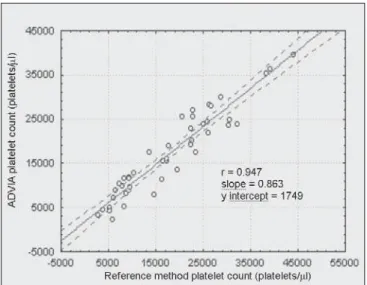
Figure 1. Comparison between the ADVIA counter and the International Committee for
Standardization in Hematology reference method in thrombocytopenic patients.
Cronkite reference method was 18,040 plate-lets per µl. As can be observed in Table 3, ADVIA and STKS showed little deviation, but H1 and T-890 exhibited greater deviation.
The linear correlation test between every counter and the reference method for throm-bocytopenic patients are shown in Figures 1, 2, 3 and 4. The ADVIA counter exhibited the highest correlation (r = 0.947).
○ ○ ○ ○ ○ ○ ○ ○ ○ ○ ○ ○ ○ ○ ○ ○ ○ ○ ○ ○
DISCUSSION
With regard to the laboratory targets for platelet counting, the different counters used indicated great accuracy and precision. How-ever, the Coulter T-890 exhibited 11 to 19.5% of the data lower than the desired target val-ues (Tables 1 and 2), similar to what was ob-tained by Lawrence16 using a counter that also
Figure 3. Comparison between the H1 counter and the International Committee for
Stand-ardization in Hematology reference method in thrombocytopenic patients.
Figure 4. Comparison between the T-890 counter and the International Committee for
employed the impedance principle. When dealing with the thrombocytopenic patient samples, comparative determinations between the automated methods and the ref-erence method suggest that the two-dimen-sional counting system employed by the ADVIA counter demonstrates higher accuracy in differentiating between platelet and non platelet particles, in comparison with the one-dimensional system used by H1, STKS and T-890 (Figures 1, 2, 3 and 4). The data herein presented are similar to those obtained by Kunicka et al.10Dickerhoff and von Ruecker4 also showed lower correlation between the H1 counter and flow cytometry (FC) with
monoclonal anti-platelet antibodies, when us-ing thrombocytopenic samples of lower than 50,000 platelets per µL. Only FC and the Brecher & Cronkite method showed signifi-cant correlation. Interestingly, these are the two reference methods recommended by the ICSH. Hanseler et al.11 using the H1 counter, claimed that for counts of less than 30,000 platelets per µL, the automated counting should be replaced by the manual chamber pro-cedure. Our data obtained with thrombocyto-penic patients also suggest the same for the one-dimensional STKS, H1 and T-890 counters.
The data from Ault6 and Kunicka et al.,10 as well as the data obtained in this
investiga-tion for H1, STKS and T-890, suggest that the one-dimensional platelet counters present a tendency to overestimate the platelet counts when other particles with the same platelet size are contaminating the sample. However, all counters showed flags in thrombocyto-penic samples.
○ ○ ○ ○ ○ ○ ○ ○ ○ ○ ○ ○ ○ ○ ○ ○ ○ ○ ○ ○
CONCLUSION
CONTEXTO: A contagem de plaquetas confiável é de grande importância para avaliar a neces-sidade da transfusão profilática em pacientes plaquetopênicos.
OBJETIVO: Avaliar, em amostras plaqueto-pênicas, a precisão e exatidão da contagem de plaquetas em quatro contadores automá-ticos em comparação com método de refe-rência de Brecher & Cronkite recomendado pelo Comitê Internacional de Estandar-dização em Hematologia.
TIPO DE ESTUDO: Avaliação da contagem automatizada de plaquetas em pacientes trombocitopênicos.
LOCAL: Hospital do Servidor Público Estadual (São Paulo - SP). Instituto Adolfo Lutz.
PARTICIPANTES: 43 pacientes tromboci-topênicos com contagens de plaquetas infe-riores a 30.000/µL.
VARIAVEIS ESTUDADAS: Método de Brecher & Cronkite como padrão de referência e qua-tro contadores automáticos.
RESULTADOS: Os contadores automáticos ADVIA-120 (Bayer), Coulter STKS, H1
○ ○ ○ ○ ○ ○ ○ ○ ○ ○ ○ ○ ○ ○ ○ ○ ○ ○ ○ ○ ○ ○ ○ ○ ○ ○ ○ ○ ○ ○ ○ ○ ○ ○ ○ ○ ○ ○ ○ ○ ○ ○
RESUMO
Raimundo Antônio Gomes Oliveira, MsPhar. Assist-ant Professor in Clinical Hematology and Biochemist-Phar-macist, Pharmacy Department and University Hospital, Universidade Federal do Maranhão, Maranhão, Brazil.
Maria Mariko Takadachi. Biochemist-Pharmacist, Hematology Laboratory, Hospital do Servidor Público Estadual de São Paulo, São Paulo, Brazil.
Kimiyo Nonoyama, MsPhar. Hematology Division, Instituto Adolfo Lutz, São Paulo, Brazil.
Orlando César de Oliveira Barretto, MD, PhD. Asso-ciate Professor, Faculdade de Medicina, Universidade de São Paulo, Laboratório de Investigação Médica-23, São Paulo, Brazil.
Sources of funding: None
Conflict of interest: None
Date of first submission: March 23, 2002
Last received: August 27, 2002
Accepted: September 30, 2002
Address for correspondence
Orlando Cesar de Oliveira Barretto Av. Pedroso de Morais, 70 São Paulo/SP - Brasil - CEP 05420-000 Tel.: (+55 11) 3813-6259
E-mail: [email protected]
COPYRIGHT©2003, Associação Paulista de Medicina
○ ○ ○ ○ ○ ○ ○ ○ ○ ○ ○ ○ ○ ○ ○ ○ ○ ○ ○ ○
Publishing information
System (Technicom-Bayer) e Coulter T-890 demonstraram boa precisão e exatidão em amostras plaquetopênicas obtidas em labo-ratório de hematologia a partir de amostras normais. Apenas o ADVIA-120, que utiliza dois princípios de contagem (volume e ín-dice de refração), demonstrou boa correla-ção com o método de referência recomen-dado pelo Comitê Internacional de Estandardização em Hematologia (ICSH, 1984/1988) para as amostras dos pacientes trombocitopênicos (r = 0,947). Entretanto, todos os aparelhos pediram nova contagem de plaquetas (flags) para as amostras trombocitopênicas.
CONCLUSÃO: A utilização do método de refe-rência de Brecher Cronkite deve ser uma con-duta rotineira e indispensável em todos paci-entes trombocitopênicos com contagens abai-xo de 30,000 plaq /µl obtidas em contadores que utilizam-se apenas do volume como prin-cípio de contagem.
PALAVRAS-CHAVE: Contagem. Plaquetas. Automação. Transfusão. Plaquetopenia.
1- Ross DW, Bentley SA. Evaluation of an automated hematology
system (Technicon H-1). Arch Pathol Lab Med 1986;110(9):803-8.
2- Cornbleet PJ, Myrick D, Judkins S, Levy R. Evaluation of the
CELL-DYN 3000 differential. Am J Clin Pathol 1992;98(6):603-14.
3- Arkin CF. Quality control and standardization in the hematology laboratory. In: Bick RL, editor. Hematology: clinical and labo-ratory practice. St Louis: Mosby; 1993.p.17-37.
4- Dickerhoff R, Von Ruecker A. Enumeration of platelets by
multiparameter flow cytometry using platelet-specific antibod-ies and fluorescent reference particles. Clin Lab Haematol 1995;17(2):163-72.
5- Jones RG, Faust AM, Matthews RA. Quality team approach in
evaluating three automated hematology analyzers with five-part differential capability. Am J Clin Pathol 1995;103(2):159-66.
6- Ault KA. Platelet counting: Is there room for improvement?
Lab Hematol 1996;2:139-43.
7- Mayer K, Chin B, Magnes J, Thaler HT, Lotspeich C, Baisley
A. Automated platelet counters: a comparative evaluation of latest instrumentation. Am J Clin Pathol 1980;74(2):135-50.
8- Ross DW, Ayscue L, Gulley M. Automated platelet counts:
accuracy, precision, and range. Am J Clin Pathol 1980;74(2):151-6.
9- Kjeldsberg CR. Principles of hematologic examination. In: Lee
GR, Bithell TC, Foerster J, et al., editors. Wintrobe’s Clinical Hematology. 9th ed. Philadelphia: Lea & Febiger; 1993.p.11-12.
10- Kunicka JE, Fischer G, Murphy J, Zelmanovic D. Improved platelet counting using two-dimensional laser light scatter. Am J Clin Pathol 2000;114:283-9.
11- Hanseler E, Fehr J, Keller H. Estimation of the lower limits of manual and automated platelet counting. Am J Clin Pathol
○ ○ ○ ○ ○ ○ ○ ○ ○ ○ ○ ○ ○ ○ ○ ○ ○ ○ ○ ○ ○ ○ ○ ○ ○ ○ ○ ○ ○ ○ ○ ○ ○ ○ ○ ○ ○ ○ ○ ○ ○ ○ ○ ○ ○ ○ ○ ○ ○ ○ ○ ○ ○ ○ ○ ○ ○ ○ ○ ○ ○ ○ ○ ○
REFERENCES
1996;105(6):782-7.
12- Klee GG. Performance goals for internal quality control of multichannel haematology analysers. Clin Lab Haematol 1990;12(Suppl 1):65-74.
13- Rowan RM. Platelet counting and the assessment of platelet function. In: Keopke JA, editor. Practical Laboratory Hematology. New York: Churchill Livingstone; 1991.p.157-70. 14- Bode AP. The use of flow cytometry in the study of blood
plate-lets. In: Riley RS, Mahin EJ, Ross W. Clinical applications of flow cytometry. New York: Igaku-Shoin; 1993.p.613-33. 15- Stanworth SJ, Denton K, Monteath J, Patton WN. Automated
counting of platelets on the Bayer ADVIA 120 analyser. Clin Lab Haematol 1999;21(2):113-7.
16- Lawrence JB, Yomtovian RA, Dillman C, et al. Reliability of automated platelet counts: comparison with manual method and utility for prediction of clinical bleeding. Am J Hematol 1995;48(4):244-50.
17- Gil-Fernández JJ, Alegre A, Fernández-Villalta MJ, et al. Clini-cal results of a stringent policy on prophylac-tic platelet trans-fusion: non-randomized comparative analysis in 190 bone mar-row transplant patients from a single institution. Bone Marmar-row Transplant 1996;18(5):931-5.
18- Sherrill JS, Corash L, Shiffer C, et al. Newer approaches to platelet transfusion therapy. In: Transfusion Medicine Educa-tion program of The American Society of Hematology, Orlando, 1996. Proceedings. Philadelphia: ASH; 1996.p.119-31. 19- Beutler E. Platelet transfusions: the 20,000/microL trigger. Blood
1993;81(6):1411-3.
20- Finazzi G. Prophylactic platelet transfusion in acute leukemia: which threshold should be used. Haematologica 1998;83(11):961-2.
21- Navarro JT, Hernández JA, Ribera JM, et al. Prophylactic platelet
transfusion threshold during therapy for adult acute myeloid leukemia: 10,000/microL versus 20,000/microL. Haematologica 1998;83(11):998-1000.
22- Gmür J, Burger J, Schanz U, Fehr J, Schaffner A. Safety of stringent prophylactic platelet transfusion policy for patients with acute leukemia. Lancet 1991;338(8777):1223-6. 23- Consensus Development Conference. Platelet transfusion
therapy. JAMA 1987;257(13):1777-80.
24- Brecher G, Cronkite EP. Morphology and enumeration of hu-man blood platelets. J Appl Physiol 1950;3:365-77. 25- Brecher G, Schneiderman MA, Cronkite EP. The
reproducibil-ity and consistency of the platelet count. Am J Clin Pathol 1953;23:15-26.
26- England JM, Rowan RM, van Assendelft OW, et al. Protocol for evaluation of automated blood cell counters. International Committee for Standardization in Haematology (ICSH). Clin Lab Haematol 1984;6(1):69-84.
27- Platelet counting by the RBC/platelet ratio method: A refer-ence method. Am J Clin Pathol 2001;115(3):460-4. 28- Tycko DH, Metz MH, Epstein EA, Grinbaum A.
Flow-cytometric light scattering measurement of red blood cell vol-ume and hemoglobin concentration. J Applied Optics 1985;24:1355-65.
29- Technicon H*1 System Information Bulletin. Technical
Publica-tion N0 TN8-8588-22. Tarrytown, NY: Technicon Instrument
Corp; December, 1988:3-4.
30- Technicon H*1 System: Operator’s Guide. International Division ed. Tarrytown, NY: Technicon Instrument Corp; 1985: 2/56-57. 31- Coulter STKS Analyser with reticulocyte analysis: product
ref-erence manual n. 4237182B. Miami: Coulter; 1995. 32- Coulter T Series with differential capabilities: product