○ ○ ○ ○ ○ ○ ○ ○ ○ ○ ○ ○ ABSTRACT○ ○ ○ ○ ○ ○ ○ ○ ○ ○ ○ ○ ○ ○ ○ ○ ○ ○ ○ ○ ○ ○ ○ ○ ○ ○ ○
INTRODUCTION
The clinical significance and management practices adopted in women with atypical sq-uamous cells (ASC) or low-grade sqsq-uamous intraepithelial lesion (LSIL) have not been uniformly accepted.1-8 Repeated cervical smears, addition of tests to detect human papillomavirus (HPV) DNA or referral of the women for immediate colposcopy are some of the different options, although they have clearly different cost-effectiveness. The pre-liminary results of the ASCUS/LSIL Triage Study (ALTS),1 an American national prospec-tive randomized trial based on 3,488 women with atypical squamous cells of undetermined significance (ASCUS) and 1,572 women with LSIL, showed that hybrid capture II (HCII) testing for high-risk HPV DNA was fre-quently positive (around 85%) in women with LSIL. This therefore demonstrated the lim-ited usefulness of the HCII assay in the man-agement of these cases, because the vast ma-jority of these women would be referred for (unnecessary) colposcopy due to the positive HPV tests. Regarding the women with AS-CUS smears, Solomon et al. (2001) concluded that HCII testing for high-risk HPV DNA types was positive in 50.6% and should be a viable option for the management of these women. HPV DNA had greater sensitivity and specificity in detecting CIN3 (cervical intraepithelial neoplasia grade 3, carcinoma in situ or severe dysplasia) or above, in com-parison with a single additional cervical test showing ASCUS or above.1
These results are not uniformly accepted, however.4,9 Recently, the Bethesda system
modified their ASCUS category to make it two-tier: ASC–US (of undetermined signifi-cance) and ASC-H (cannot exclude high-grade squamous intraepithelial lesion - HSIL).10
According to the ASCCP (American So-ciety for Colposcopy and Cervical Pathology) 2001 Consensus Guidelines for the Manage-ment of Women with Cervical Cytological Abnormalities,8 women with ASC-US should be managed using a program of two repeated cytology tests, immediate colposcopy or HPV DNA testing for high-risk HPV types. Women with ASC-H, LSIL, HSIL and atypi-cal glandular cells should be referred for im-mediate colposcopy evaluation, regardless of the result of HPV testing. However, HPV test-ing for routine clinical management of ASC-US cytological abnormalities is not yet war-ranted9-11 and continues to be an investiga-tional tool.Furthermore, evaluation of the clinical effectiveness of HPV testing combined with cervical smears needs to be completed in developing countries.
The aim of the present study was to shed further light on the issue and investigate whether HPV testing and viral load quantifi-cation are clinically useful tools for women who present with ASC or LSIL cervical smears in our settings in Brazil.
○ ○ ○ ○ ○ ○ ○ ○ ○ ○ ○ ○ ○ ○ ○ ○ ○ ○ ○ ○
MATERIAL AND METHODS
Patients
In this cross-sectional study, we analyzed a series of 119 women aged 16 to 63 (median of 31 years), referred for the Colposcopy Clinic due to an abnormal cervical smear consistent
Derchain
• Marcos Roberto Martins
• Luís Otávio Zanatta Sarian
• Edson Zangiacome Martinez
• Kari Juhani Syrjänen
predicting high-grade CIN in
women with cervical smears
showing only atypical squamous
cells or low-grade squamous
intraepithelial lesion
Department of Obstetrics and Gynecology, Universidade Estadual de
Campinas, Campinas, and Universidade de Taubaté, Taubaté, São Paulo,
Brazil, and in association with the National Health Institute, Rome, Italy
Original Ar
ticle
CONTEXT: Human papillomavirus (HPV) viral load may have an important role in predicting high-grade cervical intraepithelial neoplasia (CIN) in women with cervical smears showing atypical squamous cells or LSIL.
OBJECTIVE: To determine whether the assessment of the viral load of high-risk HPV DNA is useful in predicting the detection of high-grade cervical intraepithelial neoplasia (CIN2 and 3) in women referred because of cervical smears showing only atypical squamous cells or LSIL.
TYPE OF STUDY: Cross-sectional
SETTING: Colposcopy Clinic in a University hospital.
METHODS: A series of 119 women referred because of atypical squamous cells or LSIL between August 2000 and April 2001 were included. All women were subjected to a new cervical smear, HPV test-ing for the high-risk types ustest-ing hybrid capture II (HCII), viral load measurement in relative light units (RLU) and colposcopy, with cervical biopsies (n = 97). Cervical lesions were graded using the CIN classification.
RESULTS: Cervical biopsies revealed CIN2 or CIN3 in 11% of the cases, equally among women referred because of atypical squamous cells or LSIL. The HCII test was positive in 16% of women with atypical squamous cells and 52% of those with LSIL (OR = 5.8; 95% CI 1.4 to 26.7). There was strong corre-lation between CIN2 or CIN3 and positivity for HPV DNA when this group was compared with women with only CIN1 or normal cervix (OR = 7.8; 95% CI 1.5 to 53.4). In ROC analysis for HCII in diag-nosing CIN2 and CIN3, the area under the ROC curve was 0.784, and the viral load cutoff point of 10.0 RLU/cutoff presented 77% sensitivity and 73% specificity. Second cytology showing at least atypi-cal squamous cells did not accurately detect CIN2 or CIN3 (OR = 6.4; 95% CI 1.0 to 50.9). The sensitivities of the second cervical smear and HCII were similar, although the specificity of HCII was significantly higher than the second cervical smear.
CONCLUSIONS: The viral load of high-risk HPV types was significantly associated with the di-agnosis of CIN2 or CIN3 in women referred because of atypical squamous cells and LSIL ab-normalities in their cervical smear.
Original Ar
ticle
with ASC (n = 19) or LSIL (HPV-suggestive changes in 37 women and CIN1 in 63), be-tween August 2000 and April 2001. Women were excluded from the study if: a) they had a previous history of CIN or cervical, vaginal or vulvar cancer; b) they were referred because of an HSIL cervical smear; c) they had immuno-suppression; or d) they were pregnant. The Ethi-cal Committee of the MediEthi-cal School Hospital approved the study protocol, and all partici-pants gave their written informed consent.
All women were offered a questionnaire asking for their key sociodemographic data, and a new conventional cervical smear was taken, using the Ayre spatula and endocervi-cal brush. This was fixed in 95% ethanol and stained by means of the modified Papanico-laou method. A second specimen was obtained from the endocervix using a Dacron swab and placed in 1.0 ml of specimen transport me-dium (Digene Corporation) for DNA HPV testing using the HCII test.
All women were subjected to colposcopic examination, according to routine practice, and the results were classified according to the International Federation of Cervical Pa-thology and Colposcopy (IFCCP) classifica-tion.12 The cervix was considered colpospically normal when all of the squamous co-lumnar junction was shown, and abnormal when the cervix presented some area of acetowhite epithelium, mosaicism, punctua-tion, leukoplasia or abnormal vessels. Abnor-mal areas were classified as major or minor abnormalities. In 90 women, colposcopically targeted biopsies were taken from suspicious areas. In addition, for seven women with unsatisfactory colposcopy, a large loop exci-sion of the transformation zone (LLETZ) was done using a diathermy procedure. Twenty-two women with entirely normal colposcopy and a totally visible squamous columnar junction had no biopsy taken, and they were considered as having a normal cervix.
Cytology and histology
Referral cervical smears were available for review from all the patients. The final cyto-logical diagnoses for both the referral and the second cervical smear were obtained using the Bethesda System (2002)10 and were classified as negative, ASC, LSIL or HSIL (the second cervical smear only), based on a consensus re-view by two pathologists. Cervical biopsies were fixed in 10% phosphate-buffered forma-lin, embedded in paraffin, and stained with hematoxylin and eosin (HE). Biopsies were analyzed according to the World Health Or-ganization criteria13 and classified as negative,
CIN1, CIN2 or CIN3. Women with biopsy-confirmed cervicitis and those with normal colposcopy were classified as having a normal cervix in this study.
Hybrid capture
The specimens for HCII were tested for probe B (high-risk HPVs: types 16, 18, 31, 33, 35, 39, 45, 51, 52, 56, 58, 59 and 68), and the test was classified as positive when the relative light unit/cutoff (RLU/CO) ratio (RLU of specimen/mean RLU of two posi-tive controls) was 1 pg/ml or greater. These RLU/CO ratios also provided an estimate of the amount of HPV DNA in the specimens, i.e. the viral load in the sample. The storage of the specimens and all reagents, as well as the conduct of the tests, took place at the Medical School Hospital Laboratory, follow-ing the manufacturer’s instructions (Digene Diagnostics Inc., USA).
Statistical analysis
Mean HPV viral loads with standard de-viation (SD) were calculated in relation to cervical smears and histological results. For ethical reasons, invasive diagnostic biopsy was indicated only in cases of repeated positive cervical smears or abnormal colposcopy. Begg-corrected estimate values for the sensitivity and specificity of HCII (at standard 1.0 RLU/CO cutoffs) and second cervical smears were cal-culated.14,15 Receiver operating characteristic (ROC) analysis was used for testing the diag-nostic performance of the HCII test at cut-offs of 1.0 RLU/CO, 2.0 RLU/CO, 5.0 RLU/ CO, 10.0 RLU/CO, 50.0 RLU/CO, 100.0
RLU/CO and 500.0 RLU/CO, and above the cutoff point, in order to detect histologically-confirmed CIN2 or CIN3 lesions.
All statistical analyses were done using the SAS software, version 8.0. Odds ratios (OR) with 95% confidence interval (95% CI) were used for evaluating the association between the HPV test result, cervical smear result and disease status.
○ ○ ○ ○ ○ ○ ○ ○ ○ ○ ○ ○ ○ ○ ○ ○ ○ ○ ○ ○
RESULTS
Altogether, 23 women (19%) were found to have a normal cervix via colposcopy, 91 (76%) presented with minor abnormalities and 5 (4%) with major abnormalities. Of the women with colposcopically guided biopsy areas, 6 (5%) presented with cervicitis in the histological analysis, 78 (66%) showed CIN1 and 13 (11%) had CIN2 or CIN3 (data not shown). Considering the distribution of these results according to the first cervical smears, only CIN1 was significantly higher in women referred because of LSIL cervical smears (OR 4.14; 95% CI 1.25 to 13.9). CIN 2 or CIN3 were found in the same proportions in women with ASC or LSIL (Table 1).
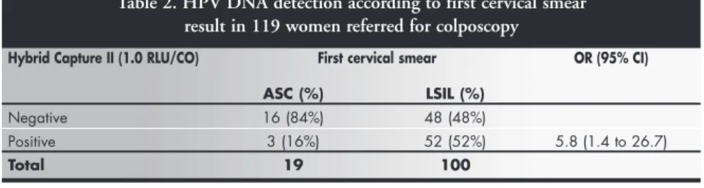
The HCII test was positive for the high-risk HPV DNA in 16% of the women (3 women) who had a cervical smear with ASC and 52% of those with LSIL. The HPV DNA detection rate was significantly higher (OR 5.8; 95% CI 1.4 to 26.7) in women with cy-tological changes consistent with LSIL than in those with ASC (Table 2).
The HCII and histology results are sum-marized in Table 3. It is noteworthy that 85%
Table 1. Histological diagnosis and first cervical smear result in 119 women referred for colposcopy
Final disease status First cervical smear OR (95% CI)
ASC (%) LSIL (%)
Normal cervix 9 (48%) 19 (19%) REF
CIN 1 8 (42%) 70 (70%) 4.14 (1.25 to 13.9)
CIN 2 or 3 2 (10%) 11 (11%) 2.1 (0.4 to 21.2)
Total 19 (100%) 100 (100%)
CIN = cervical intraepithelial neoplasia; ASC = atypical squamous cells, LSIL = low-grade squamous intraepithelial lesion; OR = odds ratio; REF= reference values for odds ratios; CI = confidence interval.
Table 2. HPV DNA detection according to first cervical smear result in 119 women referred for colposcopy
Hybrid Capture II (1.0 RLU/CO) First cervical smear OR (95% CI)
ASC (%) LSIL (%)
Negative 16 (84%) 48 (48%)
Positive 3 (16%) 52 (52%) 5.8 (1.4 to 26.7)
Total 19 100
of the women with CIN2 or CIN3 had a posi-tive HPV DNA test with a 1.0 RLU/CO cut-off point. There was a strong correlation be-tween CIN2 or CIN3 and positivity for HPV DNA when this group was compared with women with only CIN1 or normal cervix (OR = 7.8; 95% CI 1.5 to 53.4).
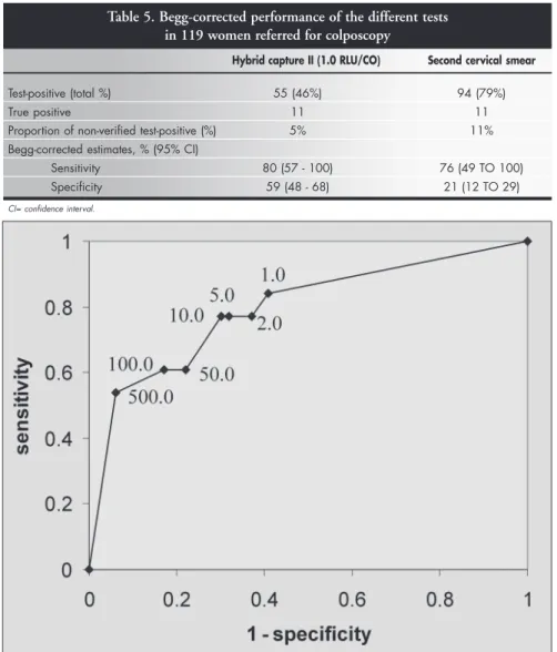
Figure 1 shows the receiver operating char-acteristic (ROC) curve constructed to describe the sensitivity and specificity of the HCII test at different cutoff points of the DNA index (RLU/CO ratio) for diagnosing CIN2 or CIN3 lesions. The area under the curve is 0.784, thus showing good performance of the HCII in the diagnosis of CIN2 or CIN3. Al-though the highest sensitivity value in the de-tection of CIN2 or CIN3 was achieved at the standard manufacturer’s recommended point of 1.0 RLU/CO, the best balance in specificity and sensitivity was reached at the cutoff point of 10.0 RLU/CO, showing a 77% sensitivity and 70% specificity. It is important to notice that a slight loss of sensitivity occurred when comparing this point to the standard 1.0 RLU/ CO cutoff, with a concurrent and more pro-nounced gain in specificity.
The second cervical smear showed ASC or SIL in 13/28 women (46%) with a nor-mal cervix; 70/78 women (89%) with histologically confirmed CIN1 and 11/13 (85%) of those with CIN2 or CIN3. The sec-ond cervical test with at least ASC did not accurately differentiate normal cervix or CIN1 from CIN2 or CIN3 (OR = 1.5; 95% CI 0.3 to 10.7) (Table 4).
Regarding the Begg-corrected perform-ances of the diagnostic tests, the sensitivities of the second cervical smear and HCII were similar, although the specificity of HCII was significantly higher than second cervical smear (Table 5).
○ ○ ○ ○ ○ ○ ○ ○ ○ ○ ○ ○ ○ ○ ○ ○ ○ ○ ○ ○
DISCUSSION
Correct identification of women with CIN2 or CIN3 or invasive cancer among those who have been referred because of ASC and LSIL cervical smear results has a major impact on the cost-effectiveness of patient management and triage.3,4,7,9,11 To assess the performance of HPV testing as a triage tool, the present cross-sectional study compared repeated cytology, HPV testing (by means of HCII) and colposcopy in a series of women referred to our clinic because of ASC and LSIL cervical smears.
In our series, the detection rate for HPV DNA by HCII was significantly higher in women referred because of LSIL (52%) than
Table 3. HPV DNA detection and the grade of the lesions in 119 women referred for colposcopy
Hybrid Capture II (1.0 RLU/CO) Final diagnosis OR (95% CI) Normal Cervix CIN 2 OR 3
CIN1 (%) (%)
Negative 62 (59%) 2 (15%)
Positive 44 (41%) 11 (85%) 7.8 (1.5 to 53.4)
Total 106 (100%) 13 (100%)
CIN = cervical intraepithelial neoplasia; OR = odds ratio; CI = confidence interval.
Figure 1. Receiver-operating characteristics (ROC) curve analysis for the performance of the hybrid capture II test in the
diagno-sis of cervical intraepithelial neoplasia grades 2 or 3 (relative light unit/cutoff, RLU/CO values).
Table 4. Second cervical smear and grade of the lesion in 119 women referred for colposcopy
Second Cervical Smear Final diagnosis OR (95% CI) Normal Cervix CIN 2 OR 3
CIN1 (%) (%)
Normal 23 (22%) 2 (15%)
ASC/LSIL/HSIL 83 (78%) 11 (85%) 1.5 (0.3 TO 10.7)
Total 106 (100%) 13 (100%)
CIN = cervical intraepithelial neoplasia; OR = odds ratio; CI = confidence interval.
Table 5. Begg-corrected performance of the different tests in 119 women referred for colposcopy
Hybrid capture II (1.0 RLU/CO) Second cervical smear
Test-positive (total %) 55 (46%) 94 (79%)
True positive 11 11
Proportion of non-verified test-positive (%) 5% 11%
Begg-corrected estimates, % (95% CI)
Sensitivity 80 (57 - 100) 76 (49 TO 100)
Specificity 59 (48 - 68) 21 (12 TO 29)
in women referred because of ASC (16%). However, in both groups the high-risk HPV detection rate was significantly lower than re-cently reported from an ongoing ALTS study in the US, in which the high-risk HPV was found in 85% of women with LSIL and in 50% of those with ASCUS.5,7 Results closer to ours were reported by Lee et al. (2001), who showed HCII-positive results in 63.8% of women with LSIL and 26.3% of women with ASCUS.2 These figures are consistent with the data re-cently reported from a large-scale screening trial in Russia, Belarus and Latvia (NIS Study), com-paring HCII testing with other screening tests.9 The results seem to be very similar if the polymerase chain reaction (PCR) is used for HPV detection, as shown by the report of Zerbini et al. (2001), who included HPV types 6, 11, 16, 18, 31, 33, and 45 in their panel and showed 27.3% and 53.5% detection rates in ASCUS and LSIL cases, respectively.16
The 11% prevalence of CIN2 or CIN3 in the present series was identical in women who were referred because of ASC and LSIL. Not unexpectedly, the detection of high-risk HPV was significantly associated with the presence of high-grade CIN lesions in the bi-opsy. Even more importantly, a significant sta-tistical association could be established be-tween the viral load and the high-grade CIN lesions. This is consonant with the data from some recent reports, suggesting that high vi-ral load of HPV16 DNA is a major risk factor for the development of carcinoma in situ (CIS), and measurements of the viral load might be of value in estimating the probabil-ity of progressive disease.17 The risk of devel-oping CIS, estimated through odds ratios, increased significantly with increasing amounts of HPV16 measured through TaqMan-PCR. Also using PCR, Zerbini et al. (2001) showed that the amount of HPV DNA in the samples varied widely among the women with biopsy-confirmed CIN1, CIN2, CIN3 or invasive cancer.16 There was also a major variation in the viral load in women within each single histological category (CIN1, CIN2 or CIN3), as well as between the different groups. These authors attributed the higher viral load in high-grade lesions to HPV16, which was the single most prevalent genotype in these patients. According to these authors, HPV16 load seems to be a specific but not a highly sensitive diagnostic marker for defining cervical disease status.16
Even more recently, Abba et al. (2003)18 analyzed the relationship between viral type and copy number of HPV DNA with respect to the grade of cervical disease. In this study,
the viral load determination was performed using low stringency PCR and these authors classified DNA concentration as 10, 20, 100 and 500 copies of HPV16 per cell, consider-ing 100 viral particles per cell in order to de-fine a cutoff between low and high viral loads. These authors found that, in samples with high-risk HPV, 45% presented high viral load whereas only 18% of samples infected with low-risk virus had a copy number above 100 particles per cell. When only samples harboring HPV16 were considered, almost 50% presented high viral load, which was sig-nificantly more than the proportion when considering other high-risk viral types (OR = 2.59; 95% CI = 1.18 to 5.70). In this same study, high-grade lesions were also significantly associated with HPV16 high viral load (OR = 8.53; 95% CI = 2.73 to 26.70).
HC tests are based on a pool of high-risk HPV types and differ from PCR in its linear dynamic range of viral load. Their results are also more influenced by sample cell count. These features make HC results more con-troversial and difficult to interpret.19 Shiffman et al. (2000), showed that women with cyto-logical abnormalities had higher viral load via HCII tests when compared with women with normal cervical smears. They showed that an analytical sensitivity of 1.0 pg/ml using the HCII assay would have permitted detection of 88.4% of high-grade lesions and invasive cancer, whereas the lower levels of detection with HCII (i.e. < 1.0 pg/ml) proved to be clinically non-specific without any gain in di-agnostic sensitivity.20 These figures are sup-ported by recent data from the NIS study, showing that the presence of high-grade his-tology was associated with HCII positivity (cutoff of 1 pg/ml) (OR = 4.8; 95% CI = 0.7 to 34.2; p = 0.047). Using this cut-off, the sensitivity of the HCII test was 96.6% (90.0 to 100), specificity 15.9% (10.6 to 21.2), posi-tive predicposi-tive value 15.1% (9.9 to 20.3) and negative predictive value (NPV) 96.8% (90.3 to 100) in detecting biopsy-confirmed high-grade lesions.9 Changing the cutoff in either direction did not significantly affect the sen-sitivity until a sensen-sitivity level of 20.0 pg/ml, and an NPV level of 500.0 pg/ml.
Although the viral load distribution in our series showed a wide range within the histological categories, a viral load of about 10.0 RLU seemed to represent the best-bal-anced specificity-sensitivity cutoff point in the ROC analysis. The best values for sensi-tivity and specificity in predicting high-grade lesions were achieved in viral load ranges of between 5.0 RLU/CO and 50.0 RLU/CO,
i.e. far above the standard manufacturer’s rec-ommendation of a cutoff point of 1.0 RLU/ CO for use in the screening of cervical HPV-related abnormalities.
Sun et al. recently reported that quantita-tive levels of high-risk HPV DNA were clearly associated with the presence of CIN2 and CIN3 or invasive carcinoma.21,22 They believed that increased DNA loads of high-risk HPV DNA, as determined by HCII in cervical specimens, could be used as the specific marker for progressive disease. For practical purposes, however, they considered that the viral load index values (RLU/CO) should be categorized into three groups: low viral loads of < 0.6 RLU/CO, intermediate viral loads of 0.6 to 10.0 RLU/CO and high viral loads of > 10.0 RLU/CO.21,22
1. Solomon D, Schiffman M, Tarone R. Comparison of three man-agement strategies for patients with atypical squamous cells of undetermined significance: baseline results from a randomized trial. J Natl Cancer Inst 2001;93(4):293-9.
2. Lee NW, Kim D, Park JT, Kim A. Is the human papillomavirus test combination with the Papanicolaou test useful for manage-ment of patients with diagnoses of atypical squamous cells of undetermined significance/low-grade squamous intraepithelial lesions? Arch Pathol Lab Med 2001;125(11):1453-7. 3. Herbst AL, Pickett KE, Follen M, Noller KL. The management
of ASCUS cervical cytologic abnormalities and HPV testing: a cautionary note. Obstet Gynecol 2001;98(5 Pt 1):849-51. 4. Paraskevaidis E, Malamou-Mitsi V, Koliopoulos G, et al.
Ex-panded cytological referral criteria for colposcopy in cervical screening: comparison with human papillomavirus testing. Gynecol Oncol 2001;82(2):355-9.
5. Sherman ME, Schiffman M, Cox JT. Effects of age and human papilloma viral load on colposcopy triage: data from the randomized Atypical Squamous Cells of Undetermined Signifi-cance/Low-Grade Squamous Intraepithelial Lesion Triage Study (ALTS). J Natl Cancer Inst 2002;94(2):102-7.
6. Hughes SA, Sun D, Gibson C, et al. Managing atypical squa-mous cells of undetermined significance (ASCUS): human papillomavirus testing, ASCUS subtyping, or follow-up cytol-ogy? Am J Obstet Gynecol 2002;186(3):396-403. 7. Human papillomavirus testing for triage of women with
cytologic evidence of low-grade squamous intraepithelial lesions: baseline data from a randomized trial. The Atypical Squamous Cells of Undetermined Significance/Low-Grade Squamous Intraepithelial Lesions Triage Study (ALTS) Group. J Natl Can-cer Inst 2000;92(5)397-402.
8. Wright TC, Cox JT, Massad LS, Twiggs LB, Wilkinson EJ. 2001
○ ○ ○ ○ ○ ○ ○ ○ ○ ○ ○ ○ ○ ○ ○ ○ ○ ○ ○ ○ ○ ○ ○ ○ ○ ○ ○ ○ ○ ○ ○ ○ ○ ○ ○ ○ ○ ○ ○ ○ ○ ○ ○ ○ ○ ○ ○ ○ ○ ○ ○ ○ ○ ○ ○ ○ ○ ○ ○ ○ ○ ○ ○ ○
REFERENCES
Consensus Guidelines for the management of women with cer-vical cytological abnormalities. JAMA 2002;287(16):2120-9. 9. Syrjänen S, Shabalova IP, Petrovichev N, et al. Human
papillomavirus testing and conventional Pap smear cytology as optional screening tools of women at different risks for cervical cancer in the countries of the former Soviet Union. J Lower Genital Tract Dis 2002;6(2):97-110.
10. Solomon D, Davey D, Kurman R, et al. The 2001 Bethesda System: terminology for reporting results of cervical cytology. JAMA 2002;287(16):2114-9.
11. Gamzu R, Almog B, Levin I, et al. Clinical and economic im-plication of adding HPV tests to the routine cytology follow-up and management of patients with histologically defined cer-vical intraepithelial neoplasia grade 1. Gynecol Oncol 2002;86(2):129-33.
12. Stafl A, Wilbanks GD. An international terminology of colpos-copy: report of the Nomenclature Committee of the Interna-tional Federation of Cervical Pathology and Colposcopy. Obstet Gynecol 1991;77(2):313-4.
13. Scully RE, Bonfiglio TA, Kurman RJ, Silverberg SG, Wilkins EJ. Histological typing of female genital tract tumors. World Health Organization - International histological classification of tumors. 2nd ed. Berlin: Springer-Verlag; 1994.
14. Begg CB, Greenes RA. Assessment of diagnostic tests when dis-ease verification is subject to selection bias. Biometrics 1983;39(1):207-15.
15. Schneider A, Hoyer H, Lotz B, et al. Screening for high-grade cervical intra-epithelial neoplasia and cancer by testing for high-risk HPV, routine cytology or colposcopy. Int J Cancer 2000;89(6):529-34.
16. Zerbini M, Venturoli S, Cricca M, et al. Distribution and viral load of type specific HPVs in different cervical lesions as
de-tected by PCR-ELISA. J Clin Pathol 2001;54(5):377-80. 17. Josefsson AM, Magnusson PK, Ylitalo N, et al. Viral load of
human papilloma virus 16 as a determinant for development of cervical carcinoma in situ: a nested case-control study. Lancet 2000;355(9222):2189-93.
18. Abba MC, Mourón SA, Gómez MA, Dulout FN, Golijow CD. Association of human papillomavirus viral load with HPV16 and high-grade intraepithelial lesion. Int J Gynecol Cancer 2003;13(2):154-8.
19. Gravitt PE, Burk RD, Lorincz A, et al. A comparison between real-time polymerase chain reaction and hybrid capture 2 for human papillomavirus DNA quantitation. Cancer Epidemiol Biomarkers Prev 2003;12(6):477-84.
20. Schiffman M, Herrero R, Hildesheim A, et al. HPV DNA test-ing in cervical cancer screentest-ing: result from women in a high-risk province of Costa Rica. JAMA 2000;283(1):87-93. 21. Sun CA, Lai HC, Chang CC, Neih S, Yu CP, Chu TY. The
significance of human papillomavirus viral load in prediction of histologic severity and size of squamous intraepithelial le-sions of uterine cervix. Gynecol Oncol 2001;83(1):95-9. 22. Sun CA, Liu JF, Wu DM, Nieh S, Yu CP, Chu TY. Viral load of
high-risk human papillomavirus in cervical squamous intraepithelial lesions. Int J Gynaecol Obstet 2002;76(1):41-7. 23. Lorincz AT, Castle PE, Sherman ME, et al. Viral load of human papillomavirus and risk of CIN3 or cervical cancer. Lancet 2002;360(9328):228-9.
24. Muñoz N, Bosch FX, de Sanjosé S, et al. Epidemiologic classi-fication of human papillomavirus types associated with cervical cancer. N Engl J Med 2003;348(6):518-27.
25. Schelecht NF, Trevisan A, Duarte-Franco E, et al. Viral load as a predictor of the risk of cervical intraepithelial neoplasia. Int J Cancer 2003;103(4):519-24.
○ ○ ○ ○ ○ ○ ○ ○ ○ ○ ○ ○ ○ ○ ○ ○ ○ ○ ○ ○
CONCLUSIONS
In the studied population, referred for ex-amination due to an ASC or LSIL cytological
smear, HCII testing for high-risk HPV types was a sensitive tool in detecting CIN2 or CIN3, with better diagnostic performance than repeated cervical smears. As the
Carga viral do papilomavírus humano como fa-tor preditivo de neoplasia intra-epitelial de alto grau em mulheres com células esca-mosas atípicas ou lesão escamosa intra-epite-lial de baixo grau na colpocitologia
CONTEXTO: A determinação da carga viral do papilomavírus humano (HPV) pode ter im-portante papel na detecção de neoplasia intra-epitelial cervical (NIC) de alto grau em mu-lheres com colpocitologia apresentando cé-lulas escamosas atípicas ou sugestivas de le-são escamosa de baixo grau.
OBJETIVO: Avaliar se a determinação da carga viral do DNA HPV é útil para predizer a detecção da neoplasia intra-epitelial de alto grau (NIC2 e 3) em mulheres referidas por colpocitologias mostrando apenas células escamosas atípicas ou lesão intra-epitelial de baixo grau.
TIPO DE ESTUDO: Transversal
LOCAL: Serviço de colposcopia de hospital uni-versitário.
MÉTODOS: Foram incluídas 119 mulheres en-caminhadas por células escamosas atípicas e lesão intra-epitelial de baixo grau entre agos-to de 2000 e abril de 2001. De agos-todas as mu-lheres foi coletada nova colpocitologia, espé-cime para teste de HPV usando captura de híbridos II (CHII), carga viral medida em unidades relativas de luz (URL). Foi realiza-da colposcopia com biópsia cervical em 97 mulheres. As lesões cervicais foram classifi-cadas usando a classificação NIC. Para diag-nóstico final, a colposcopia normal ou a pre-sença de cervicite confirmada por biópsia
fo-ram classificadas como colo normal. Para análise estatística foram calculados o odds ratio (OR), com intervalo de confiança em 95%, e foi traçada uma curva “receiver operator characteristic” (ROC).
RESULTADOS: As biópsias cervicais mostraram NIC2 ou 3 em 11% dos casos, igualmente distribuídas entre as mulheres encaminhadas por causa da presença de células escamosas atípicas ou lesão intra-epitelial de baixo grau. A CHII foi positiva em 16% das mulheres com células escamosas atípicas e em 52% daquelas com lesão intra-epitelial de baixo grau (OR = 5,8; IC 95% 1,4 a 26,7). Entre as mulheres com CHII positiva, 7% tinham cérvice normal, 73% NIC 1 (OR = 6,3; IC 95% 1,8 a 23,8) e 20% tinham NIC2 ou 3 (OR = 33,0; IC 95% 4,2 a 347,8). Na análi-se da curva ROC para CH II, diagnostican-do NIC2 e 3, a área sob a curva foi de 0,784 e o ponto de corte da carga viral de 10.0 URL mostrou sensibilidade de 77% e especifi-cidade de 70%. A segunda colpocitologia mostrando ao menos células escamosas atípicas não apresentou boa performance na detecção NIC 2 ou 3 (OR = 6,4%; IC 95% 1,0 a 50,9).
CONCLUSÕES: A carga viral do DNA-HPV de alto risco oncológico foi significativa-mente associada com o diagnóstico de NIC2 e 3 em mulheres encaminhadas por detecção de células escamosas atípicas e le-são intra-epitelial escamosa de baixo grau na colpocitologia.
PALAVRAS-CHAVE: Papilomavírus humano. Carga viral. Neoplasia intra-epitelial cervical.
○ ○ ○ ○ ○ ○ ○ ○ ○ ○ ○ ○ ○ ○ ○ ○ ○ ○ ○ ○ ○ ○ ○ ○ ○ ○ ○ ○ ○ ○ ○ ○ ○ ○ ○ ○ ○ ○ ○ ○ ○ ○
RESUMO RESUMORESUMO RESUMORESUMO
○ ○ ○ ○ ○ ○ ○ ○ ○ ○ ○ ○ ○ ○ ○ ○ ○ ○ ○ ○
PUBLISHING INFORMATION
ACKNOWLEDGMENTS: The skillful technical assistance of Denise da Rocha Pitta Lima de Moraes, Lúcia Maria Fagian de Carvalho and Elisabete Aparecida Campos is grate-fully acknowledged.
André Luis Ferreira Santos. MSc in Gynecology, Universidade de Taubaté, Taubaté, São Paulo, Brazil.
Sophie Françoise Mauricette Derchain. Associate pro-fessor, Department of Obstetrics and Gynecology, Universidade Estadual de Campinas, Campinas, São Paulo, Brazil.
Marcos Roberto Martins. MSc in Gynecology, Universidade de Taubaté, Taubaté, São Paulo, Brazil.
Luís Otávio Zanatta Sarian. MSc in Gynecology, Universidade Estadual de Campinas, Campinas, São Paulo, Brazil.
Edson Zangiacome Martinez. PhD in Statistics, Universidade Estadual de Campinas, Campinas, São Paulo, Brazil.
Kari Juhani Syrjänen. Associate professor, National In-stitute of Health, Rome, Italy.
Conflict of interest: Not declared
Sources of funding: Fundação Amparo a Pesquisa do Estado de São Paulo (FAPESP), grant number 99/11264-0, and Conselho Nacional de Desenvolvimento Científico e Tecnológico (CNPq), grant number 300354/01-0.
Date of first submission: April 22, 2003
Last received: June 30, 2003
Accepted: August 7, 2003
Address for correspondence:
Luís Otávio Zanatta Sarian R. Alexander Fleming, 848
Campinas/SP — Brasil — CEP 13092-340 Tel. (+55 19) 3252-4948
E-mail: [email protected]