Corona Mortis
: anatomical and surgical description on 60 cadaveric
hemipelvises
Corona Mortis
: descrição anatômica e cirúrgica em 60 hemipelvis cadavéricas
Túlio Fabianode oliveira leiTe1; lucas alves sarmenTo Pires2; Kiyoshi GoKe3; Júlio Guilherme silva4; carlos alberTo arauJo chaGas2.
INTRODUCTION
T
he obturator artery (OA) has a very variable origin, usually originating from the anterior wall of the in-ternal iliac artery (IIA). It runs anteriorly and inferiorly on the pelvic wall below the obturator nerve (ON), perfora-ting the obturator fascia and reaching the obturator fo-ramen (OF)1-3. In its trajectory the OA distributes severalcollateral branches: two muscular branches (for the iliac and internal obturator muscles), a pubic branch (which runs on the posterior surface of the pubis body to anas-tomose with the ipsilateral branch), a bladder branch (to the posterior face of the urinary bladder) and an anas-tomotic branch1,2. This latter deserves special attention
due to its trajectory, as it crosses the upper branch of the pubis (UBP) perpendicularly and anastomoses with the inferior epigastric artery (IEA)1,2. After exiting the
pelvis, the OA is divided into two terminal branches, an internal branch, with path at the inner border of the OF giving branches to the external obturator, pectin, and gracile muscles, and an external branch, which runs at
the OF outer border to form the cruciform anastomo-sis1.
The IEA, on the other hand, is a branch of the external iliac artery (IEA). Stemming a few millimeters above the inguinal ligament (LI), it runs horizontally and superiorly to the transverse fascia and runs anteriorly towards the arcuate line, between the rectus abdominis muscle and a posterior layer of its sheath. The IEA then anastomoses with an upper epigastric artery, a branch of the internal thoracic artery. During its trajectory, the IEA gives a branch to the spermatic cord, a suprapubic branch and an anastomotic branch (for the OA)1,2,4.
Corona mortis (CM), or death crown, is de-fined as an arterial or venous connection between the anastomotic branches of the obturator artery and the inferior epigastric artery over the superior branch of the pubis5-9. This anatomical variant is of clinical and
sur-gical interest, as it is susceptible to iatrogenic lesions during hernia repairs, gynecological and orthopedic procedures, and may also be damaged in fractures of the pubis or acetabulum. The literature also reports the
1 - Medical School, University of São Paulo, Institute of Radiology, São Paulo, SP, Brazil. 2 - Fluminense Federal University, Department of Mor-phology, Niterói, RJ, Brazil. 3 - Estácio de Sá University, Department of Anatomy, Rio de Janeiro, RJ, Brazil. 4 - Federal University of Rio de Janeiro, Department of Physiotherapy, Rio de Janeiro, RJ, Brazil.
A B S T R A C T
Objective: to report the prevalence of arterial corona mortis and to describe its surgical and clinical applicabilities. Methods: We dissected 60 hemipelvises (50 men and 10 women) fixed in a 10% formalin solution for the purpose of gathering information on corona mortis. We measured the caliber and length of the obturator artery and its anastomotic branch with the aid of a digital caliper and submitted the data to statistical analyzes and comparisons with the GraphPad Prism 6 software. Results: arterial corona mortis was present in 45% of the studied sample. The most common origin of the obturator artery was the internal iliac artery; however, there was one exceptional case in which it originated from the femoral artery. The caliber of the anastomotic branch was on average 2.7mm, whereas the caliber of the obturator artery was 2.6mm. Conclusion: the vascular connections between the obturator, internal iliac, external iliac and inferior epigastric arterial systems are relatively common over the upper pubic branch. The diameter and a trajectory of the anastomotic artery may vary. Thus, iatrogenic lesions and pelvic and acetabular fractures can result in severe bleeding that puts the patient’s life at risk.
difficulty in performing CM hemostasis and the fact that this anatomical variation determines collateral cir-culation between EIA and the IIA5-10.
This work aims to address the surgical and anatomical aspects of this arterial connection in 60 ca-daveric hemipelvis.
METHODS
We dissected 60 hemipelvises of adult corpses (50 men and 10 women) fixed in 10% formalin solution to analyze the vascular pattern of the pelvic region, spe-cifically the origins and anastomoses of the OA. Among the pelvis dissected, 32 were left and 28, right. The ca-davers used in this study belonged to the Anatomy Labo-ratory of the Gama Filho University.
After the dissection and analysis of the OA ori-gin, we measured its trajectory (form origin to OF) and its caliber (transverse diameter) with the aid of a digital caliper. If the anastomotic branch of OA was present, we also evaluated this vessel’s length, caliber and dis-tance from the upper branch of the pubis to the pubic symphysis. We performed the statistical analysis with the GraphPad Prism 6 software. We report morphometric data as mean ± standard deviation (SD). We compared the length and caliber of the anastomotic branch of both genders and sides using the Mann-Whitney-U test, con-sidering a p-value <0.05 as significant).
This work followed the norms of the 1995 Hel-sinki Declaration (revised in Edinburgh, 2000).
RESULTS
The most common origin for OA was the IIA (45%) (Figure 1), followed by a common trunk with IEA from the EIA (36.68%) (Figure 2). There was one case where the OA originated from the femoral artery (1.66%). The percentage of all origins can be verified in table 1.
Of the 60 hemipelvises, 27 (45%) had arte-rial CM: 21 were men (77.77%) and six were women (22.23%) (Table 2). The Mann-Whitney U test did not reveal a statistically significant difference of the length and caliber of the anastomotic branch between genders or sides (p>0.05).
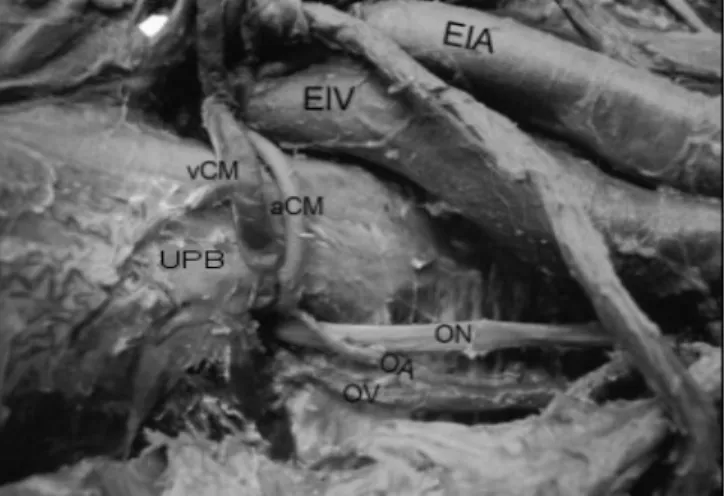
Figure 1. Dissection of a right hemipelvis. Anterior view. The Corona Mortis can be seen above the upper branch of the pubis.
External iliac artery (EIA), external iliac vein (EIV), venous Corona Mortis (vCM), arterial Coronal Mortis (aCM), upper pubic branch (UPB), obturator nerve (ON), obturator artery (OA), obturator vein (OV).
Figure 2. Dissection of a left hemipelvis. Anterior view. The common trunk between the inferior epigastric artery and the obturator artery can be seen.
External iliac artery (EIA), external iliac vein (EIV), common trunk between obtura-tor and inferior epigastric arteries (CT), inferior epigastric artery (IEA), upper pubic branch (UPB), obturator artery (OA).
Table 1. Origins of the obturator artery.
Origin Number of Cases %
Internal Iliac Artery 27 45%
Common trunk with the
Inferior Epigastric Artery 22 36.38%
Superior Gluteal Artery 6 10%
Inferior Epigastric Artery 4 6.66%
Femoral Artery 1 1.66%
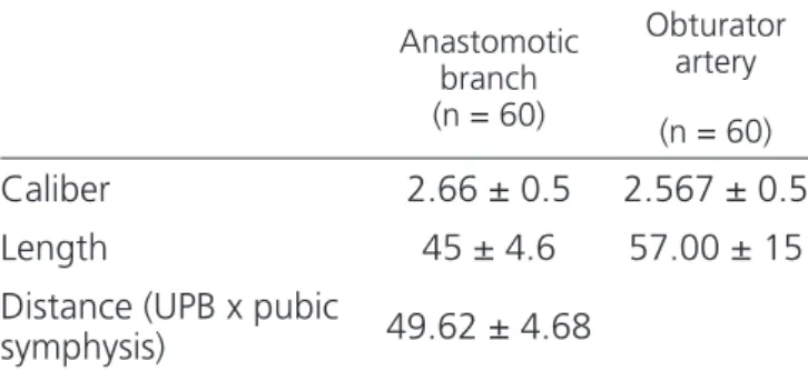
The mean OA caliber was 2.56±0.5mm. The mean OA length was 57±15mm. The anastomotic bran-ch had a total length of 45±4.6mm, a mean caliber of 2.66±0.5mm and distance between the UBP and the pubic symphysis was on average 49.62±4.68mm. We summarize these results in table 3.
selection would also imply differences in the caliber of the OA and the IEA, causing the variation known as CM.
Anatomical variations of the OA origin are des-cribed in detail in the literature: it may originate from the EIA, from an ischial-pudendal trunk (formed by the in-ternal pudendal and lower gluteal arteries), the upper or lower gluteal arteries, the internal pudendal artery, the femoral artery and from two distinct roots (one from the EIA and another from the IIA)1-3,10,11. The OA can also
ori-ginate from the IEA, ilium-lumbar artery, lower bladder artery, vaginal artery, accessory hemorrhoidal artery, ex-ternal pudendal artery, accessory pudendal artery, prosta-tic artery, and internal pudendal artery1,14.
The IEA, on the other hand, has a varied origin in relation to its position, as it can originate as much as 6cm above the inguinal ligament1-3. This vessel may
origi-nate in the femoral artery and ascend to the pelvis throu-gh the femoral ring, from the deep femoral artery, from a common trunk with the deep circumflex artery of the ilium or from the OA itself and, in addition, there was a described case where the IEA originated from two distinct roots (from the EIA and IIA)1,2. Unusual branches of the
IEA may be the dorsal artery of the penis (or clitoris), the superficial epigastric artery, the deep circumflex artery of the ilium, the medial femoral circumflex artery, and the accessory external pudendal artery. The suprapubic and funicular branches may be absent1,3.
According to Testut and Latarjet1, when the
OA branches from the EIA, it can reach the OF through two distinct trajectories: 1) it can descend vertically throu-gh the lateral wall of the femoral vein or 2) it can enter the OF obliquely and inferiorly when crossing the supe-rior wall of the femoral vein. The authors report that the second possibility is dangerous during hernia surgeries, due to its proximity to the hernial sac3. Goss2 states that
the most dangerous moment of this second trajectory is when the OA crosses the lacunar ligament. Our results showed that the OA originated along with the IEA (or from it) in 36.68% of the cases, a significant percenta-ge. In one of the hemipelvises studied, the OA originated from the femoral artery: a fact reported only once in a study by Sañudo et al.11.
Although the anatomy books describe the usu-al presence of the anastomosis between the OA and IEA anastomotic branches, they do not use the term Corona Table 2. Prevalence of arterial Corona Mortis (CM).
Men Women Total
Arterial CM 21
(77.77%) 6 (22.23%) 27 (45%)
Absent - - 33 (55%)
Table 3. Morphometric data of the obturator artery and its anastomotic branch.
Anastomotic branch (n = 60)
Obturator artery
(n = 60)
Caliber 2.66 ± 0.5 2.567 ± 0.5
Length 45 ± 4.6 57.00 ± 15
Distance (UPB x pubic
symphysis) 49.62 ± 4.68
Results are described as mean ± standard deviation. UPB = upper pu-bic branch.
DISCUSSION
Vascular formation during the embryonic pe-riod consists of the appearance of vessels and anasto-moses that may or may not persist during ontogenesis10.
Two arterial plexuses are formed through the dorsal root of the umbilical artery: the abdominal plexus and the pelvic plexus. During the fifth week of development, the umbilical arteries form a new connection to the fifth pair of lumbar segmental arteries (which form the pelvic ple-xus), and then form the IIA, the EIA, and consequently the common iliac artery. The OA is formed through the IIA, whereas all other arteries of the lower limbs develop like branches from the EIA, for example, the IEA10-12. Due
Mortis, and few books cite its clinical and surgical im-portance. The term consists of two Latin words: “coro-na” (used in anatomy to design structures in the form of a crown or circular form), and “mortis”, which comes from the term “mors”, meaning death4,15. It definition
is intriguing because some authors believe that it is any form of anastomosis between the IEA and the OA or be-tween the IIA and the EIA6,8,9,16,17, while others believe
that CM is only the anastomosis of the OA and IEA anas-tomotic branches5.
Authors like Gilroy et al.16, Mahato18 and
Ju-soh et al.13 use terms such as “aberrant”, “accessory”
or “anomalous” to refer to CM. Gilroy16 states that the
prevalence of CM is high, and therefore should not be called by such terms. We share this opinion.
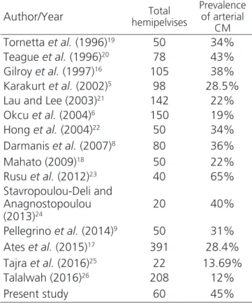
The prevalence of arterial CM (with or without venous CM) has been reported in numerous studies, ran-ging from 12 to 65%5,6,8,9,16-26. The results of other
stu-dies are summarized in table 4. These numbers should draw the attention of surgeons and anatomists becau-se the number of studies reporting a prevalence greater than 20% is higher, indicating that this variation in the arterial form is not so unusual.
Tabela 4. Prevalence of corona mortis (CM) according to the literature.
Author/Year hemipelvisesTotal
Prevalence of arterial
CM
Tornetta et al. (1996)19 50 34%
Teague et al. (1996)20 78 43%
Gilroy et al. (1997)16 105 38%
Karakurt et al. (2002)5 98 28.5%
Lau and Lee (2003)21 142 22%
Okcu et al. (2004)6 150 19%
Hong et al. (2004)22 50 34%
Darmanis et al. (2007)8 80 36%
Mahato (2009)18 50 22%
Rusu et al. (2012)23 40 65%
Stavropoulou-Deli and Anagnostopoulou (2013)24
20 40%
Pellegrino et al. (2014)9 50 31%
Ates et al. (2015)17 391 28.4%
Tajra et al. (2016)25 22 13.69%
Talalwah (2016)26 208 12%
Present study 60 45%
Reports of the arterial CM’s length vary: 62mm according to Tornetta et al.19, 52mm
accor-ding to Hong et al.22, 68mm according to Darmanis et
al.8 and 52.4mm according to Stavropoulou-Deli and
Anagnostopoulou24. The CM’s caliber is has on
avera-ge between 2.6mm8,22 and 3mm24. The present study
revealed similar results, the length being 49.6±4.6mm, and the caliber, 2.6±0.5mm. The caliber should alarm surgeons and clinicians as this vessel can cause signifi-cant bleeding should it rupture.
The IEA and OA are susceptible to iatrogenic lesions during procedures due to their variable natu-re7,15,27,28, as seen previously. The CM may be injured
during laparoscopic approaches for repair of inguinal and femoral hernias, preperitoneal or extraperitoneal repair at the moment of attachment of the mesh to the pectineum ligament (Cooper’s), which may lead to un-controllable bleeding, pseudoaneurysms and formation of retroperitoneal hematomas29,30.
Traditional (Stoppa) accesses used for fractu-res of the anterior pelvic region and the anterior aceta-bular column are related to a high risk of hemorrhage and additional damage to adjacent structures, such as soft tissues and neurovascular structures27. The
difficul-ty in repairing these fractures is in identifying a safe place to position the implant, especially in just-articular and quadrilateral fractures27.
In pubic osteotomies, procedures with relati-vely high complication rates, the CM can be injured due to a limitation of the surgical field. Since the presence of CM on the UBP posed a potential risk of injury in or-thopedic surgeries, some authors suggest preoperative imaging to identify possible vascular anatomical varia-tion to minimize complicavaria-tions31.
Ates et al.17 evaluated the risk of vascular
in-jury in CM during extraperitoneal repairs and concluded that to prevent this complication careful dissection is necessary on the posterior face of UBP and to apply clips or hemostatic clamps on the pectine ligament near the pubic symphysis.
by compression of the urinary tract by a pelvic hema-toma32. An apparently benign UBP fracture associated
with thermodynamics instability should raise suspicion of CM rupture, especially in elderly and anticoagulated patients. Avulsion of CM can be identified by an an-giotomography4,15. Alternative treatments, such as CM
embolization, may be used to stop bleeding7.
Burch’s colposuspension, introduced in 1961, was the gold standard for the treatment of stress uri-nary incontinence. Recently, this procedure has fallen into disuse due to the emergence of new minimally in-vasive techniques, such as the retropubic sling (introdu-ced in 1998) or the transobturator sling (2002). These new methods, though minimally invasive, are not free of complications, since CM can still be injured24,33.
Prostatic artery embolization has shown pro-mising results for the treatment of benign prostatic hyperplasia. The study of these vessels’ anatomy should be necessary, since the prostatic artery may be an OA branch. Thus, understanding the arterial anatomy is es-sential for the interventional radiologist to perform the procedure safely and adequately14.
One of the interesting aspects of CM is the ability to function as a collateral circulation path10. In
a case described by Khandari et al.34, CM played a key
role after the patient underwent avascular acetabular necrosis due to an inadequate treatment of a trans-verse fracture. In this situation the arterial CM parti-cipated as a collateral circulation to supply the lower limb, avoiding amputation. This aspect of CM, though extremely important, is of little emphasis in the litera-ture.
We observed that the vascular connections between the obturator, internal and external iliac and inferior epigastric systems are relatively common over the UBP. The diameter and trajectory of this anasto-motic artery may vary. Iatrogenic lesions and pelvic and acetabular fractures can result in severe bleeding that puts the patient’s life at risk. On the other hand, this anastomosis has a considerable role as a pathway of collateral circulation in peripheral arterial obstructi-ve disease. Thus, we note the importance of studying this anatomical variation, since we do not consider it as unusual as previously thought.
REFERENCES
1. Testut L, Latarjet A. Tratado de anatomía humana. Barcelona: Salvat; 1958.
2. Goss CM, editor. Gray’s anatomy of the human body. Philadelphia: Lea & Febiger; 1973.
3. Bergman R, Thompson S, Afifi A, Saadeh F. Compendium of human anatomic variation: text,
atlas, and world literature. Baltimore: Urban & Schwarzenberg; 1988.
4. Garrido-Goméz J, Pena-Rodríguez C, Martín-Noguerol T, Hernández-Cortes P. Corona mortis artery avulsion due to a stable pubic ramus fracture. Orthopedics. 2012;35(1):e80-2.
5. Karakurt L, Karaca I, Yilmaz E, Burma O, Serin E. Corona mortis: incidence and location. Arch Orthop
Objetivo: relatar a prevalência da corona mortis arterial e descrever suas aplicabilidades cirúrgicas e clínicas. Métodos: sessenta hemi-pelvises (50 homens e 10 mulheres) fixadas em uma solução de formalina a 10% foram dissecadas com o propósito de obter informa-ções sobre a corona mortis. Medidas do calibre e comprimento da artéria obturatória e seu ramo anastomótico foram mensuradas com o auxílio de um paquímetro digital e submetidas a análises e comparações estatísticas no programa GraphPad Prism 6. Resultados: a
corona mortis arterial esteve presente em 45% da amostra estudada. A origem mais comum da artéria obturatória foi da artéria ilíaca interna, porém, houve um caso excepcional no qual a artéria obturatória se originou da artéria femoral. O calibre do ramo anastomótico foi em média 2.7mm, enquanto que o calibre da artéria obturatória foi 2.6mm. Conclusão: as conexões vasculares entre os sistemas obturatório, ilíacos interno e externo e epigástrico inferior são relativamente comuns sobre o ramo superior da pube. O diâmetro e a trajetória dessa artéria anastomótica podem variar. Assim, lesões iatrogênicas, fraturas pélvicas e acetabulares podem resultar em he-morragias graves que colocam a vida do paciente em risco.
Descritores: Corona Mortis. Variação Anatômica. Anatomia. Cirurgia Geral.
Trauma Surg. 2002;122(3):163-4.
6. Okcu G, Erkan S, Yercan HS, Ozic U. The incidence and location of corona mortis: a study on 75 cadavers. Acta Orthop Scand. 2004;75(1):53-5. 7. Lorenz JM, Leef JA. Embolization of postsurgical
obturator artery pseudoaneurysm. Semin Intervent Radiol. 2007;24(1):68-71.
8. Darmanis S, Lewis A, Mansoor A, Bircher M. Corona mortis: an anatomical study with clinical implications in approaches to the pelvis and acetabulum. Clin Anat. 2007;20(4):433-9.
9. Pellegrino A, Damiani GR, Marco S, Ciro S, Cofelice V, Rosati F. Corona mortis exposition during laparoscopic procedure for gynecological malignancies. Updates Surg. 2014;66(1):65-8. 10. Goke K, Pires LAS, Tulio TFO, Chagas CAA. Rare
origin of the obturator artery from the external iliac artery with two obturator veins. J Vasc Bras. 2016;15(3):250-3.
11. Sañudo J, Mirapeix R, Rodriguez-Niedenführ M, Maranillo E, Parkin IG, Vázquez T. Obturator artery revisited. Int Urogynecol J. 2011;22(10):1313-8. 12. Schoenwolf GC, Bleyl SB, Brauer PR, Francis-West
PH. Larsen’s human embryology. 5th ed: Churchill Livingstone; 2014.
13. Jusoh AR, Rahman NA, Latiff AA, Othman F, Das S, Ghafar NA, et al. The anomalous origin and branches of the obturator artery with its clinical implications. Rom J Morphol Embryol 2010;51(1):163-6.
14. Garcia-Monaco R, Garategui L, Kizilevsky N, Peralta O, Rodriguez P, Palacios-Jaraquemada J. Human cadaveric specimen study of the prostatic arterial anatomy: implications for arterial embolization. J Vasc Interv Radiol. 2014;25(2):315-22.
15. Kong WM, Sun CK, Tsai IT. Delayed presentation of hypovolemic shock after a simple pubic ramus fracture. Am J Emerg Med. 2012;30(9):e2091-4. 16. Gilroy AM, Hermey DC, DiBenedetto LM, Marks
SC Jr, Page DW, Lei QF. Variability of the obturator vessels. Clin Anat 1997;10(5):328-32.
17. Ates M, Kinaci E, Kose E, Soyer V, Sarici B, Cuglan S, et al. Corona mortis: in vivo anatomical knowledge and the risk of injury in totally extraperitoneal inguinal hernia repair. Hernia. 2016;20(5):659-65. 18. Mahato NK. Retro-pubic vascular anomalies: a
study of abnormal obturator vessels. Eur J Anat. 2009;13(3):121-6.
19. Tornetta P 3rd, Hochwald N, Levine R. Corona mortis. Incidence and location. Clin Orthop Relat Res. 1996;(329):97-101.
20. Teague DC, Graney DO, Routt ML Jr. Retropubic vascular hazards of the ilioinguinal exposure: A cadaveric and clinical study. J Orthop Trauma. 1996;10(3):156-9.
21. Lau H, Lee F. A prospective endoscopic study of retropubic vascular anatomy in 121 patients undergoing endoscopic extraperitoneal inguinal hernioplasty. Surg Endosc. 2003;17(9):1376-9. 22. Hong HX, Pan ZJ, Chen X, Huang ZJ. An anatomical
study of corona mortis and its clinical significance. Chin J Traumatol. 2004;7(3):165-9.
23. Rusu MC, Cergan R, Motoc AG, Folescu R, Pop E. Anatomical considerations on the corona mortis. Surg Radiol Anat. 2010;32(1):17-24.
24. Stavropoulou-Deli A, Anagnostopoulou S. Corona mortis: anatomical data and clinical considerations. Aust N Z J Obstet Gynaecol. 2013;53(3):283-6. 25. Tajra JBM, Lima CF, Pires FR, Sales L, Junqueira D,
Mauro E. Variability of the obturator artery with its surgical implications. J Morphol Sci. 2016;33(2):96-8.
26. Al Talalwah W. A new concept and classification of corona mortis and its clinical significance. Chin J Traumatol. 2016;19(5):251-4.
27. Balbachevsky D, Pires RES, Faloppa F, Reis FB. Treatment of pelvic and acetabular fractures through modified Stoppa port. Acta Ortop Bras. 2006;14(4):190-2.
28. Leite TFO, Chagas CAA, Pires LAS, de Paula RC, Babinski MA. De Garengeot’s hernia in an 82-year-old man: a case report and clinical significance. J Surg Case Rep. 2016;2016(7):pii: rjw120.
29. Poelman MM, van den Heuvel B, Deelder JD, Abis GS, Beudeker N, Bittner RR, et al. EAES Consensus Development Conference on endoscopic repair of groin hernias. Surg Endosc. 2013;27(10):3505-19. 30. Ramser M, Messmer AS, Zbinden I, Von Holzen
rju081.
31. Wada K, Goto T, Tezuka F, Tamaki S, Hamada D, Tsutsui T, et al. Variations in the obturator artery around the obturator foramen assessed by three-dimensional computed tomographic angiography and prevention of vascular-related complications in rotational acetabular osteotomy. Int Orthop. 2016;41(1):133-9.
32. Theodorides AA, Morgan BW, Simmons D. Haemodynamic instability resulting from a low energy pubic ramus fracture in a 78-year-old woman. A case report and review of the literature. Injury. 2011;42(7):722-4.
33. Rehder P, Glodny B, Pichler R, Mitterberger MJ. Massive retropubic hematoma after minimal invasive mid-urethral sling procedure in a patient with a
corona mortis. Indian J Urol. 2010;26(4):577-9. 34. Kandhari VK, Desai MM, Bava SS, Wade RN.
Avascular necrosis of acetabulum: the hidden culprit of resistant deep wound infection and failed fixation of fracture acetabulum - a case report. J Orthop Case Rep. 2015;5(4):36-9.
Received in: 05/04/2017
Accepted for publication: 20/07/2017 Conflict of interest: none.
Source of funding: none.
Mailing address:
Túlio Fabiano de Oliveira Leite