Risk factors associated with complications of acute appendicitis
Fatores de risco associados às complicações de apendicite aguda
AnA PAulA MArconi iAMArino1; YArA JuliAno1; otto MAuro rosA1; neil FerreirA novo1; Murillo de liMA FAvAro1; MArcelo Augusto Fontenelle ribeiro Júnior, tcbc-sP1.
INTRODUCTION
T
he acute inflammatory abdomen encompasses the major conditions seen by surgeons working in emer-gency services around the world. It is a clinical picture ranging from simple, self-limiting, benign diagnoses to those that threaten life and require rapid surgical inter-vention. About 6.5% of emergency room visits are due to abdominal pain1.Acute appendicitis (AA) represents the most common surgical condition in the abdomen. It presents an incidence of 48.1 per 10,000 inhabitants per year, and its peak incidence occurs in patients between ten and 20 years of age. The overall lifetime risk is estimated between 5% and 20%, being 8.6% for men and 6.7% for women2,3. It affects approximately 250,000 patients
per year in the United States and is responsible for at least 40,000 hospital admissions per year in England1.
The signs and symptoms are usually anorexia, periumbi-lical colic, nausea and vomiting, followed by moderate fever (38° C) and signs of peritoneal inflammation in the lower right quadrant of the abdomen4,5. Many of these
findings, however, may occur in other clinical or surgical conditions, such as mesenteric lymphadenitis, intrape-ritoneal hemorrhage, acute salpingitis, endometriosis, Meckel’s diverticulitis, among others. Diagnosis is made based on clinical evaluation and confirmed by leukocyte counting, ultrasonography (US) and radiographic studies of the abdomen2,6. Incorrect diagnosis is more frequent
in children, in women, and in the elderly6. The accuracy of a good anamnesis, combined with a well-performed physical examination, is 95% in patients who present a classic clinical picture7. The complications resulting from
the evolution of the acute inflammatory process, such as suppuration, perforation with or without hemorrha-ge, and gangrene of the appendix are serious, making early surgery fundamental to contain the evolution of the condition5.
The treatment of acute appendicitis is appen-dectomy, conventional or laparoscopic. However, anti-biotic therapy alone, with drugs against Gram negative and anaerobic bacteria, has been used, since it has the potential to considerably reduce the costs associated with surgery8,9. Studies suggest that non-surgical
the-1 - Santo Amaro University and Grajaú General Hospital, General Surgery, São Paulo, SP, Brazil.
A B S T R A C T
Objective: to identify the main risk factors associated with the development of complications in patients with acute appendicitis. Methods:
we conducted a case-control study of 402 patients with acute appendicitis hospitalized in a secondary hospital, divided into two groups: the control group, with 373 patients who progressed without postoperative complications (Group 1) and the study group, with 29 patients who presented complications (Group 2). We evaluated demographic data, signs and symptoms of the disease, imaging tests and hospital-ization data. Results: factors associated with complications were fever, radiological and sonographic changes, abrupt positive decompres-sion and diarrhea. Migration of pain, nausea, vomiting and abrupt positive decompresdecompres-sion were the findings that were significantly more frequent in both groups (p = 0.05). The duration of signs and symptoms in days in group 2 was significantly higher than in group 1, with a median of three days for the group with complications (p = 0.05). Conclusion: alterations in imaging, fever, diarrhea, positive abrupt decompression, duration of symptoms and lower age are associated with a higher frequency of complications in acute appendicitis, which reinforces the importance of anamnesis, physical examination and indication of complementary exams in the approach of these patients.
rapy is safe, provided that the patient has an adequate follow-up and can undergo operative treatment if ne-cessary8.
But despite the technological progress in diag-nosis and therapy, acute appendicitis continues to be an important cause of morbidity and mortality, especially in the extremes of age, in which signs and symptoms may not have a classic clinical presentation. This study aims to evaluate the main risk factors associated with the development of complications in patients with acute appendicitis.
METHODS
We conducted a case-control study by means of data analysis of the medical records of hospitalized patients diagnosed with acute appendicitis in the year 2013 at the Grajaú General Hospital (HGG - Instituto de Responsabilidade Sírio Libanês) and at the Santo Amaro University. We obtained data from the Inpatient Manage-ment System and included all patients with acute appen-dicitis in this period, regardless of age.
Patients with suspected acute appendicitis were submitted to clinical and laboratory evaluation ac-cording to the institutional protocol of abdominal pain. In the presence of clinical findings suggestive of appen-dicitis, imaging examinations (abdominal radiographs, ul-trasonography and/or computed tomography) followed. With the diagnosis established, the surgery was perfor-med through an incision in the right iliac fossa.
We evaluated demographic data, signs and symptoms, imaging and hospitalization data, as well as the following postoperative complications: intra-abdomi-nal abscess, sepsis and wound infections.
We used the Cochran G, Chi-square, Fisher’s exact, Mann-Whitney, and Kendall concordance tests in the statistical analysis10.
The present work was submitted to and appro-ved by the Ethics in Research Committee of the Santo Amaro University, under the opinion of number 624735.
RESULTS
We studied 402 patients, divided into two groups: control group (Group 1), with patients who
presented no postoperative complications (n=373), and study group (Group 2), composed of patients presenting with complications (n=29). Of the 373 patients in group 1, 220 (59%) were male and 153 (41%) were female. The pediatric population (up to 12 years of age) corres-ponded to 31%, or 116 patients. In group 2, 15 (52%) were male and 14 (48%) female. The pediatric popula-tion were 19 patients (65%).
In group 2 the postoperative complications observed were: intra-abdominal abscess in 19 cases (65%), wound infections in seven (24%), and sepsis in six (21%), and three patients had two simultaneous complications. Drainage of the peritoneal cavity was performed in 62% of patients in group 2.
Computed Tomography (CT) was not perfor-med in 21 patients (72%) of group 2 and in 256 pa-tients (68%) of group 1, because the diagnosis had been confirmed by other methods.
The mean age of group 1 was 21.9 years (1 to 65 years) and the mean length of hospital stay was 3.05 days. In group 2, the mean age was 16.9 years (2 to 45 years) and the mean length of hospital stay was 13.1 days.
Regarding the evolutionary phases of appen-dicitis, according to the surgical description, group 1 had 55 (15%) patients in the edematous stage, 140 (38%) in the phlegmonous phase, 99 (26%) in the gan-grenous phase, 75 (20%) in perforated phase and four (1%) patients had normal appendices (tactical appen-dectomy). Group 2 had three (10%) patients in the ede-matous phase, five (17%) in the phlegmonous phase, eight (28%) in the gangrenous phase and 13 (45%) in the perforated phase.
From the Cochran G test, the factor fre-quencies for the two groups were compared and the following factors were associated with appendicitis: pain migration, nausea and vomiting, and painful de-compression (PD+) were significantly (p=0.05) in both groups (Table 1).
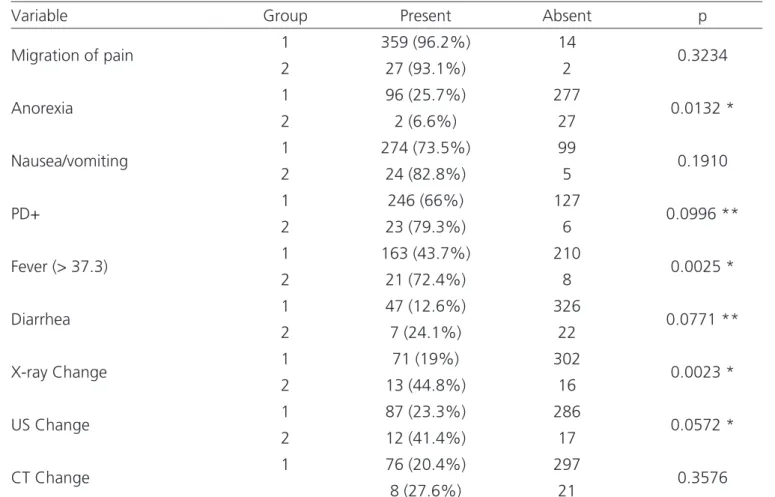
For the comparison between the control and the study groups, we used the chi-square test or the Fisher’s exact test (Table 2).
(2>1) and alteration in Ultrasound (2>1). PD+ and diar-rhea suggested differences between groups (2>1 for
both). The other factors did not present significant dif-ferences between groups.
Table 1. Comparison of the findings between the groups with and without complications.
Main Findings
Patients
No Complication Complication
n
Frequency
n
Frequency
Migration of pain 359 96% 27 93%
Nausea and vomiting 274 73% 24 83%
PD+ 300 80% 27 93%
Fever (> 37.3) 163 44% 21 72%
X-ray Change 71 19% 13 45%
US Change 87 23% 12 41%
CT Change 76 20% 8 27%
Anorexia 96 26% 2 0,07%
Diarrhea 47 13% 7 24%
G Test G = 1166.32 (p=0.0000) G = 91.20 (p=0.0000)
PD+: sudden painful decompression; US: ultrasound; CT: computed tomography.
Table 2. Comparison of frequencies of the factors associated with appendicitis between the two groups.
Variable Group Present Absent p
Migration of pain 1 359 (96.2%) 14 0.3234
2 27 (93.1%) 2
Anorexia 1 96 (25.7%) 277 0.0132 *
2 2 (6.6%) 27
Nausea/vomiting 1 274 (73.5%) 99 0.1910
2 24 (82.8%) 5
PD+ 1 246 (66%) 127 0.0996 **
2 23 (79.3%) 6
Fever (> 37.3) 1 163 (43.7%) 210 0.0025 *
2 21 (72.4%) 8
Diarrhea 1 47 (12.6%) 326 0.0771 **
2 7 (24.1%) 22
X-ray Change 1 71 (19%) 302 0.0023 *
2 13 (44.8%) 16
US Change 1 87 (23.3%) 286 0.0572 *
2 12 (41.4%) 17
CT Change 1 76 (20.4%) 297 0.3576
8 (27.6%) 21
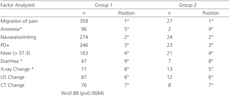
We ordered the frequencies of the associated factors in descending order and applied the Kendall test to analyze the concordance between groups, according to table
Table 4. Duration of the main complaint among groups.
Duration of Symptoms
Group 1 Group 2
Median 1.5 3
Average 2.5 3.5
Z=3.68 (p=0.0002)
3, which suggests agreement in six of the nine factors analy-zed. There was disagreement between the groups in only three factors: anorexia, diarrhea and radiological changes.
Table 3. Associated factors between groups.
Factor Analyzed Group 1 Group 2
n Position n Position
Migration of pain 359 1° 27 1°
Anorexia* 96 5° 2 9°
Nausea/vomiting 274 2° 24 2°
PD+ 246 3° 23 3°
Fever (> 37.3) 163 4° 21 4°
Diarrhea * 47 9° 7 8°
X-ray Change * 71 8° 13 5°
US Change 87 6° 12 6°
CT Change 76 7° 8 7°
W=0.88 (p=0.0684)
* discordant factors; PD+: sudden painful decompression; US: ultrasound; CT: computed tomography.
To evaluate the duration of the main complaint (abdominal pain) and to compare the control and study groups, we used the Mann-Whitney test, with which we could observe that the duration of the signs and symp-toms in days of group 2 was significantly higher than group 1 (p=0.05), as seen in table 4.
DISCUSSION
Studies have shown that the worst progno-sis in acute appendicitis occurs in elderly patients with associated comorbidities, as well as a longer time of disease evolution and the occurrence of appendicular perforation11. The complications found in patients
un-dergoing appendectomy are usually related to the sta-ge at which the disease is diagnosed and treated. Stu-dies by Petroianu et al.6 with regard to the appendicitis
morphological classification indicated that among 170 patients studied, 23 were in the catarrhal phase, 99 in the fibrinopurulent phase, 31 in gangrenous, and 17, in the perforation phase. This study confirmed the relationship between complications and appendicitis phase, since 45% of the patients in the complications group had appendicitis perforation. And in the control group (without complications) the phlegmonous pha-se predominated (38%). As expected, the study group had a considerably longer hospital stay than the con-trol group, 13.2 days, as observed in our cases.
According to Fischer et al.12, in a total of 272
appendectomies evaluated, of which 88 (32.3%) in the catarrhal phase, 79 (29%) in the phlegmonous phase, 70 (25.3%) in the suppurative phase and 35 (12,8%) in gangrenous phase, the mean time of hospitalization was 4.3 days (2 to 36 days). Reis et al.7 analyzed the
anatomopathological evolution of 300 cases of acute appendicitis and observed that the phlegmonous form predominated (71.3%). In 63 cases, characteristic per-foration of the gangrenous form occurred.
Mendoza et al.13 studied 113 patients
du-ration of symptoms was 12h in 22.1%, 12 to 24h in 31.8%, 24 to 48h in 33.6%, 48 to 72h in 10.6% and more than 72h in 1.7%. They observed 19 patients in the edematous stage, 41 patients in the phlegmo-nous one, 22 in the gangrephlegmo-nous phase, four in the perforated stage and 6 in the perforated phase with peritonitis. The remaining 21 had normal appendices.
Petroianu et al.6 identified that the
radio-graphic sign of fecal accumulation in the cecum was present in 165 of the 170 patients with acute appen-dicitis. The radiographic signal sensitivity for acute appendicitis was 97% and its specificity was 85.3%. The positive predictive value of this signal for acute appendicitis was 78.9%, while its negative predictive value stands out with 98%. Another study, however, showed that simple x-ray of the abdomen should not be required, since it has low specificity and sensitivity, while US has sensitivity of 75 to 90% and specificity of 86 to 100%, but it depends on a qualified opera-tor1. Studies with US showed that its sensitivity ranged
from 68 to 96%, and specificity, from 46.7 to 95.9%, with PPV between 82.2 and 94% and accuracy from 65.7 to 87%14-17. CT has sensitivity and specificity of
90 to 100% and 91 to 99%, respectively. Studies showed its sensitivity ranging from 91.2 to 98.5%, specificity from 62.5 to 98%, positive predictive value (PPV) from 92.1 to 98% and accuracy of 90%16-20. CT
findings consist of appendix lumen dilation, thicke-ning of the wall, presence of fecalites and inflamma-tion1. In our sample, 72% of the patients in the study
group and 68% of the patients in the control group were not submitted to CT, since it was possible to confirm the diagnosis by other methods such as sim-ple radiographs and US, which, when positive, were considered risk factors associated with complications. Although the literature highlights CT as a method of choice in the diagnosis of appendicitis, this tool is not always available.
Lima et al.14 observed a higher prevalence of
appendicitis in young adults (60%), with a predomi-nance of males. The mean length of hospital stay was seven days, with no significant differences between genders. The most frequent evolutionary phase was phase II with 34.3%. Of the patients diagnosed in sta-ge IV, 65.8% were men. The hospitalization time was
higher in this phase, with a mean of 12.4 days, with a significant difference between phase I and phase IV (p=0.001). Eighty-one patients used drains for an average of 4.8 days and the mean length of hospi-tal stay was 10.4 days. Of the patients studied, 196 were submitted to amoxycillin/clavulanate antibiotic prophylaxis only in 64.3% of the cases. These patients had shorter hospitalization time compared to those who did not undergo prophylaxis. Thirty-eight pa-tients (5.9%) developed postoperative complications, with wound infection (52.6%) and wound dehiscen-ce (26.3%) being the most frequent. There were also complications due to intra-abdominal abscess, sepsis and fistula. Seventeen patients died (2.7%). Among them, the majority were male, mean age was 38.4 ye-ars, 70.6% had complicated AA and 47% were diag-nosed in stage IV, with a direct correlation between the evolutionary stage and death. Regarding death causes, 53% were due to septic shock and 47% to unknown or indeterminate causes.
Despite new and better antibiotics, advances in imaging and supportive care, a large number of pa-tients with acute appendicitis develop serious compli-cations and have morbid and prolonged recoveries8.
Silva et al.2 considered surgical wound infections and
intraabdominal abscesses as the main morbidity fac-tors and that the perforated phase contributed to the increase of such complications. The main risk factors for complications after appendectomies were: female gender, necrotic or perforated appendicitis and cavity drainage. A recent study showed that the perforation rate of patients with appendicitis was 16%. The mean duration from onset of symptoms to hospital admis-sion was 4.4 days. The factors that contributed to the appendix perforation included a diagnostic error and initial patient approach (56%), delayed hospitaliza-tion (11%) and use of analgesics (9%)21.
REFERENCES
1. Edelmuth RCL, Ribeiro Júnior MAF. Afecções abdominais inflamatórias. Emerg Clin. 2011;6(29):43-9.
2. Silva SM, Almeida SB, Lima OAT, Guimarães GMN, Silva ACC, Soares AF. Fatores de risco para as complicações após apendicectomias em adultos. Rev Bras Coloproct. 2007;27(1):31-6.
3. Tan WJ, Acharyya S, Goh YC, Chan WH, Wong WK, Ooi LL, et al. Prospective comparison of the Alvarado Score and CT Scan in the evaluation of the suspected appendicitis: a proposed algorithm to guide CT use. J Am Coll Surg. 2015;220(2):218-24.
4. Matos B, Santana C, Souza D, Rodrigues E, Gonçalves E, Dias F, et al. Apendicite aguda. Rev Med Minas Gerais. 2011;21(2 Supl 4):S1-S113.
5. Freitas RG, Pitombo MB, Maya MCA, Leal PRF. Apendicite aguda. Rev Hosp Univ Pedro Ernesto. 2009;8(1):38-51.
6. Petroianu A, Alberti LR, Zac RI. Importância do sinal radiográfico de acúmulo fecal no ceco para o diagnóstico diferencial de apendicite aguda. Acta Med Port. 2007;20 (2):151-6.
7. Reis JM, Oliveira DCN, Luccatto TM, Reis Júnior WB. Diagnóstico e tratamento de 300 casos de apendicite aguda em crianças e adolescentes atendidos em
um hospital universitário. Rev Med Minas Gerais. 2008;18 (1):11-5.
8. Gomes N, Bridi TL, Ribeiro MAF Jr. Existe lugar para o tratamento clínico de apendicite aguda? Emerg Clin. 2010;5(25):118-21.
9. Smink D, Soybel DI. Management of acute appendicitis in adults [Internet]. Waltham (MA): UpToDate Inc; c2017 [cited 2017 Jun 25]. Available f r o m : h t t p s : / / w w w . u p t o d a t e . c o m / c o n t e n t s / management-of-acute-appendicitis-in-adults
10. Siegel S, Castellan Júnior NJ. Estatística não paramétrica para ciências do comportamento. 2ª ed. Artmed: Porto Alegre; 2006.
11. Almeida MWR, João AT, Oliveira FS, Mattos HC, Silva AR, Silva MCGB. Influência da idade no tempo de internação e no grau evolutivo das apendicites agudas. Rev Col Bras Cir. 2006;33(5):294-7.
12. Fischer CA, Pinho MSL, Ferreira S, Milani CAC, van Santen CR, Marquardt RA. Apendicite aguda: existe relação entre o grau evolutivo, idade e o tempo de internação? Rev Col Bras Cir. 2005;32(3):136-8. 13. Mendoza JDV, Rodriguez CG, Guerrero MAV.
Evaluación prospectiva de la Escala de Alvarado em el diagnóstico de apendicits aguda. Cir Gen. 2010;32(1):17-23.
14. Lima AP, Vieira FJ, Oliveira GPM, Ramos PS, Avelino ME, Prado FG, et al. Perfil clínico-epidemiológico anamnesis, physical examination and complementary
methods in the diagnosis of acute appendicitis, espe-cially in the presence of risk factors for complications:
patients below 12 years of age, presence of fever, PD+, diarrhea, imaging exams alterations, as well as the long duration of signs and symptoms.
Objetivo: identificar os principais fatores de risco associados ao desenvolvimento de complicações em pacientes portadores de apendici-te aguda. Métodos: estudo caso controle de dados dos prontuários de 402 pacientes internados com apendicite aguda em um hospital de nível secundário, separados em dois grupos: grupo controle, com 373 pacientes que evoluíram sem complicações pós-operatórias (Grupo 1) e grupo estudo, com 29 pacientes que apresentaram complicações (Grupo 2). Foram avaliados dados demográficos, sinais e sintomas da doença, exames de imagem e dados da internação. Resultados: os fatores associados às complicações foram febre, alte-rações radiológicas e ultrassonográficas, descompressão brusca positiva e diarreia. Migração da dor, náuseas, vômitos e descompressão brusca positiva foram os achados significativamente mais frequentes nos dois grupos (p=0,05). Já a duração dos sinais e sintomas, em dias, no grupo 2 foi significativamente maior que no grupo 1, com mediana de três dias para o grupo com complicações (p=0,05).
Conclusão: alterações nos exames de imagem, febre, diarreia, descompressão brusca positiva, tempo de duração de sintomas e menor faixa etária estão associados à maior frequência de complicações na apendicite aguda, o que reforça a importância da anamnese, do exame físico e da indicação de exames complementares na abordagem desses pacientes.
Descritores: Apendicite. Apendicectomia. Complicações Intraoperatórias. Diagnóstico.
da apendicite aguda: análise retrospectiva de 638 casos. Rev Col Bras Cir. 2016;43(4):248-53.
15. Nutels DBA, Andrade ACG, Rocha AC. Perfil das complicações após apendicectomia em um hospital de emergência. ABCD Arq Bras Cir Dig. 2007;20(3):146-9.
16. Ozkan S, Duman A, Durukan P, Yildirim A, Ozbakan O. The accuracy rate of Alvarado score, ultrasonography, and computerized tomography scan in the diagnosis of acute appendicitis in our center. Niger J Clin Pract. 2014;17(4):413-8.
17. Shogilev DJ, Duus N, Odom SR, Shapiro NI. Diagnosing appendicitis: evidence-based review of the diagnostic approach in 2014. West J Emerg Med. 2014;15(7):859-71.
18. Yildirim E, Karagülle E, Kirbas I, Türk E, Hasdoğan B, Teksam M, et al. Diagn Interv Radiol. 2008;14(1):14-8.
19. Çağlayan K, Günerhan Y, Koç A, Uzun MA, Altinli E, Köksal N. The role of computerized tomography in the diagnosis of acute appendicitis in patients
with negative ultrasonography findings and a low Alvarado score. Ulus Travma Acil Cerrahi Derg. 2010;16(5):445-8.
20. Nanjundaiah N, Mohammed A, Shanbhag V, Ashfaque K, Priya SA. A comparative study of RIPASA score and ALVARADO score in the diagnosis of acute appendicitis. J Clin Diagn Res. 2014;8(11): NC03-5.
21. Öztürk A, Korkmaz M, Atalay T, Karaköse Y, Akinci ÖF, Bozer M. The role of doctors and patients in appendicitis perforation. Am Surg. 2017;83(4):390-3.
Received in: 23/05/2017
Accepted for publication: 20/07/2017 Conflict of interest: none.
Source of funding: none.
Mailing address:
Tulio Fabiano de Oliveira Leite