Brief hospitalization protocol for pressure ulcer surgical treatment:
outpatient care and one-stage reconstruction
Protocolo de internação breve para tratamento cirúrgico de lesões por pressão:
preparo ambulatorial e cobertura em tempo único
Dimas anDré milcheski1; rogério rafaelDa silva menDes1; fernanDo ramosDe freitas1; guilherme Zaninetti1; aralDo ayres moneiro
Júnior1; rolf gemperli, tcBc-sp1.
INTRODUCTION
T
reatment of complex wounds is part of the job of the Plastic Surgeon. Population aging and compli-cations of diseases such as diabetes and neurological disturbances are relevant to the occurrence of such wounds. Among several causes, pressure ulcers/sores may be highlighted, resulting from continuous ische-mia over bone prominences1,2. Usually, they are obser-ved in paralysed or unconscious patients that cannot feel or respond to periodic need of repositioning3. Pre-ventive measures include identification of high risk pa-tients and their constant clinical evaluation, program-med repositioning, use of mattress that relief pressure and of barriers to local moisture, and correct nutrition. Prevention of such lesions directly reflects hospital care and multidisciplinary coordinated approach of health teams4.The National Pressure Ulcer Advisory Panel (NPUAP), in 1989, proposed a classification of pressure ulcers that was revised in 20075. It considers the depth
of tissue damage, exposition of deep structures (mus-cles, tendons, bones) and the presence of contamina-tion and slough. The classificacontamina-tion ranges from I to IV and the highest score is related to lesions with loss of total tissue thickness, exposing muscles and bones.
At Clinicas Hospital of the University of São Paulo (HC-FMUSP) (São Paulo, Brazil), the Group for treatment of complex wounds is part of Plastic Surgery Department and attends patients with pressure wou-nds at the Emergency Room, Hospital or Ambulatory. Over the last years, it has been observed an increase of referred patients to our Ambulatory with pressure ulcers. In response to that, a protocol for pre-operatory care of patients was developed by the plastic surgery team for the treatment of pressure lesions with good clinical conditions that could be taken care with a short period of hospitalization, in order to provide a one-sta-ge surone-sta-gery for closure of wounds.
The objective of the present study was to evaluate a brief hospitalization protocol for the treat-ment of pressure wounds proposed by the Complex
1 - Faculty of Medicine, University of São Paulo, Plastic Surgery Division, Hospital das Clínicas, São Paulo, SP, Brasil.
A B S T R A C T
Objective: to evaluate a brief hospitalization protocol for the treatment of pressure ulcers, proposed by the Complex Wound Group of Clinical Hospital of University of Sao Paulo Medical School, particularly in regard to selection of patients, hospitalization time, cutaneous covering, complications and sore recurrence. Methods: retrospective cohort of 20 consecutive patients with 25 pressure lesions Grade IV. All patients were ambulatorily prepared and were hospitalized for surgical one time procedure for pressure lesion closing. Results: in total, 27 flaps were performed to close 25 wounds. Three patients showed minor dehiscence (11.1%). There was no recurrence during the post-surgical follow-up period. No patient suffered a new surgery and no flap showed partial or total necrosis. Median time of hospitaliza-tion was 3.6 days (2-6 days) and median follow-up was 91 months (2-28 months). All patients maintained their lesions closed, and there was no recurrence during follow-up. Conclusion: the brief hospitalization protocol was considered adequate for the resolution of pressure wounds, showing an average time of hospitalization of 3.6 days and rate of minor surgical wound dehiscence of 11.1%.
Wound Group of HC-FMUSP with emphasis on the patients selection, period of hospitalization, type of cutaneous covering, complications and recurrence of wounds.
METHODS
This was a retrospective cohort study that evaluated 20 consecutive patients with 25 grade IV le-sions. They were treated from 2016 to 2017 at ambu-latory and admitted according to the brief hospitaliza-tion protocol for surgical treatment, with the following selection criteria: Grade IV pressure wounds; Albumin >3.0; Hemoglobin >10.0; Controlled spasticity; Clean wound, with borders showing regression; Absence of clinical signals and/or image exams suggesting oste-omyelitis; Familial or caregiver support.
Patients were followed-up at ambulatory after discharge for evaluation of: healing/absence of dehis-cence (up to 30 days) or recurrence (more than 30 days). Minor dehiscence was considered when it was possible to treat with ambulatory dressings and it was conside-red major when it was necessary a new surgery. Patients that did not match the inclusion criteria were excluded as well as those with loss of postoperative follow-up. Patients with comorbidities or who smoked were not excluded.
Regarding surgical technique, it followed a standard procedure: extended bursectomy and debri-dement until a clinically viable bone was reached; the-se were followed by immediate reconstruction with a fasciocutaneous loco-regional flap. An exception was made to trochanteric lesions that were covered with a miocutaneous fascia lata tensor flap (Figure 1). The per-formed flaps had about 20% to 40% of their extent de-epidermised and they were inserted inside the wou-nd for bone protection awou-nd filling of the dead space. All patients were drained and samples of deep bone tissue were sent to pathological analysis, in order to rule out the presence of chronic osteomyelitis and to guide post -operatory antibiotic therapy.
Patients with pressure lesions in more than one location were treated according to the possibility of alternate decubitus during post-operatory period for adequate recovery and avoidance of new wounds.
Figure 1. Trochanteric pressure ulcer.
(Patient 3)- Trochanteric pressure lesion. A) marking of fascia lata tensor miocu-taneous flap; B) extensive cavity defect after bursectomy; C) partial resection of trochanter; D) the flap was resected and de-epidermised for filling the defect; E) immediate post-operatory with covering and filling the defect; F) two months post-operatory.
RESULTS
Median age of patients was 38.1 years (22-75 years old). Seventeen patients (85%) were male and three (15%) female. Fifteen patients (75%) were paraple-gic, three tetraplegic (15%) and two (10%) presented a chronic degenerative disease that caused immobilization.
Median hemoglobin level was 13.1g/dl (10.3 to 15.5), and median albumin was 4.0g/dl (3.1 to 4.7). Five patients used drugs to control spasticity (25%). Control-led spasticity is related to the ability of patients to change and maintain a different decubitus. No patients had been previously submitted to surgical treatment of their wou-nds. Epidemiological data of each operated patient are listed at table 1.
Results of surgical procedures are listed at ta-ble 2.
In total, 27 flaps were made to cover 25 wounds. Among these, 3 (11.1%) showed minor dehiscence (tre-ated only with ambulatory dressings) and one hematoma (3.7%) without the need of surgical drainage. There were no recurrences at follow up period. No patient suffered a new surgery and no flap presented partial or total necrosis (Figure 2).
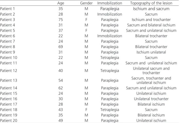
Table 1. Epidemiological data of operated patients.
Age Gender Immobilization Topography of the lesion Patient 1 35 M
Paraplegia
Ischium and sacrum
Patient 2 28 M Immobilization Sacrum
Patient 3 75 F Paraplegia Ischium and trochanter Patient 4 31 M Paraplegia Sacrum and bilateral ischium Patient 5 37 F Paraplegia Sacrum and unilateral ischium Patient 6 22 M Immobilization Bilateral trochanter Patient 7 24 M Paraplegia Sacrum Patient 8 69 M Paraplegia Bilateral trochanter Patient 9 31 M Paraplegia Ischium unilateral Patient 10 22 M Tetraplegia Sacrum
Patient 11 24 M Paraplegia Sacrum and unilateral ischium
Patient 12 40 M Tetraplegia Unilateral sacrum and trochanter
Patient 13 54 M Paraplegia Sacrum, trochanter and unilateral ischium Patient 14 62 M Paraplegia Sacrum and unilateral ischium Patient 15 24 M Paraplegia Unilateral ischium Patient 16 30 M Paraplegia Unilateral trochanter Patient 17 28 M Paraplegia Bilateral ischium Patient 18 43 F Tetraplegia Sacrum Patient 19 35 M Paraplegia Bilateral ischium Patient 20 49 M Paraplegia Unilateral ischium
Table 2. Surgical procedures.
Lesion Flap
Patient 1 Ischium Posterior thigh VY Patient 2 Sacrum Gluteus VY
Patient 3 Ischium and Unilateral Trochanter Posterior thigh VY + fascia lata tensor Patient 4 Bilateral Ischium Bilateral posterior thigh VY Patient 5 Sacrum Bilateral gluteus VY Patient 6 Trochanter Unilateral Trochanter Fascia Lata tensor Patient 7 Sacrum Gluteus VY
Patient 8 Bilateral Ischium Bilateral posterior thigh VY Patient 9 Ischium Posterior thigh VY Patient 10 Sacrum Gluteus VY Patient 11 Sacrum Bilateral gluteus VY Patient 12 Sacrum Gluteus VY Patient 13 Sacrum Gluteus VY Patient 14 Ischium Posterior thigh VY Patient 15 Ischium Posterior thigh VY Patient 16 Unilateral Trocanter Fascia Lata tensor Patient 17 Bilateral Ischium Bilateral posterior thigh VY Patient 18 Sacrum Gluteus VY
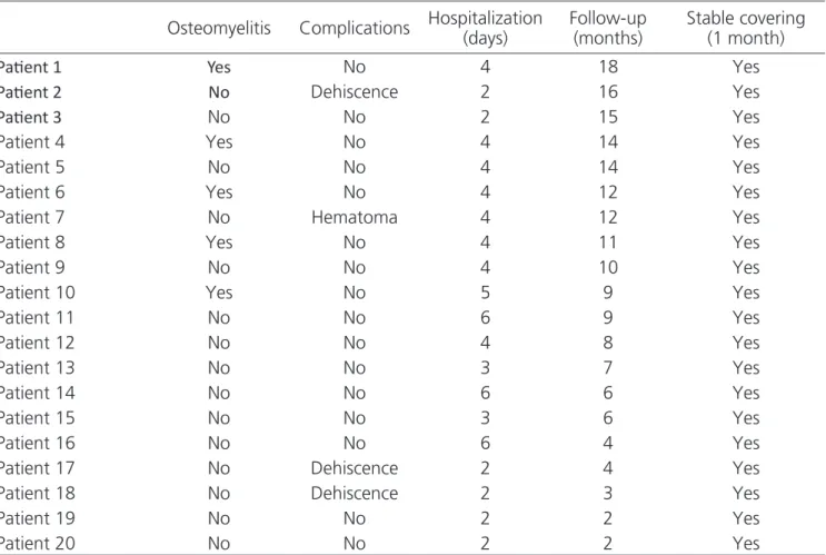
Medium time of hospitalization was 3.6 days (2-6 days), medium follow-up was 9.1 months (2-18 months). All patients maintained their wound closed and none of them showed recurrence during the follow-up period. Outcome data are described at table 3.
Figure 2. Ischiatic pressure lesion.
(Patient 9)- Ischiatic pressure lesion. A) marking of posterior fasciocutaneous thi-gh flap; B) defect after bursa resection; C) the flap was dissected and partially de-epidermised; D) inserted flap fixed at ischium periosteum for filling and pro-tection; E) immediate post-operatory; F) one month of post-operatory with stable covering.
DISCUSSION
Over the last years it was observed an increase of referred patients to our ambulatory with pressure le-sions; this can be explained by many reasons, such as no resolution of pressure sore during acute phase of hospi-talization, recurrence of previous operated wounds, fail to follow guidelines of local care and change of decu-bitus, low socio-economic level of patients and absen-ce of a caregiver to help with local care and change of decubitus.
Pressure wounds have different prevalence ac-cording to countries and regions, maybe due to health local systems, HDI, per capita income, culture, etc. Howe-ver, several location data show a relatively constant and convergent prevalence. In developed countries, risk pa-tients have a prevalence of 1% to 50% (inpapa-tients) and 8.3% (at home). In Germany, 21.2% of inpatients and 8.3% at home have pressure wounds. In the U.S.A., pre-valence varies from 2% to 28% (medium 11%)6.
Table 3. Outcome data.
Osteomyelitis Complications Hospitalization (days)
Follow-up (months)
Stable covering (1 month)
Paient 1 Yes No 4 18 Yes
Paient 2 No Dehiscence 2 16 Yes
Paient 3 No No 2 15 Yes
Patient 4 Yes No 4 14 Yes
Patient 5 No No 4 14 Yes
Patient 6 Yes No 4 12 Yes
Patient 7 No Hematoma 4 12 Yes
Patient 8 Yes No 4 11 Yes
Patient 9 No No 4 10 Yes
Patient 10 Yes No 5 9 Yes
Patient 11 No No 6 9 Yes
Patient 12 No No 4 8 Yes
Patient 13 No No 3 7 Yes
Patient 14 No No 6 6 Yes
Patient 15 No No 3 6 Yes
Patient 16 No No 6 4 Yes
Patient 17 No Dehiscence 2 4 Yes Patient 18 No Dehiscence 2 3 Yes
Patient 19 No No 2 2 Yes
In Brazil, pressure lesions prevalence is 16.9% for risk patients that rises to 39.4% for those with more than 60 years old. A study by the Federal University of Minas Gerais (UFMG) involving hospitals all over the country, studied 473 patients (251 men and 222 women) with 18 to 103 years old (medium 58.4 years); it observed pressure lesions in 80 patients (16.9%) with 137 ulcers. Among those patients, 47.4% had nutrition deficiency and 52.6% some gra-de of malnutrition7. Another aggravating factor is the immobilization grade of patient. The lowest the ability to move the higher the probability of a more severe lesions. Among tetraplegics and paraplegics, preva-lence of pressure ulcers is 20% to 60%. Around 85% of patients with spinal cord injury may develop pres-sure lesions during treatment8,9.
Most affected sites by pressure lesions are sacral and trochanteric regions. In a study by the Ins-titute of Orthopedics and Trauma from HC-FMUSP in-cluding 45 patients, 32.5% of pressure lesions were sacral, 32.5% trochanteric, 15.5% involved ischium and 19.5% other regions. UFMG cited study included 137 lesions, and 66 (48.1%) were sacral, 30 (21.9%) trochanteric, 22 (16%) calcaneus and 21 (15.3%) in-volved other sites10.
In our cohort, 46.8% of pressure sores were ischiatic, mainly in male patients (84%), adults at working age (medium 38.8 years) and paraplegics (73.5%). These data may represent a selection bias of the brief hospitalization protocol for patients with is-chiatic lesions with better pre-operatory conditions. In general, two distinct epidemiologic population with pressure lesions are observed. One aged, with severe comorbidities (cardiopathies and neuropathies), with low level of conscience and lesions related to horizon-tal decubitus position (Figure 3). The other includes young patients, victims of spinal cord injury, usually paraplegics, with pressure lesions related to seated orthostatic position (wheelchairs). The first popula-tion presents mainly ulcers at sacrum and trochanter, and the other, at ischium (uni or bilateral).
Initial treatment of these lesions include lo-cal pressure relief, improvement of spasms, usually frequent in these patients, enzymatic or surgical de-bridement, and maintenance of a clean and
moistu-re environment that allows for granulation and moistu- re-e-pithelization of the wound bed11. Approximately 70 to 90% of pressure ulcers are superficial and heal by second intention with these cares. Lesions Grades III and IV, deeper and occasionally associated with os-teomyelitis, usually need surgical treatment, require covering with flaps for definitive treatment. In those patients, it is important to optimize home care (chan-ge of decubitus, local care) and nutritional status in order to increase surgical success12.
Figure 3. Sacral pressure ulcer.
(Patient 10)- Sacral pressure lesion. A) wound; B) defect after bursa resection; C) part resected; D) dissection and advance of gluteus fasciocutaneous V-Y flap; E) immediate post-operatory with covering of defect; F) six months of post-operatory with stable covering.
Our department chose to perform predo-minantly fasciocutaneous flaps routinely, since they provide adequate covering, good filling when de-epi-dermised and full irrigation of the wound, aiding in-fection control. Classically, the first option would be miocutaneous flaps, however, more recent papers showed similar quality of covering13. On the other side, muscular tissue is more sensitive to ischemia, in-creases operatory morbidity and prevents future use in case of recurrence14.
3.0g/dl, that reflect a good nutritional status related to lower rate of dehiscence of surgical wound15,16.
The presence of spasticity in patients with pressure lesions is another important aspect17, since it difficult or prevents change of decubitus. These pa-tients are at higher risk of presenting pressure ulcers, or recurrence in operated or healed areas. Usually, therapeutic options include baclofen associated or not with benzodiazepines. In the studied cohort, five patients were controlled with drugs.
The absence at pre-operatory of osteomye-litis was also observed for inclusion of patients at the short hospitalization protocol, since it lowers surgi-cal success rate18. In case of clinical (presence in in-flammatory signs, fever, purulence) or laboratory (increase of leucocytes and PCR) suspicion, nuclear magnetic resonance was performed to confirm or rule out osteomyelitis. If positive, the patient was refer-red to Orthopedic Department for multidisciplinary treatment. If negative, the patient was included. At present, magnetic resonance is considered the best study for diagnosis of osteomyelitis19. However, the surgical protocol included partial resection of bone prominence to attenuate the pressure point that was sent to pathological analysis. Five samples were posi-tive for osteomyelitis at post-operatory. The use and period of use of antibiotics according to sensitivity va-ried from 14 to 28 days, and, interestingly, no patient with positive culture had complications or presented signs of osteomyelitis during ambulatory follow-up. It is possible that complete debridement and partial resection of bone prominences until viable bone were important for these results, as well as a vascularized flap that provided oxygen and nutrients needed for the treatment of this condition.
The presence of a family member or care-giver during treatment of pressure lesions is vital for success16. Therefore, this was another inclusion crite-ria included in the present study. It is critical for the patient to change position (dorsal to lateral, lying to seated) and this must be done very carefully with the aid of another person during the first four weeks of
post-operatory. One of the three dehiscence lesions was observed on the seventh day of post-operatory, due to mechanical trauma caused directly by change of position. Therefore, during pre-operatory visit, it was reinforced the need of familial involvement or the constant presence of a caregiver. Indirectly, it was also observed if the patient came to pre-operatory consulta-tion with the wound in good condiconsulta-tions (clean, correct dressing, regression borders) and if hygiene conditions were adequate. If positive, the patient was considered eligible for the protocol.
In view of the growing number of non-ope-rated pressure lesions and the logistic difficulty to admit and operate these patients, it was proposed this short hospitalization protocol to treat pressure lesions grade IV. The patient is prepared at ambulatory and selected if fulfilled the inclusion criteria. Three goals were anticipated: 1) Lowering of hospitalization time; 2) Lowering of post-operatory dehiscence and wound complications; 3) Obtaining a stable covering with lower recurrence. In view of the obtained results (3.6 days of hospitalization, 11.1% of minor dehiscence and ab-sence of recurrence) it is possible to affirm that goals were met. Literature historical data are in accordance to these results. Sameem et al.20 in a systematic re-view of 55 published studies, observed with fasciocu-taneous flaps 11.7% of complications (5.1% of flap necrosis), 6.9% of post-operatory infection and a me-dium of 11.2% of recurrence (13 to 31%).
Regarding medium follow-up of 9.1 months, this time interval may be considered relatively short for an accurate analysis. It must be observed that the-se patients have reduced mobility and depend on fa-milial members and transport to attend ambulatory visits. Therefore, many patients are lost for follow-up, when their wounds are healed. It is important to men-tion that time interval to observe recurrence is higher than the one here reported, around 12 to 24 months.
REFERENCES
1. Milcheski DA, Ferreira MC, Nakamoto H, Tuma Jr P, Gemperli R. Tratamento cirúrgico de ferimentos descolantes nos membros inferiores - proposta de protocolo de atendimento. Rev Col Bras Cir. 2010;37(3):195-203.
2. MC Ferreira. Complex Wounds. Clinics.
2006;61(6):571-8.
3. Phillips T, Stanton B, Provan A, Lew R. A study of the impact of leg ulcers on quality of life: financial, social, and psychologic implications. J Am Acad Dermatol. 1994;31(1):49-53.
4. Pokorny ME, Rose MA, Watkins F, Swanson M,
Kirkpatrick MK, Wu Q. The relationship between pressure ulcer prevalence, body mass index, and braden scales and subscales: a further analysis. Adv Skin Wound Care. 2014;27(1):26-30.
5. National Pressure Ulcer Advisory Panel. Pressure ulcer definition and stages [Internet]. Washington: NPUAP; 2007. [cited 2007 Apr 13]. Available from: http://www.npuap.org/
6. Alves P, Mota F, Ramos P, Vales L. Epidemiologia das úlceras de pressão: interpretar dados epidemiológicos como indicador de qualidade [Internet]. Berlin: ResearchGate; 2007. [cited 2007 Apr 25] Available from: https://www.researchgate. net/publication/257140360
7. Brito PA, de Vasconcelos Generoso S, Correia MI. Prevalence of pressure ulcers in hospitals in Brazil and association with nutritional status--a multicenter, cross-sectional study. Nutrition. 2013;29(4):646-9.
8. Eslami V, Saadat S, Habibi Arejan R, Vaccaro AR, Ghodsi SM, Rahimi-Movaghar V. Factors associated with the development of pressure ulcers after spinal cord injury. Spinal Cord. 2012;50(12):899-903. 9. Byrne DW, Salzberg CA. Major risk factors for
pressure ulcers in the spinal cord disabled: a literature review. Spinal Cord. 1996;34(5):255-63.
10. Costa MP, Sturtz G, Costa FPP, Ferreira MC, Barros Filho TEP. Epidemiologia e tratamento das úlceras de pressão: experiência de 77 casos. Acta Ortop Bras. 2005;13(3):124-33.
11. Alvarez OM, Fernandez-Obregon A, Rogers RS, Bergamo L, Masso J, Black M. Chemical debridement of pressure ulcers: a prospective, randomized, comparative trial of collagenase and papain/urea formulations. Wounds. 2000;12(2):15-25.
12. Quassem A, Humphrey LL, Forciea MA, Starkey M, Denberg TD. Treatment of pressure ulcers: a clinical practice guideline from the American College of Physicians. Ann Intern Med. 2015;162(5):370-9. 13. Barreiro GC, Millan LS, Nakamoto H, Montag E, Tuma
Jr PT, Ferreira MC. Reconstruções pelveperineais com uso de retalhos cutâneos baseados em vasos perfurantes: experiência clínica com 22 casos. Rev Bras Cir Plást. 2011;26(4):680-4.
14. Yamamoto Y, Ohura T, Shintomi Y, Sugihara T, Nohira K, Igawa H. Superiority of the fasciocutaneous flap in reconstruction of sacral pressure sores. Ann Plast Surg. 1993;30(2):116-21.
15. Sugino H, Hashimoto I, Tanaka Y, Ishida S, Abe Y, Nakanishi H. Relation between the serum albumin level and nutrition supply in patients with pressure Objetivo: avaliar o protocolo de internação breve para tratamento de lesões por pressão, em vigência no Grupo de Feridas Complexas do Hospital das Clínicas da Faculdade de Medicina da Universidade de São Paulo, com ênfase na seleção do paciente, no tempo de internação, na cobertura cutânea realizada, nas complicações e nas recidivas das lesões. Métodos: coorte retrospectiva de 20 pacientes consecutivos com 25 lesões por pressão Grau IV. Todos os pacientes foram preparados em ambulatório e foram internados para fecha-mento cirúrgico da lesão por pressão em cirurgia única. Resultados: no total foram confeccionados 27 retalhos para cobertura de 25 feridas abordadas. Foram verificados três casos (11,1%) de deiscências menores. Não foi observada recidiva no período de seguimento pós-operatório. Nenhum paciente foi reoperado e nenhum retalho sofreu necrose parcial ou total. O tempo de internação médio foi de 3,6 dias (dois a seis dias) e o seguimento foi de 9,1 meses, em média, oscilando entre dois e 18 meses. Todos os pacientes permanece-ram com a lesão fechada e nenhum deles apresentou recidiva da lesão durante o seguimento. Conclusão: o protocolo de internação breve para resolução de lesões por pressão foi considerado adequado, com curto tempo de internação e baixos índices de deiscência de ferida cirúrgica.
Descritores: Lesão por Pressão. Ferimentos e Lesões. Retalhos Cirúrgicos. Cirurgia Plástica.
ulcers: retrospective study in an acute care setting. J Med Invest. 2014;61(1-2):15-21.
16. Thomas DR. Role of nutrition in the treatment and prevention of pressure ulcers. Nutr Clin Pract. 2014;29(4):466-72.
17. Ricci JA, Bayer LR, Orgill DP. Evidence-based medicine: the evaluation and treatment of pressure injuries. Plast Reconstr Surg. 2017;139(1):275e-286e.
18. Ubbink DT, Brölmann FE, Go PMNYH, Vermeulen H. Evidence-based care of acute wounds: a perspective. Adv Wound Care. 2015;4(5):286-94.
19. Pineda C, Espinosa R, Pena A. Radiographic imaging in osteomyelitis: the role of plain radiography, computed tomography, ultrasonography, magnetic resonance imaging, and scintigraphy. Semin Plast Surg. 2009;23(2):80-9.
20. Sameem M, Au M, Wood T, Farrokhyar F, Mahoney J. A systematic review of complication and recurrence rates of musculocutaneous, fasciocutaneous, and perforator-based flaps for treatment of pressure sores. Plast Reconstr Surg. 2012;130(1):67e-77e.
Received in: 18/07/2017
Accepted for publication: 23/08/2017 Conflict of interest: none.
Source of funding: none.
Mailing address:
Dimas André Milcheski