28 Sao Paulo Med J. 2014; 132(1):28-35
ORIGINAL ARTICLE
DOI: 10.1590/1516-3180.2014.1321594Cold ischemia or topical-ECMO for lung preservation:
a randomized experimental study
Preservação pulmonar por isquemia fria ou ECMO-tópico:
um estudo aleatório experimental
Alessandro Wasum Mariani
I, Israel Lopes Medeiros
II, Paulo Manuel Pêgo-Fernandes
III, Flavio Guimarães Fernandes
IV,
Fernando Do Vale Unterpertinguer
IV, Lucas Matos Fernandes
V, Paulo Francisco Cardoso
VI, Mauro Canzian
VII, Fabio Biscegli Jatene
VIIIDepartment of Experimental Surgery, Instituto do Coração (InCor), Hospital das Clínicas (HC), Faculdade de Medicina da Universidade
de São Paulo (FMUSP), and Laboratório de Pesquisa em Cirurgia Torácica, Faculdade de Medicina da Universidade de São Paulo (LIM
61), São Paulo, Brazil
ABSTRACT
CONTEXT AND OBJECTIVE: Lung preservation remains a challenging issue for lung transplantation groups. Along with the development of ex vivo lung perfusion, a new preservation method known as topical-ECMO (extracorporal membrane oxygenation) has been proposed. The present study compared topical-ECMO with cold ischemia (CI) for lung preservation in an ex vivo experimental model.
DESIGN AND SETTING: Randomized experimental study, conducted at a public medical school. METHOD: Fourteen human lungs were retrieved from seven brain-dead donors that were considered unsuitable for transplantation. The lung bloc was divided and each lung was randomized to be preserved by means of topical-ECMO or CI (4-7 °C) for eight hours. These lungs were then reconnected to an ex vivo perfusion system for functional evaluation. Lung biopsies were obtained at three times. The functional variables assessed were oxygenation capacity (OC) and pulmonary artery pressure (PAP); and the histologi-cal variables were lung injury score (LIS) and apoptotic cell count (ACC).
RESULTS: The mean OC was 468 mmHg (± 81.6) in the topical-ECMO group and 455.8 (± 54) for CI (P = 0.758). The median PAP was 140 mmHg (120-160) in the topical-ECMO group and 140 mmHg (140-150)for CI (P = 0.285). The mean LIS was 35.57 (± 4.5) in the topical-ECMO group and 33.86 (± 6.1) for CI (P = 0.367). The ACC was 25.00 (± 9.34) in the topical-ECMO group and 24.86 (± 10.374) for CI (P = 0.803). CONCLUSIONS: The present study showed that topical-ECMO was not superior to cold ischemia for up to eight hours of lung preservation.
RESUMO
CONTEXTO E OBJETIVO: A preservação pulmonar permanece um desaio para os grupos transplantado-res. Com o desenvolvimento da perfusão pulmonar ex vivo, foi proposto um novo método de preservação chamado de tópico (oxigenação de membrana extracorpórea). O presente estudo compara ECMO-tópico com isquemia fria (IF) para preservação pulmonar em um modelo experimental ex vivo.
TIPO DE ESTUDO E LOCAL: Estudo experimental randomizado, conduzido em uma faculdade de medi-cina pública.
MÉTODO: Quatorze pulmões humanos foram retirados de sete doadores de morte cerebral considera-dos não aptos a transplante. O bloco pulmonar foi dividido e cada um foi aleatorizado para preservação por ECMO-tópico ou IF (4-7 °C) durante oito horas. Esses pulmões foram então re-conectados a um sis-tema de perfusão ex vivo para avaliação funcional. Biópsias pulmonares foram obtidas em três tempos. As variáveis funcionais avaliadas foram: capacidade de oxigenação (CO) e pressão de artéria pulmonar (PAP). As variáveis histológicas estudadas foram escore de lesão pulmonar (ELP) e contagem de células apoptóticas (CCA).
RESULTADOS: A média da CO foi de 468 mmHg (± 81.6) no grupo ECMO-tópico e 455.8 (± 54) no grupo IF (P = 0,758); a PAP média foi de 140 mmHg (120-160) para ECMO-tópico e 140 mmHg (140-150) para IF (P = 0,285); o ELP médio foi 35,57 (± 4,5) no ECMO-tópico e 33,86 (± 6,1) no IF (P = 0,367). A CCA foi 25,00 (± 9,34) no grupo ECMO-tópico e 24,86 (± 10,374) no IF (P = 0,803).
CONCLUSÕES: O presente estudo demonstrou que o ECMO-tópico não é superior a IF para oito horas de preservação pulmonar.
IMD, PhD. Attending Physician, Department of
Thoracic and Cardiovascular Surgery, Instituto do Coração (InCor), Hospital das Clínicas (HC), Faculdade de Medicina da Universidade de São Paulo (FMUSP), São Paulo, Brazil.
IIMD, PhD. Attending Physician, Department
of Thoracic Surgery, Hospital de Messejana, Fortaleza, Brazil.
IIIMD, PhD. Full Professor of Thoracic Surgery,
Instituto do Coração (InCor), Hospital das Clínicas (HC), Faculdade de Medicina da Universidade de São Paulo (FMUSP), São Paulo, Brazil.
IVMedical Student, Faculdade de Medicina da
Universidade de São Paulo (FMUSP), São Paulo, Brazil.
VMD. Attending Physician, Department of Thoracic and
Cardiovascular Surgery, Instituto do Coração (InCor), Hospital das Clínicas (HC), Faculdade de Medicina da Universidade de São Paulo (FMUSP), São Paulo, Brazil.
VIMD, PhD. Professor, Faculdade de Medicina da
Universidade de São Paulo (FMUSP), São Paulo, Brazil.
VIIMD, PhD. Attending Physician, Department of
Pathology, Instituto do Coração (InCor), Hospital das Clínicas (HC), Faculdade de Medicina de Universidade de São Paulo (FMUSP), São Paulo, Brazil.
VIIIMD, PhD. Full Professor of Cardiovascular Surgery,
Head of Thoracic and Cardiovascular Surgery Departament, Instituto do Coração (InCor), Hospital das Clínicas (HC), Faculdade de Medicina da Universidade de São Paulo (FMUSP), São Paulo, Brazil.
KEY WORDS:
Organ preservation. Reperfusion injury. Lung transplantation. Transplantation, homologous. Thoracic surgery.
PALAVRAS-CHAVE:
Cold ischemia or topical-ECMO for lung preservation: a randomized experimental study | OR IG IN A L A R T IC L E
INTRODUCTION
Lung transplantation has been established as a treatment option for end-stage lung disease.1 However, the low tolerance of lungs
to ischemia is notorious and can lead to grat dysfunction that impacts on the recipient’s outcome.2 Despite the variety of
pres-ervation techniques proposed, such as topical cooling,3
autoper-fusion with extracorporeal circulation4 and donor core cooling,5
pulmonary artery lush perfusion with cold preservation solu-tion has endured the test of time and has remained the most frequently used technique because of its practicality and efec-tiveness.6 Extracellular preservation solutions such as Perfadex
(VitroLife AB, Gothenburg, Sweden) and Celsior (Sang Stat, Lyon, France) have been used frequently for lung preservation.7
Concomitantly to the introduction of ex vivo lung perfusion, a method of preservation named topical-ECMO (extracorporeal membrane oxygenation) has been proposed by Steen et al. It has been tested experimentally8 and used clinically.9,10 his method
was designed to preserve the lungs ater ex vivo assessment and consists of immersion of the lung in a semi-inlated state in Steen solution diluted in Perfadex inside the ex vivo box where the
lungs were placed ater reconditioning. Although topical-ECMO has been proposed and used clinically, the method has not been compared with cold ischemia ater single-lush perfusion for preservation so far.
OBJECTIVE
he aim of this study was to evaluate whether topical-ECMO can provide better preservation quality than shown by regular cold ischemia, as methods for lung preservation.
METHODS
his was a randomized experimental study approved by our university hospital’s ethics committee (CAPPESQ 0212/08). Between December 2009 and August 2010, lungs retrieved from brain-dead donors that were refused for transplantation based on current clinical criteria were used in this study. he organs were included in the protocol if all eforts to recover the lungs failed. Written consent was obtained from family members of the donors to permit the use of the organs in this study. he lungs were perfused through the pulmonary artery with cold Perfadex (Vitrolife, Gothenburg, Sweden) and harvested in the usual fashion at the time of multiorgan retrieval. he organs were immersed in cold Perfadex, stored in a cooler and transported to our laboratory.
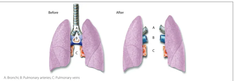
Upon arrival, the lung block was dissected out, and the let and right lungs were separated by sectioning the let atrium, main pulmonary artery and the tracheal carina (Figure 1). Separation of the lungs allowed each lung to be subjected to a diferent forms of preservation. he lungs were then randomly
assigned for topical-ECMO or cold ischemia (CI). he random-ization was done by means of numbered envelopes containing computer-generated random sequences of numbers.
Topical-ECMO was carried out by means of complete lung immersion in solution (Steen Solution; VitroLife AB, Gothenburg, Sweden) within a containment box (VitroLife AB, Gothenburg, Sweden) (Figure 2). he solution was contin-uously circulated through this box by means of a centrifugal pump (Braile Biomedica, São José do Rio Preto, Brazil) with a low of 4 l/min and through a membrane oxygenator (Braile Biomedica, São José do Rio Preto, Brazil) receiving oxygen at 5 l/min in order to achieve oxygenation. he average tempera-ture was maintained by means of a heat exchanger with a tar-get of 8 °C. he lung temperature was monitored continuously using a thermometer placed inside the pulmonary vein.
CI lungs were stored in a bag containing Perfadex solution and were immersed in a second plastic bag containing saline solution at 4 °C. he bags were then stored in a refrigerator at a temperature of 4-7 °C.
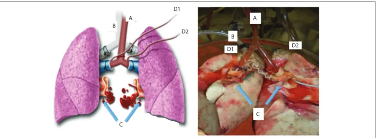
Ater eight hours of preservation, the let and right lungs were reconnected in parallel, by means of two Y-shaped cannu-lae: one in the trachea and the other in the pulmonary artery. he pulmonary veins remained separated. his reconnection technique made it possible for the topical-ECMO and CI lungs to undergo reperfusion and ventilation in the ex vivo perfusion
system simultaneously, with the same reperfusion solution and exactly the same ventilation parameters. Since the pulmonary veins remained independent, it enabled sampling for blood gas analysis separately (Figure 3).
he system used during the reperfusion phase consisted of the XVIVO box (VitroLife AB, Gothenburg, Sweden), attached to the centrifugal pump, heat exchanger, membrane oxygenator and a venous reservoir (Braile Biomedica, São José do Rio Preto, Brazil), as we previously described.11 he Y cannula for the pulmonary
arteries had two separate probes connected to pressure transduc-ers that allowed continuous and independent monitoring of pul-monary artery pressure. he right and let lung pulpul-monary vein eluents were collected directly into the XVIVO box, where they were mixed and drained into the venous reservoir by gravity. We elected to keep the atria open in order to facilitate assembly of the system, by eliminating the need for special atrial cannulae. his is feasible during short-term perfusion, whereas in cases requiring long-term perfusion, a closed system is preferred so as to avoid pulmonary edema.12 he system was illed with 1,500 ml of Steen
ORIGINAL ARTICLE | Mariani AW, Medeiro s I L , P ê g o-Fernandes PM, Fernandes FG, Unterpertinguer FDV, Fernandes LM, Cardoso PF, Canzian M, Jatene FB
30 Sao Paulo Med J. 2014; 132(1):28-35
using a formula based on the size of the donor (CO = 3 * body sur-face area, assuming target cardiac index = 3). his low was suicient to assess the lungs in the system and low enough to avoid pulmonary edema formation.
A steady state was usually reached ater 60 minutes of perfu-sion, and perfusate gases were collected and hemodynamics were recorded at this time. he variables assessed were pulmonary arte-rial partial oxygen pressure (PvO2), pulmonary partial oxygen pressure of the eluent from the pulmonary veins (PaO2) and par-tial carbon dioxide (PaCO2), pulmonary artery pressure (PAP) and pulmonary vascular resistance (PVR) [PVR = PAP/pulmonary artery low * 80 (dynes/sec/cm5)].
Lung tissue samples from the middle lobe and ligulae were col-lected before harvesting, ater the topical-ECMO or cold ischemia
Figure 2. Containment box for topical-ECMO, with lung immersed in Steen Solution.
period and ater ex vivo lung perfusion (EVLP). he samples were
ixed in 10% bufered formalin for 24 hours, embedded in paraf-in, sectioned at thicknesses of 5 mm and stained with hematoxy-lin and eosin (Figure 4). For all cases, semiquantitative scoring was performed by an experienced lung pathologist using the follow-ing histology parameters: interstitial edema, intra-alveolar edema, arteriolar thickening, vascular thrombosis, intra-alveolar hemor-rhage, intra-alveolar ibrin deposition, necrosis, inlammatory cell iniltrate, pleural iniltrate, peribronchiolar inlammatory reaction, organizing pneumonia, peribronchiolar ibrosis, ibroblast foci, peribronchiolar muscular hypertrophy, pleural plaques, interstitial ibrosis, alveolar lining cell hyperplasia, alveolar macrophages, pig-mented macrophages, denudated bronchiolar epithelium, vasculi-tis and emphysema. he severity of these indings was determined using a four-grade scale: absent = 0; minimal = 1; moderate = 2; and intense = 3. he sum of each parameter resulted in the Lung Injury Score (LIS), with values ranging from 0 to 66.13
Apoptosis was assessed by means of in situ terminal deoxy-nucleotidyl transferase (TdT)-mediated deoxyuridine triphos-phate nick end labeling (TUNEL), using the In Situ Cell Death Detection Kit (Roche, Basel, Switzerland). he TUNEL method is based on the enzymatic ability of TdT to catalyze a template-independent addition of deoxyribonucleotide triphosphate to the 3=-OH ends of double- or single-stranded DNA. he sections were deparainized and rehydrated. Protein digestion was done by applying proteinase K to the slides for 15 minutes at room tem-perature, which was followed by four washes in distilled water for two minutes each. Equilibration bufer was applied to the sec-tions, which were then incubated in a humidiied chamber for three minutes. Following this, the sections were incubated with TdT in a humidiied chamber at 37 °C for 1 hour. Fluorescein-labeled antidigoxigenin antibody was applied to the sections, and they were incubated in a humidiied chamber for 30 minutes at
Before After
C C
B A
B A
Cold ischemia or topical-ECMO for lung preservation: a randomized experimental study | ORIGINAL ARTICLE
room temperature. he sections were then washed with phos-phate-bufered saline.
Immediately ater completing the protocol, the slides were viewed using a luorescence microscope and photographed using a charge-coupled device digital camera with a 590-nm emission ilter. To localize apoptotic cells, randomly selected specimens of lung tissue were photographed under the luorescence optical microscope at high magniication (original magniication 400 x). Apoptotic cells appeared as bright green (Figure 5). Counts were obtained from ive randomly chosen ields per slide, representing a total area of 0.1 mm2. An independent, blinded examiner
per-formed the cell counting.
Immunohistochemical analysis was performed using the polyclonal rabbit antibody for CD3 (1:100, Abcam, Cambridge, United Kingdom). he sections were deparainized and a 0.3% hydrogen peroxide solution was applied for 35 min to inhibit endogenous peroxidase activity. Antigen retrieval was performed using citrate solution for 45 minutes. he sections were incubated with the primary antibody overnight at 48 °C. he streptavidin-biotin complex (LSAB+; DakoCytomation, Carpinteria, CA, United States) was used as the secondary antibody and 3,3-diami-nobenzidine (DAB) (Sigma Chemical Co, St. Louis, MO, United States) was used as the chromogen. he sections were counter-stained using Harris hematoxylin. For negative controls, the primary antibody was replaced with phosphate-bufered saline (PBS). he inlammation index was determined semiquantita-tively based on the presence of inlammatory cells in lung tissue, using a three-grade scale: minimal inlammation = 1, moderate inlammation = 2 and marked inlammation = 3.
All histological and immunohistochemical parameters were evaluated and measured by a pulmonary pathologist who was blinded to any information regarding the cases.
Lungs were weighed immediately ater harvesting, at the end of the preservation time and ater reperfusion. hey were also subsequently dried out for 48 hours at 70 °C and weighed again in order to obtain the wet/dry weight ratio.
he normality tests applied were Kolmogorov-Smirnov and Shapiro-Wilk. To compare functional and histological data from before to ater the procedure, Student’s paired-samples test was performed. he Wilcoxon signed rank test was used for vari-ables that were not normally distributed. Repeated-measurement ANOVA was used to analyze the diferences in measurements that evolved over time in pairs of groups. For qualitative vari-ables in 2 x 2 tvari-ables, we used the chi-square test, or Fisher’s exact test when the expected value was less than ive. In tables larger
Figure 4. Histological appearance of the lung after ex vivo perfusion (original magniication 100 x; hematoxylin-eosin).
B A
C
A
C B
D1 D2
D1
D2
ORIGINAL ARTICLE | Mariani AW, Medeiros IL, Pêgo-Fernandes PM, Fernandes FG, Unterpertinguer FDV, Fernandes LM, Cardoso PF, Canzian M, Jatene FB
32 Sao Paulo Med J. 2014; 132(1):28-35
than 2 x 2, the likelihood function was used because the expected value was less than ive in all cases. he results were expressed as the mean and standard error of the mean, or as the median and interquartile range for variables that were not normally dis-tributed. he statistical analyses were performed using the SPSS (Statistical Package for the Social Sciences) 18.0 software (SPSS Inc, Chicago, IL) with a conidence interval of 95% and a signiicance level of 0.05.
RESULTS
Seven sets of lungs were assessed. he demographics are summa-rized in Table 1. During reperfusion, the oxygenation capacity was estimated from the PaO2 measured in the eluent perfusate
from the pulmonary veins of the lungs. For the lungs preserved by means of topical-ECMO and CI, respectively, the capacities were 468 ± 81.6 mmHg and 455.86 ± 54 mmHg, with no sta-tistically signiicant diferences between the groups (P = 0.758). he mean partial oxygen pressure of the deoxygenated perfusate inlow during EVLP (PvO2) was 91.94 ± 24.4 mmHg. he mean arterial PaCO2 in lungs preserved by means of topical-ECMO and CI were 17.45 ± 3.6 mmHg and 17.02 ± 3.1 mmHg respec-tively, with no statistically signiicant diferences between the groups (P = 0.617).
Hemodynamics showed that the median PAP in topical-ECMO lungs was 140 mmHg (120-160) and in CI lungs, 140 mmHg (140-150); P = 0.285. he median PVR also showed no statisti-cally signiicant diferences between the groups: topical-ECMO = 459 dynes/sec/cm5 and CI = 474.50 dynes/sec/cm5; P = 0.285.
he mean weight changes of the lungs between the groups over time (pre-ischemic, post-ischemic and post-reperfusion times) did not show any signiicant diferences, as depicted in
Figure 6. he wet-to-dry ratios in the topical-ECMO and CI groups were 2.77 ± 0.93 and 3.21 ± 1.85, respectively, with no statistically signiicant diference (P = 0.358). he lung his-tology, studied by means of LIS and the apoptotic cell count (ACC), did not show any signiicant diferences between the groups over time (LIS P = 0.531 and ACC P = 0.803), as shown in Figures 7 and 8.
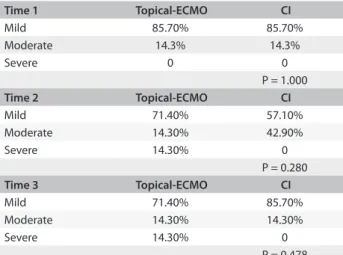
he degree of tissue inlammation was assessed by detect-ing the presence of inlammatory cells. Iniltration of CD3+ T-lymphocytes into the lung tissue was evaluated at three times and did not signiicantly difer between the groups, as shown in Table 2.
DISCUSSION
Our experiments showed that there were no diferences between topical-ECMO and CI in terms of preservation quality, hemody-namic performance and histology of the lungs assessed.
Although topical-ECMO was created and has been used as a method for lung preservation at Lund University in Sweden, it has not been tested for eicacy or safety. In the irst paper in which this method appears, 12 pigs were used as non-heartbeat donors and lung function was assessed ex vivo ater
harvest-ing. Ater the ex vivo evaluations had been completed, the lungs
were cooled and preserved using topical-ECMO at a tempera-ture of 12 °C, until transplantation.8 he next paper described
the irst human case that received a lung ater ex vivo evaluation.
Ater perfusion, the let lung was stored using topical-ECMO at 8 °C and remained there for nine hours and 30 minutes, until transplantation.9
In 2009, the same group published six human cases of trans-plantation ater ex vivo lung perfusion. Once again, ater the ex vivo perfusion had been completed, the lungs were cooled down
Figure 5. Photomicrograph demonstrating apoptotic cells (green) under a luorescence optical microscope.
Donor data Results
Gender
Male 4 (57.1%)
Female 3 (42.8%)
Age (years) 53.86 ± 16.7
Time on mechanical ventilation (days) 4.86 ± 3.93
PaO2 (mmHg) 192.2 ± 92.2
PCO2 (mmHg) 33.1 ± 9.6
Cause of death
Hemorrhagic stroke 5 (71.4%)
Head trauma 2 (28.6%)
Reason for rejection for transplantation
PaO2 < 300 mmHg 6 (85.7%) Bilateral contusion 1 (14.3%) Side of topical-ECMO
Right 4 (57.1%)
Left 3 (42.9%)
Table 1. Demographics and clinical characteristics of the donors
Cold ischemia or topical-ECMO for lung preservation: a randomized experimental study | ORIGINAL ARTICLE
and stored using topical-ECMO until transplantation. he mean duration of use of topical-ECMO was ive hours and 53 minutes for the irst implanted lung and 8 hours and 46 minutes for the sec-ond lung.10 Despite the good results in all these papers, it cannot
be airmed whether the use of topical-ECMO interfered with lung preservation or with lung function, either positively or negatively. Since the time when the topical-ECMO method was designed, this is the irst comparative study evaluating it against any other preservation strategy. Our use of rejected human lungs in an experimental study was motivated by the aim of testing the method in a realistic situation that would also be more represen-tative of the clinical lung transplantation scenario. he study was designed such that it would optimize the use of rejected donor lungs and, likewise, would reduce variability by using both lungs from the same donor, i.e. one lung for each of two diferent pres-ervation techniques.
he diference between the topical-ECMO described by Steen et al. and the one used here was the solution. While the Swedish group8-10 used a mixture of Steen Solution, Perfadex and a
vari-able quantity of red blood cells in order to reach a hematocrit level of around 5%, pure Steen Solution was used in the present work.
he concept of splitting the double-lung block and shar-ing both the airway and the inlow perfusion connections had been tested previously in a pilot study in our laboratory and was proven to be feasible without signiicant functional deterioration of the lungs.14
he oxygenation capacity during the ex vivo perfusion can
be considered to be the most important parameter for functional assessment, since it relects both gas exchange and grat perfor-mance. Previous studies have shown that PaO2 values correspond more reliably to the quality of lung preservation than do other parameters such as those from pathology or radiology.15 In the
Time 1 Topical-ECMO CI
Mild 85.70% 85.70%
Moderate 14.3% 14.3%
Severe 0 0
P = 1.000
Time 2 Topical-ECMO CI
Mild 71.40% 57.10%
Moderate 14.30% 42.90%
Severe 14.30% 0
P = 0.280
Time 3 Topical-ECMO CI
Mild 71.40% 85.70%
Moderate 14.30% 14.30%
Severe 14.30% 0
P = 0.478
Table 2. Degree of CD3 T-lymphocyte iniltration into lung tissue at three times
ECMO = extracorporal membrane oxygenation; CI = cold ischemia. Figure 6. Lung weight gain between groups over time comparing topial ECMO (extracorporal membrane oxygenation) and CI (cold ischemia).
700
600
500
400
300
200
100
0
G
r
ams
T1 450.000
422.29
442.86
593.43 p = 0.282
521.14
408.57
T2 T3
Topical-ECMO Cold Ischemia
Figure 7. Comparison of lung injury scores between groups over time comparing topical ECMO (extracorporal membrane oxygenation) and CI (cold ischemia).
35
30
25
20
15
10
5
0
S
co
re
T1 7.571
6.268 7.714
p = 0.531
8.714
9.571 7.857
T2 T3
Topical-ECMO Cold Ischemia
Figure 8. Comparison of apoptotic cell counts between groups over time comparing topical ECMO (extracorporal membrane oxygenation) and CI (cold ischemia).
30.00
25.00
20.00
15.00
10.00
5.00
0.00
A
popt
otic c
ell c
oun
t
T1 18.57
16.71
20.71
17.57
25.00 24.86 p = 0.803
T2 T3
ORIGINAL ARTICLE | Mariani AW, Medeiros IL, Pêgo-Fernandes PM, Fernandes FG, Unterpertinguer FDV, Fernandes LM, Cardoso PF, Canzian M, Jatene FB
34 Sao Paulo Med J. 2014; 132(1):28-35
present study, the absence of signiicant diferences between the groups demonstrates that oxygen capacity and CO2 clearance were similar in both groups.
he parameter of weight variation has been used experimen-tally as a reliable measurement of pulmonary edema in the set-ting of lung preservation. he process of ischemia and reperfu-sion results in increased vascular permeability and disruption of the alveolar-capillary barrier, which ultimately causes water extravasation. he amount of edema can be therefore consid-ered to be inversely proportional to the quality of preservation.16
Despite the usefulness of weight variation in determining edema, its accuracy is debatable since it is inluenced by other factors such as alveolar hemorrhage, which also renders the method less accurate for detection of milder degrees of edema. In our study, the weight gain ater reperfusion was similar in both groups and possibly relected the degree of edema inherent to the ex vivo
reperfusion, which may become an adverse factor during lon-ger reperfusions, as described previously.12 On the other hand,
the wet-to-dry ratio has been found to be a more reliable mea-surement of the amount of water in the specimens at the end of reperfusion.17 herefore, the higher the value is, the greater the
edema will be. In our study, the wet-to-dry ratio was similar in both groups, thus suggesting that similar amounts of edema were present regardless of the preservation strategy used.
here are two major limitations regarding the histologi-cal changes found in the lungs: the degree of the histologihistologi-cal changes does not show any linear correlation with the func-tional outcome; and the presence of pulmonary parenchymal lesions before harvesting, induced by pro-inlammatory agents secondary to brain death, may be great enough to cause pro-found changes to the pulmonary tissue.18 Nevertheless,
histologi-cal evaluation remains a powerful indicator of the quality of lung preservation.19 he injury score used in this study (ELP) was
sim-ilar between the groups, both ater the period of preservation and ater reperfusion. his inding shows that the two preservation methods yielded comparable preservation quality.
ACC performed by means of an immunohistochemical technique (TUNEL) has been studied in human lung transplant cases.20 Fischer et al. found that there was a signiicant increase
in the number of apoptotic cells two hours ater grat reperfu-sion.21 hese studies demonstrated that there was a signiicant
correlation between the percentage of necrotic cells and dete-rioration of grat function, as estimated by means of PaO2 ater implantation. In our study, the ACC was equivalent in the two preservation techniques.
Immunohistochemistry was used to stain CD3+ cells. he lymphocytic iniltrate was quantiied in order to assess the mag-nitude of the inlammatory iniltrate, and we found no diferences between the groups. his was indicative that the two preservation
methods were comparable with regard to inlammation. On the other hand, the shorter period of ex vivo reperfusion in our study (eight hours) may have played a role in these indings. Cypel et al. used a similar method for comparing lung preservation by means of an ex vivo reperfusion system, with cold ischemia for twelve
hours. heir study showed that ex vivo reperfusion presented lower levels of lymphocyte iniltrate, thus indicating lower inten-sity of inlammation and therefore better lung preservation.22
The present study has several limitations. The small num-ber of cases and the presence of lung injury prior to harvest-ing, plus the fact that no lungs were transplanted and reper-fused, are among the most prominent of these. Because of the great difficulty in obtaining human lung donors for experi-mental research (small numbers of cases, non-acceptance by the relatives and logistic difficulties), we had to base our sam-ple on opportunity sampling (with no prior samsam-ple calcula-tion). The high variability between cases of lung donors that may exist in such investigations was the reason for develop-ing the “split lung block technique”.14 Our attempt to minimize
bias by pairing each set of lungs from the same donor may have mitigated some of the factors, but many aspects remain unclear and will require future studies. The small number of cases makes it impossible for us to extrapolate the results from this study to the general population.
he implications from this study for lung transplantation practice lie in the fact that the topical-ECMO technique, which is more complex and expensive, does not seem to bring any ben-eits regarding lung preservation. Within the ield of research, this work may contribute through providing additional data on the use of ex vivo lung perfusion systems, in ischemia-reperfu-sion studies.
CONCLUSION
he results show that topical-ECMO does not seem to improve lung preservation, compared with cold ischemia, for up to eight hours of preservation.
REFERENCES
1. Christie JD, Edwards LB, Kucheryavaya AY, et al. The Registry of the
International Society for Heart and Lung Transplantation:
Twenty-eighth Adult Lung and Heart-Lung Transplant Report--2011. J Heart
Lung Transplant. 2011;30(10):1104-22.
2. de Perrot M, Keshavjee S. Lung preservation. Ann Thorac Surg.
2002;74(2):629-31.
3. Unilateral lung transplantation for pulmonary ibrosis. Toronto Lung
Transplant Group. N Eng J Med. 1986;314(18):1140-5.
4. Hardesty RL, Griith BP. Autoperfusion of the heart and lungs for
preservation during distant procurement. J Thorac Cardiovasc Surg.
Cold ischemia or topical-ECMO for lung preservation: a randomized experimental study | ORIGINAL ARTICLE
5. Fraser CD Jr, Tamura F, Adachi H, et al. Donor core-cooling provides
improved static preservation for heart-lung transplantation. Ann
Thorac Surg. 1988;45(3):253-7.
6. Colquhoun IW, Kirk AJ, Au J, et al. Single-lush perfusion with modiied
Euro-Collins solution: experience in clinical lung preservation. J Heart
Lung Transplant. 1992;11(4 Pt 2):S209-14.
7. Van Raemdonck D. Thoracic organs: current preservation technology
and future prospects; part 1: lung. Curr Opin Organ Transplant.
2010;15(2):150-5.
8. Steen S, Liao Q, Wierup PN, et al. Transplantation of lungs from
non-heart-beating donors after functional assessment ex vivo. Ann
Thorac Surg. 2003;76(1):244-52; discussion 252.
9. Steen S, Ingemansson R, Eriksson L, et al. First human transplantation
of a nonacceptable donor lung after reconditioning ex vivo. Ann
Thorac Surg. 2007;83(6):2191-4.
10. Ingemansson R, Eyjolfsson A, Mared L, et al. Clinical transplantation of
initially rejected donor lungs after reconditioning ex vivo. Ann Thorac
Surg. 2009;87(1):255-60.
11. Pêgo-Fernandes PM, de Medeiros IL, Mariani AW, et al. Ex vivo lung
perfusion: early report of Brazilian experience. Transplant Proc.
2010;42(2):440-3.
12. Cypel M, Yeung JC, Hirayama S, et al. Technique for prolonged
normothermic ex vivo lung perfusion. J Heart Lung Transplant.
2008;27(12):1319-25.
13. Canzian M, de Matos Soeiro A, de Lima Taga MF, et al. Semiquantitative
assessment of surgical lung biopsy: predictive value and impact on
survival of patients with difuse pulmonary iniltrate. Clinics (Sao
Paulo). 2007;62(1):23-30.
14. Mariani AW, Medeiros IL, Pêgo-Fernandes PM, et al. Modelo
experimental ex vivo com bloco pulmonar dividido [Ex vivo
experimental model: split lung block technique]. J Bras Pneumol.
2011;37(6):791-5.
15. Wang LS, Yoshikawa K, Miyoshi S, et al. The efect of ischemic time
and temperature on lung preservation in a simple ex vivo rabbit
model used for functional assessment. J Thorac Cardiovasc Surg.
1989;98(3):333-42.
16. de Perrot M, Keshavjee S. Lung preservation. Semin Thorac Cardiovasc
Surg. 2004;16(4):300-8.
17. Wittwer T, Franke UF, Fehrenbach A, et al. Experimental lung
transplantation: impact of preservation solution and route of delivery.
J Heart Lung Transplant. 2005;24(8):1081-90.
18. Orens JB, Garrity ER Jr. General overview of lung transplantation and
review of organ allocation. Proc Am Thorac Soc. 2009;6(1):13-9.
19. Nakajima D, Chen F, Yamada T, et al. Reconditioning of lungs donated
after circulatory death with normothermic ex vivo lung perfusion. J
Heart Lung Transplant. 2012;31(2):187-93.
20. Fischer S, Cassivi SD, Xavier AM, et al. Cell death in human lung
transplantation: apoptosis induction in human lungs during ischemia
and after transplantation. Ann Surg. 2000;231(3):424-31.
21. Fischer S, Maclean AA, Liu M, et al. Dynamic changes in apoptotic
and necrotic cell death correlate with severity of
ischemia-reperfusion injury in lung transplantation. Am J Respir Crit Care Med.
2000;162(5):1932-9.
22. Cypel M, Rubacha M, Yeung J, et al. Normothermic ex vivo perfusion
prevents lung injury compared to extended cold preservation for
transplantation. Am J Transplant. 2009;9(10):2262-9.
Acknowledgements: The authors thank the Organ Procurement Organization of Hospital das Clínicas, Faculdade de Medicina da
Universidade de São Paulo; the Organ Procurement Organization of
Irmandade da Santa Casa de Misericórdia de São Paulo; and the Health
Department of the State of São Paulo
Sources of funding: Fundação de Amparo à Pesquisa do Estado de São
Paulo (FAPESP), grant number 2007/58857-3; Braile Biomédica (São José
do Rio Preto, São Paulo, Brazil); Farmoterápica (São Paulo, Brazil); and
Vitrolife, Gothenburg, Sweden
Conlict of interest: None
Date of irst submission: September 1, 2012 Last received: May 6, 2013
Accepted: May 15, 2013
Address for correspondence:
Alessandro Wasum Mariani
Rua Treze de Maio, 1.217 - apto 31
São Paulo (SP) — Brasil
CEP 01327-000
Tel. (+55 11) 2661-5248