Efects of respiratory physiotherapy on
intracranial pressure and cerebral perfusion
pressure in severe traumatic brain injury patients
Efeitos da isioterapia respiratória na pressão
intracraniana e pressão de perfusão cerebral
no traumatismo cranioencefálico grave
INTRODUCTION
Brain injury (BI) is worldwide the primary cause of mortality and func-tional disability. Every year about 1.5 million people die and hundreds of mil-lions require emergency treatment.(1) Recognition and treatment of intracranial
hypertension (ICH) must be immediate.(2,3) Secondary cerebral injuries result
from systemic and intracranial causes and may lead to development of func-tional, psychological, behavioral and cognitive sequels, with an important onus on rehabilitation and diiculty for psychosocial and familial reintroduction of these patients.(4,5)
Cassia Toledo1, Cinthia Garrido2,
Eliane Troncoso3, Suzana Margareth
Lobo4.
1. Physiotherapist from the
Improvement Program of the Faculdade de Medicina de São José do Rio Preto – FAMERP - São José do Rio Preto (SP), Brazil.
2. Physiotherapist from the
Improvement Program of the Faculdade de Medicina de São José do Rio Preto - FAMERP - São José do Rio Preto (SP), Brazil.
3. Physiotherapist from the Physiotherapy Department of the Hospital de Base – São José do Rio Preto – FAMERP - São José do Rio Preto (SP), Brazil.
4. Professor from the Faculdade de Medicina de São José do Rio Preto – FAMERP - São José do Rio Preto (SP), Brazil.
ABSTRACT
Objective: After brain injury intra-cranial hypertension is the major cause of mortality, in addition to the possibi-lity of functional, behavioral and cogni-tive sequels. Scarcity of studies on the efects of respiratory physiotherapy on these patients may lead to contradictory performances. his study aimed to assess the efects of customary respiratory phy-siotherapy maneuvers on intracranial and cerebral perfusion pressures in pa-tients with severe brain injury.
Methods: Clinical, prospective trial with patients with severe traumatic brain injury, mechanically ventilated and with a continued measurement of intracranial pressure. he efects of manual vibro-compression maneuvers and intratrache-al aspiration with or without sintratrache-aline infu-sion on the measurements of intracranial and cerebral perfusion pressures, betwe-en the irst and third day after cerebral injury were evaluated.
Results: Data were collected from 11 patients, 41 years of age (median) and APACHE II of 19.5 ± 5. he
manu-al vibrocompression maneuver did not cause an increase of intracranial pressure on any of the days assessed. Intracranial pressure signiicantly increased after in-tratracheal aspiration maneuvers in re-lation to the basal measurement (day1, 9.5 ± 0.9 mm Hg vs 18.0 ± 3.2 mm Hg; day 2, 10.6 ± 1.7 mm Hg vs 21.4 ± 3.8 mm Hg; day 3, 14.4 ± 1.0 vs 24.9 ± 2.7 mm Hg; p<0.05 for all). However, these elevations were transient (about 27 se-conds) and accompanied by compensa-tory increases of the cerebral perfusion pressure.
Conclusion: he manual vibrocom-pression maneuver did not increase in-tracranial pressure or cerebral perfusion pressure in patients with severe brain injury. Intratracheal aspiration induced a signiicant and transient increase of the intracranial and cerebral perfusion pres-sures.
Keywords: Physical therapy modali-ties/methods; Intracranial hypertension/ therapy; Intracranial pressure; Respira-tory therapy; Intubation, intratracheal; Brain injuries
Received from Improvement Program of the Faculdade de Medicina de São José do Rio Preto - FAMERP - São José do Rio Preto (SP), Brazil.
Submitted on 22 April, 2008 Accepted on 26 November, 2008
Author for correspondence:
Suzana Margareth Lobo
Faculdade de Medicina de São José do Rio Preto - Serviço de Terapia Intensiva do Hospital de Base e Laboratório de Sepse.
Avenida Brigadeiro Faria Lima, 5544 CEP 15090-000 - São José do Rio Preto (SP), Brazil.
In the intensive care unit (ICU), the main objective of care for neurological patients is to avoid secondary injury by maintaining hemodynamic, metabolic and respiratory stability to ensure an adequate ofer of oxygen and nu-trients to the cerebral tissue.(5) In adult patients IHC is
deined as presence of intracranial pressure (ICP) over 20 mm Hg, persisting for more than 20 minutes.(6,7). Values of
ICP lower than10 mm Hg (tolerated up to 20 mm Hg), cerebral perfusion pressure (CPP) over 70 mm Hg and mean arterial pressure (MAP) from 70 to110 mm Hg are considered normal or desirable values.(8) Increases of ICP
may cause decrease of CPP, if there is no concomitant in-crease of MAP. (7) his decrease results in circulatory
im-pairment with cerebral hypoxia and increase of cerebral edema, that in the more severe cases can lead to encephalic death.(1,6) Monitoring of ICP is indicated for all patients
with BI with a possibility of neurological recovery, with values of the Glasgow Coma Scale (GCS) from 3 to 8 and with abnormal indings at computerized tomography (CT).(7,8)
During recovery of neurological injury some of the fundamental procedures are mechanical ventilation and deep sedation. In these conditions there is a major risk of pulmonary complications. Respiratory physiotherapy be-longs to the therapeutic armamentarium with a signiicant role in preventing mechanical and infectious complica-tions. Earlier studies suggest that, in insuicient sedation conditions, maneuvers of respiratory physiotherapy may determine signiicant increases in cardiac rate, arterial pres-sure, cardiac output, oxygen consumption and production of CO2.(9) Systemic alteration of arterial pressure (AP),
of intrathoracic pressure and of cough relex, induced by respiratory physiotherapy, must have some impact on CPP. It was reported that respiratory physiotherapy may be safely used in patients with a ICP lower than 30 mm Hg, but that the intratracheal aspiration (ITA) maneuver signiicantly rises the ICP.(10,11) he objective of our study
was to assess the efects of manual vibrocompression ma-neuvers and of ITA, on the measurements of ICP and CPP at the acute stage of BI.
METHODS
Prospective clinical, interventional, non-randomized and uncontrolled trial carried out from September, 2007 to January, 2008. he study was approved by the Research Ethics Committee of the Faculdade de Medicina de São José do Rio Preto – FAMERP (N°241/2007). Free written consent was signed by next of kin. Patients admitted at the ICU with BI, with age ≥18 years, admitted for less than
24 hours, monitored for ICP and mechanically ventilated were included. Brain death or life expectancy of less than 3 day were exclusion criteria.
Results of 330 ICP and MAP measurements from 11 patients obtained between the irst and third day after cere-bral injury were evaluated. he CPP was calculated using the formula CPP = MAP – ICP. Monitoring of ICP was carried out by subarachnoideal catheter with monitor and Kit Micro Externo Ventura – KOMPACTO®. MAP was monitored by means of invasive arterial pressure (AP).
he respiratory physiotherapy protocol comprised bilateral and manual vibrocompression maneuvers and ITA with and without saline infusion , performed in the morning for about 20 minutes. All physiotherapeutic in-terventions were performed by the same physiotherapist (CT) for all patients, who were in dorsal decubitus, with a 300 inclination of the headrest during the procedure. he
ICP and the MAP were monitored at the following times: immediately before beginning the procedure (TO), at the end of the manual vibrocompression maneuver (T1), im-mediately after the irst ITA (T2), imim-mediately after the second ITA, with saline infusion (T3) and after end of the entire procedure (T4).
he results are presented as the mean ± SD. Statistical signiicance was determined with a t test or an analysis of variance for repeated measurements. A Bonferroni ad-justment was used for multiple comparisons. A P value of < 0.05 was considered statistically signiicant.
RESULTS
Data from 11 patients were collected, 10 of the male gender and one of the female gender, with 41 years of age (median). GCS values, Acute Physiology and Chronic Health Evaluation (APACHE) II index and the main ind-ings of the CT images are described in table 1. Values for GCS and APACHE II were 5.4 ± 1.4 and 19.5 ± 5, respectively.
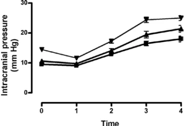
he manual vibrocompression maneuver did not lead to ICP increase in any of the days assessed. On the con-trary, at day 3 a signiicant drop of ICP after the vibro-compression maneuver was observed when compared to the basal measurement (14.5 ± 1.0 mm Hg vs 11.6 ± 1.6 mm Hg, p<0.05) (Figure 1).
24.9 ± 2.7 mm Hg vs 14.4 ± 1.0; p<0.05 for all). At the end of the physiotherapeutic procedure the ICP was above 20 mm Hg in 9 of the 11 patients, with a median of 27 mm Hg (minimum, 22 mm Hg; maximum, 47 mm Hg). Nevertheless a rapid return to the basal levels was observed (median, 27 seconds; minimum, 10 seconds; maximum, 180 seconds). he MAP rose signiicantly after the ICA maneuver (T2 and T3) on day 1 and, after ITA with tra-cheal saline infusion (T3) at every day (Figure 2). MAP at the end of the procedure (T4) was higher that the basal every day (day 1, 110 ± 5 mm Hg vs 99 ± 3 mm Hg; day 2, 104 ± 6 mm Hg vs 93 ± 3 mm Hg; day 3, 113 ± 4 mm Hg vs 97 ± 2 mm Hg vs; p<0.05 for all) (Figure 2).
CPP did not change on day 1 and 2 but was signii-cantly higher at the end of the procedure (89 ± 3 mm Hg) in comparison to the initial measurement (82 ± 3 mm Hg) (p<0,05) (Figure 3).
Figure 2 – Mean arterial pressure measurements at diferent ti-mes. At baseline (T0), at vibrocompression ((T1), after intratra-cheal aspiration (ITA) without traintratra-cheal saline infusion (T2), after ICA with tracheal saline infusion (T3),and endpoint (T4), on days 1(), 2 () e 3 (). P<0.05 for T4 vs T0, T3 vs T0, T2 vs T0 on days 1 and 3; T4 vs T0ande T3 vs T0 on day 2
Figure 3 Cerebral perfusion pressure measurements at dife-rent times baseline (T0), after vibrocompression (T1), after ITA without tracheal saline infusion (T2), after ITA with tra-cheal saline infusion (T3), and endpoint (T4), on days 1(), 2 () e 3 (). *: P<0.05 for T4 vs T0 on day 3.
Figure 1- ICP measurements at diferent times. Baseline mo-ment (T0) after vibrocompression (T1), after intratracheal as-piration (ITA) without tracheal saline infusion (T2), after TIA with tracheal saline infusion (T3), and endpoint (T4) on days 1(), 2 () e 3 (). P<0.05 forT1 vs T0 on day 1; T4 vs T0, T3 vs T0 and T2 vs T0 on days 1, 2 and 3.
Table 1- Demographic, imaging and severity data
Patients Gender Age Weight GCS APACHE II Cerebral tomography
1 M 28 70 7 17 EDH SAH
2 M 42 65 7 12 Frontal Contusion
3 M 58 75 3 21 SDH, SAH, Temporal contusion
4 M 76 80 6 27 SDH temporal contusion
5 M 31 70 6 16 EDH, SAH
6 M 41 85 6 27 Temporal and mastoid bone fracture
7 M 19 80 3 16 SDH
8 M 57 130 4 21 SDH, DAÍ
9 M 27 75 6 16 EDH, SAH, DAI, pneumoencephalus, fracture of the cranial base
10 M 22 70 6 17 SDH DAI, temporal fracture
11 F 51 65 5 25 SDH
DISCUSSION
It has been reported that respiratory physiothera-peutic interventions commonly performed in critical-ly ill patients may influence cerebral oxygen transport due to its adverse effects on cardiac output (CO). (12,13)
Theoretically, respiratory physiotherapy measures ap-plied on the chest, increase intrathoracic pressure (ITP) with drop of cerebral venous return and im-plies increase of ICP.(3,10)
Results of this study disclosed that the manual vibrocompression maneuver did not determine an increase of ICP or CPP on any of the assessed days. On the contrary, on the third day of intervention a significant drop of ICP followed the maneuver. How-ever, the ICT maneuver, especially with tracheal saline infusion caused a significant increase of ICP. Yet, this increase was transitory and followed by compensatory increase of CPP and none of the patients presented with ICH.
Our findings differ from those of other authors on some points and must portray differences in the meth-odology used or in the population studied. Thiesen et al. reported increased ICP after the ICA maneuver in patients with severe BI, albeit with no increase of MAP and maintenance of CPP at adequate levels. (10)
While Nemer et al. reported that unilateral chest com-pression as well as ITA may significantly change the ICP in patients with BI and stroke.(11) In that study
there were no reports of CPP nor about the time when ICP remained elevated, hindering comparison with our study.
In general, findings of these studies suggest that ITA causes small and temporary increases of ICP; if MAP increases in response to the maneuver, the CPP also increases. (9) It is desirable to maintain CPP levels
above 70 mm Hg to assure adequate oxygen transport to the brain.(7) Increases of MAP, of jugular venous
oxygen tension, velocity of the middle cerebral artery flow are described, which together suggest a protec-tive compensatory response, maintaining the cerebral oxygen delivery during and after ITA.(12,13) Increase
of arterial pressure apparently is a compensatory re-sponse to the procedure-induced hypoxia.(10) Other
authors suggest that the ITA may increase ICP as a response to the cough reflex and to hypercapnia with consequent cerebral vasodilatation.(11,14) Contrariwise,
a study that assessed the effects of using positive end expiratory pressure (PEEP) on ICP reported decreases of MAP related to lower levels of central venous
pres-sure (CVP) suggesting that in hypovolemic patients, increase of pressure on the chest may have a signifi-cant hemodynamic effect.(3)
The reason for the decline of ICP after the manual vibrocompression maneuver, perceived on the third day, remains unknown. It may possibly be the out-come of amelioration of pulmonary ventilation and drop of PaCO2 caused by the intervention. (9) We know
that elevation of ICP may be attenuated by small peri-ods of hyperventilation, prior to aspiration.(12,15,16) This
mechanism must be better assessed in future studies.
CONCLUSIONS
he manual vibrocompression maneuver did not in-duce increase ICP or CPP in patients with severe BI. How-ever, ITA led to a signiicant and transient ICP increase, accompanied by parallel increase of CPP. Such maneu-vers are safe in BI patients, as long as they are adequately performed and under surveillance. Other studies with a higher number of patients are required to corroborate our results.
RESUMO
Objetivos: Após um traumatismo cranioencefálico, a
hiper-tensão intracraniana representa a maior causa de mortalidade, além da possibilidade de seqüelas funcionais, comportamentais e cognitivas. A escassez de estudos sobre os efeitos da isioterapia respiratória nestes pacientes pode levar à condutas contraditó-rias. O objetivo deste estudo foi avaliar os efeitos de manobras usuais de isioterapia respiratória sobre a pressão intracraniana e a pressão de perfusão cerebral em pacientes com traumatismo cranioencefálico grave.
Métodos: Ensaio clínico, prospectivo, em pacientes com
traumatismo cranioencefálico, ventilados mecanicamente e com medida contínua da pressão intracraniana. Foram avaliados os efeitos das manobras de vibrocompressão manual e aspiração intratraqueal sem e com instilação de soro isiológico, sobre as medidas de pressão intracraniana e de pressão de perfusão cere-bral, entre o primeiro e o terceiro dia após a lesão cerebral.
Resultados: Foram obtidos os dados de 11 pacientes com
Conclusões: A manobra de vibrocompressão manual não de-terminou aumento da pressão intracraniana ou da pressão de perfu-são cerebral em pacientes com traumatismo cranioencefálico grave. A aspiração intratraqueal levou a aumento signiicativo e transitório da pressão intracraniana e da pressão de perfusão cerebral.
Descritores: Modalidades de isioterapia/métodos; Hiper-tensão intracraniana/terapia; Pressão intracraniana; Terapia res-piratória; Intubação intratraqueal; Traumatismos encefálicos
REFERENCES
1. Perel P, Edwards P, Wentz R, Roberts I. Systematic review of prognostic models in traumatic brain injury. BMC Med Inform Decis Mak. 2006;6:38. Review.
2. Zanier ER, Ortolano F, Ghisoni L, Colombo A, Losappio S, Stocchetti N. Intracranial pressure monitoring in inten-sive care: clinical advantages of a computerized system over manual recording. Crit Care. 2007;11(1):R7. Comment in: Crit Care. 2007;11(1):117.
3. Georgiadis D, Schwarz S, Baumgartner RW, Veltkamp R, Schwab S. Inluence of positive end-expiratory pressure on intracranial pressure and cerebral perfusion pressure in pa-tients with acute stroke. Stroke. 2001;32(9):2088-92. 4. Maldaun MVC, Zambelli HJL, Dantas VP, Fabiani RM,
Martins AM, Brandão MB, et al. Análise de 52 pacientes
com traumatismo de crânios atendidos em UTI pediátrica: considerações sobre o uso da monitorização da pressão
in-tracraniana. Arq Neuropsiquiatr.2002;60(4):967-70.
5. Helmy A, Vizcaychipi M, Gupta AK. Traumatic brain injury: intensive care management. Br J Anaesth. 2007;99(1):32-42. Review.
6. Enrione MA. Current concepts in the acute management of severe pediatric head trauma. Clin Pediatr Emerg Med. 2001;2:28-40.
7. Guidelines for the management of severe traumatic brain injury. J Neurotrauma. 2007; 24 Suppl 1:S1-106. 8. Jantzen JP. Prevention and treatment of intracranial
hyper-tension. Best Pract Res Clin Anaesthesiol. 2007;21(4):517-38.
9. Stiller K. Physiotherapy in intensive care: towards an
evi-dence-based practice. Chest. 2000;118(6):1801-13. 10. hiesen RA, Dragosavac D, Roquejani AC, Falcão ALE,
Araújo S, Dantas Filho VP, et al. Inluência da
isiotera-pia respiratória na pressão intracraniana em pacientes com traumatismo cranioencefálico grave. Arq Neuropsiquiatr. 2005;63(1):110-3.
11. Nemer SN, Machado ST, Caldeira JB, Azeredo LM, Clipes T, Gago R, et al. Efeitos da isioterapia respiratória e da mobilização passiva sobre a pressão intracraniana. Fisioter Bras. 2005;6(6):437-43.
12. Kerr ME, Weber BB, Sereika SM, Darby J, Marion DW, Orndof PA. Efect of endotracheal suctioning on cerebral
oxygenation in traumatic brain-injured patients.Crit Care
Med. 1999; 27(12):2776-81. Comment in: Crit Care Med. 1999;27(12):2843-4.
13. Gemma M, Tommasino C, Cerri M, Giannotti A, Piazzi B, Borghi T. Intracranial efects of endotracheal suctioning in the acute phase of head injury. J Neurosurg Anesthesiol. 2002;14(1):50-4.
14. Celik SA, Kanan N. A current conlict: use of isotonic so-dium chloride solution on endotracheal suctioning in cri-tically ill patients. Dimens Crit Care Nurs. 2006;25(1):11-4.
15. Nekludov M, Bellander BM, Mure M. Oxygenation and cerebral perfusion pressure improved in the prone posi-tion. Acta Anaesthesiol Scand. 2006;50(8):932-6. Com-ment in: Acta Anaesthesiol Scand. 2007;51(4):514-5. 16. Mascia L, Grasso S, Fiore T, Bruno F, Berardino M, Ducati