*Correspondence: Trishna Bal. Department of Pharmaceutical Sciences, Birla Institute of Technology, 835215-Mesra-Ranchi-Jharkhand, India. E-mail: [email protected]
A
vol. 49, n. 4, oct./dec., 2013
Formulation and evaluation of carvedilol microcapsules using
Eudragit NE30D and sodium alginate
Trishna Bal
1,*, Shubhranshu Sengupta
2, Padala Narasimha Murthy
31Department of Pharmaceutical Sciences, Birla Institute of Technology, Jharkhand, India, 2Department of Horticulture, Birsa Agriculture University, Jharkhand, India, 3Royal College of Pharmacy and Health Sciences, Orissa, India
Inclusion complexes of carvedilol(CR) with hydroxyl propyl beta-cyclodextrin (HPBCD) was prepared using co-grinding technique. Then, the inclusion complex was microencapsulated using combinations of
Eudragit NE30D (EU) and sodium alginate (SA) utilizing oriice gelation technique. The formulations
were analysed by using Scanning electron microscopy (SEM), Fourier Transform Infrared spectroscopy (FTIR), Differential scanning Calorimetry (DSC) and X-ray diffractometer (XRD) and also evaluated for
particle size, encapsulation eficiency, production yield, swelling capacity, mucoadhesive properties, zeta
potential and drug release. The microcapsules were smooth and showed no visible cracks and extended drug release of 55.2006% up to 12 hours in phosphate buffer of pH 6.8, showing particle size within the
range of 264.5-358.5 µm, and encapsulation eficiency of 99.337±0.0100-66.2753±0.0014%.The in vitro release data of optimized batch of microcapsules were plotted in various kinetic equations to understand
the mechanisms and kinetics of drug release, which followed irst order kinetics, value of “n” is calculated to be 0.459 and drug release was diffusion controlled. The mice were fed with diet for inducing high
blood pressure and the in vivo antihypertensive activity of formulations was carried out administering the optimized formulations and pure drug separately by oral feeding and measured by B.P Monwin IITC Life Science instrument and the results indicated that the bioavailability of carvedilol was increased both in vitro and in vivo with the mucoadhesive polymers showing primary role in retarding the drug release.
Uniterms: Carvedilol/microcapsules/evaluation. Carvedilol/microcapsules/formulation. Eudragit
NE30D. Oriice Gelation technique.
Prepararam-se complexos de carvedilol (CR) com hidroxipropil beta-ciclodextrina (HPBCD), utilizando a técnica de co-moagem. O complexo de inclusão foi microencapsulado empregando-se associações
de Eudragit NE30D (EU) e alginato de sódio (AS), utilizando a técnica de geliicação de orifício. As
formulações foram analisadas utilizando-se microscopia eletrônica de varredura (SEM), espectroscopia no infravermelho com Transformada de Fourier, calorimetria diferencial de varredura (DSC) e difratometria de
raios X (XDR) e, também, avaliadas por tamanho de partícula, eiciência de encapsulação, rendimento de
produção, capacidade de inchamento, propriedades mucoadesivas, potencial zeta e liberação do fármaco.
Obtiveram-se microcápsulas lisas e sem fendas visíveis, com liberação prolongada do fármaco de 55,2006% em 12 horas em tampão fosfato pH 6,8, com tamanho de partículas na faixa de 264,5-358,5 mm e eiciência de encapsulação de 99,3337±0,0100-66,2753±0,0014%. Os dados de liberação in vitro de lote otimizado de microcápsulas foram plotados em várias equações cinéticas para se entender os mecanismos e a cinética
de liberação do fármaco, que é de primeira ordem, o valor de “n” foi de 0,459 e a liberação do fármaco foi por difusão controlada. Os camundongos foram alimentados com dieta para induzir pressão sanguínea
alta e a atividade anti-hipertensiva in vivo das formulações foi obtida por administração de formulações otimizadas e fármaco puro, separadamente, por via oral e medida pelo equipamento BP Monwin IITC Life Science. Os resultados mostraram que a biodisponibilidade do carvedilol aumentou tanto in vitro quanto in vivo com os polímeros mucoadesivos, mostrando papel principal no retardamento da liberação do fármaco.
Unitermos: Carvedilol/microcápsulas/avaliação. Carvedilol/microcápsulas/formulação. Eudragit NE30D.
INTRODUCTION
The most desirable and convenient method of drug administration is the oral route (Patil et al., 2009).
Carvedilol (CR) is a nonselective β-adrenergic blocking agent with α1-blocking activity. It is well absorbed from the gastrointestinal tract but subjected to considerable
irst-pass metabolism in the liver (Tanwar, Chauhan, Sharma,
2009)and its oral bioavailability in humans is only 20%
(Guarve et al., 2009). The drug has a short half-life of 2.2
± 0.3 h. The drug is formulated with a series of polymers
to improve the absorption and prolong the half life, thereby preventing degradation in gastric region. Since the drug has low bioavailability due to poor water solubility and slow dissolution rates (Hirlekar, Kadam, 2009)several methods are used to improve the solubility profile of CR, of which complexation with cyclodextrins has been widely used to improve the solubility and dissolution rate of poorly soluble drugs (Wang et al., 2006; Archontaki
et al., 2002).Cyclodextrins (CDs) are macrocyclic oligosaccharides with six to eight D-glucose units called
α-Cyclodextrin, β-Cyclodextrin and γ-Cyclodextrin.
The most important property of CDs is that they have hydrophobic central cavities capable of forming stable complexes with properly sized drug molecule (Vyas, Saraf, Saraf2008; Shewale et al., 2008; Bhutani et al., 2007).
Among the various CDs, hydroxypropyl beta-cyclodextrin (HPBCD) is most successful in improving the dissolution rate (Pitha et al., 1986)of poorly soluble drugs.
Polymeric microparticles are recommended for oral, nasal or pulmonary administration. These systems are able to promote extended release of a bioactive compound and also can protect the drug from degradation
and physiological metabolism (Salsa, Veiga, Pina1997).
In order to develop microcapsules, several polymers both from natural and synthetic sources can be used (Deasy
1984).
The present work is mainly focussed on improving the solubility characteristics of CR by use of CDs, mainly HPBCD by formation of binary systems of CR/ HPBCD in a 1:1 ratio (w/w ratio) and then formulating this
binary system in the form of microcapsules using Oriice Gelation technique, by the use of some mucoadhesive
hydrophilic polymers like Sodium alginate and an aqueous dispersion of Eudragit NE30D for oral use.The optimized microcapsules were then evaluated in vitro to determine the drug release and in vivo to determine the eficacy of
the formulations.
MATERIAL AND METHODS
Material
Carvedilol(CR) was kindly gifted by Glenmark
Pharmaceuticals (Mumbai,India), hydroxylpropyl
beta-cyclodextrin (HPBCD) was kindly gifted by Roquette (Lestrem, France), sodium alginate (SA) was purchased from Loba Chemicals(India); Eudragit NE30D (EU) was a gift sample from Evonik Degussa Rohm Pharma (Mumbai, India). All other chemicals used were of analytical reagent grade purity.
Preparation of Microcapsules
The method used for the preparation of microcapsules
adopted was Oriice Gelation technique (Chowdary. Rao,
2003). Binary system of HPBCD/CR was prepared by
co-grinding technique (Hirlakar, Kadam 2009) in which
mixing of CR in a ratio of 1:1(w/w ratio) with HPBCD in a glass mortar for 30 minutes was done , and stored in a desiccator. Then this binary system was mixed with SA and EU separately for formulating different batches of microcapsules as stated in Table I. The physical mixtures (PM) (of the CR along with HPBCD, SA and Eudragit NE30D) were analyzed separately for any possible
drug-TABLE I - Composition of formulations
Formulation Code
Drug (carvedilol): hydroxypropyl beta-cyclodextrin (HPBCD)
(mg)
sodium alginate(SA) (mg)
Eudragit NE30D(EU) (mg)
CRSAEU1 1:1 600 200
CRSAEU2 1:1 600 400
CRSAEU3 1:1 600 600
CRSAEU4 1:1 200 600
CRSAEU5 1:1 600 1050
---excipient interactions. The binary system of HPBCD/ CR was triturated with SA and then mixed with Eudragit NE30D in a glass mortar pestle and dispersed this mixture
in milipore water (obtained from Elix milipore water ilter)
using magnetic stirrer for 30 min at 200 rpm and allowed
to form uniform slurry named as “CRV”. A 3%w/v calcium chloride solution was prepared and iltered separately and to this solution, the slurry “CRV” was added dropwise
through a 10 mL syringe (Dispovan) having needle of
size no.26G .The microcapsules formed were allowed
to remain in the calcium chloride solution for 30 min to complete the curing reaction. The formed microcapsules
were iltered and washed with millipore water to remove
any traces of calcium chloride from the microcapsule surfaces and dried the microcapsules in open air and kept in a desiccator.
Evaluation of microcapsules
Determination of Yield of Production
The production yields (Ranjha, Khan, Nazeem, 2010)of microspheres of various batches were calculated
using the weight of inally dried microspheres with respect
to the initial total quantity of the drug and polymer used for preparation. Percent production yields were calculated as per the formula below:
Determination percentage encapsulation efficiency
Percentage encapsulation eficiency is the percentage
of drug encapsulated in the microcapsules related to the initial quantity of the drug used in the formulation. 100 mg of microcapsules were taken and crushed in a glass
mortar-pestle. In a 100 mL volumetric lask, the grounded
microcapsule powder was mixed with methanol to make up the volume up to 100 mL and placed the whole system in a sonicator for 30 min to get the maximum extraction
of CR in the solvent. The sample so obtained were iltered
to obtain clear solution and assayed for the drug content spectrophotometrically at 242 nm. Percent encapsulation
eficiency (Ranjha, Khan, Nazeem, 2010)was determined by using the formula below.
• Particle Size
The Particle size of the dried microcapsules was measured using a stage micrometer scale by optical microscopy method. This study was done in triplicate. 100 nos. of dry microcapsules were placed on a clean glass
slide and a few drops of liquid parafin was added and
covered with a glass slide and observed under a compound microscope using stage and ocular micrometer (Dhaliwal
et al., 2008; Patil et al., 2009).
• Determination of bulk density
The bulk density of the formulations was determined by using the following formula (Ranjha, Khan, Nazeem, 2010). This study was done in triplicate.
• Determination of Tapped density
Tapped Density is used to investigate packing properties of microcapsules into capsules. The Tapped density was measured by employing the conventional tapping method using a 10ml measuring cylinder and the
number of tapings was 100 as suficient to bring a plateau
condition. This study was done in triplicate. Tapped density was calculated by using the following formula (Ranjha, Khan, Nazeem, 2010):
• Determination of Carr’s consolidation Index It is indirect measurement of bulk density, size and shape, surface area, moisture content and cohesiveness of
materials since all of them can inluence the consolidation
index. It is also called as Compressibility index (Ci). This study was done in triplicate. It is denoted by Ci and
is calculated using the formula below (Ranjha, Khan, Nazeem, 2010):
A Carr’s index less than 15% is referred to as very
good flow,16-26% is good, 27-35% is fairly good and
℘35% are considered as poor (Carr, 1965).
• Determination of Hausner’s ratio
It is another parameter for measuring lowability of
the microcapsules. This study was done in triplicate. It is calculated using the following formula (Ranjha, Khan, Nazeem, 2010):
• Determination of Angle of repose
Angle of repose (θ=tan-1 h/r) of the microcapsules
the glass funnel on a horizontal surface (Ranjha, Khan, Nazeem, 2010). This study was done in triplicate .The height (h) of the heap formed was measured and the radius (r) of the cone base was also observed and calculated.
• Percentage of swelling of Microcapsules
Swelling rate of the microcapsules was measured as a function of water uptake. The formulations were placed in phosphate buffer of pH 6.8 at room temperature for a time period of 10hours. At different time intervals, the microcapsules were taken out and very gently pressed with a tissue paper to remove the excess liquid and then weighed. These studies were done in triplicate. The percentage swelling of the microcapsules was determined by using the following formula as below (Patil Sanjay, Sawant Krutika, 2009):
• Mucoadhesion properties of the microcapsule Bioadhesive strength of the microcapsules was measured on a modified physical balance using the
method described by Gupta, Garga and Khar(1992). Carbopol 934P which was taken as standard to compare
the mucoadhesivity of microcapsules. The microcapsules were sandwiched between two mucosal surfaces of rat intestine. The intestine were placed on two oppositely placed platforms of two slides ,one hanged to the left pan of balance and other placed on a water bath at the base. Then weights were placed to the right pan of the balance in ascending order starting from lower weights and after each addition of weights, allowing to stand for 1min, and then added the next weight and this process was continued till the two mucosal surfaces detached from one another on the left side of pan and thus the detachment force required to separate two glass slides was measured. This study was done in triplicate.
In vitro Drug release of microcapsules
900 mL of phosphate buffer of pH6.8 (Hirlekar,
Kadam, 2009) was taken as dissolution medium for in vitro drug release in USP Type-I dissolution apparatus. 100mg of the microcapsules were taken and filled in a hard gelatin capsule and placed in the basket and started
the dissolution at 75 rpm and continued the study for a
period of 13 hours. 5ml of sample was withdrawn after
every 0.5,1,2,3,4,5,6,7,8,9,10,11,12,13 hours and analyzed
spectrophotometrically at 241 nm and calculated the cumulative drug release and calculated the drug release
kinetics. This study was done in triplicate. The statistical ANOVA analysis of the in vitro release studies is also plotted.
Data obtained from in vitro release studies were itted
to various kinetic equations to ind out the mechanisms of
the drug release. The kinetic models used were Zero order
equation, irst order Equation, Higuchi equation, Hixson
Crowell Equation, Peppas-Korsmeyer Equation.
Phyisicochemical evaluations of microcapsules
• FTIR spectroscopy (Ranjha, Khan, Nazeem, 2010) Binary System (HPBCD/ CR)–Polymers interactions were studied by FTIR spectroscopy (FTIT Shimadzu 8400S). Also the spectra for pure drug and drug loaded microcapsules were recorded. Samples were prepared in KBr disks (2 mg sample in 200 mg of KBr).The scanning was 400-4000 cm-1 and the resolution was 2 cm-1.
• X-ray powder diffractometry
This technique was carried out to investigate the effect of polymers and complexing agent HPBCD on the characteristics of the drug after formulation. Powdered samples of pure drug, polymers, HPBCD and microcapsules were irradiated with monochromatized
X-rays (Cu-kα) of 30kV and 15mA current in a Rigaku analytical XRD (Model Minilex, Japan).The scanning
rate employed was 0.20 min-1 of 2θ.The X-ray powder
diffractometry (X-RD) patterns of the dug and drug loaded microcapsules were recorded (Ranjha, Khan, Nazeem, 2010).
• DCS studies of the microcapsule (Ranjha, Khan, Nazeem, 2010;
5 mg weight of samples(Pure drug, Polymers, optimized microcapsule) were taken to carry out tests in DSC using Aluminium sample pans at a scanning speed of 10 oC per min form 10 oC-200 oC to detect any interaction
between drug and polymers.
Morphological studies of microcapsules
The optimized batch of microcapsules used for determination of surface morphology were coated by gold sputtering technique and observed using the Scanning electron microscope (Ranjha, Khan, Nazeem, 2010)
(Model Jeol Japan; JSM-6390LV).
Determination of zeta potential of microcapsules
Krutika, 2009). Also the zeta potential of the individual
polymers and the drug were measured for a comparative study.
In vivo animal studies
• Animals
Male swiss albino mice weighing from 100-120 g were housed in a temperature and light controlled room
(23±2 oC; 12 hours light/dark cycle), with free access to
water and food. All the procedures are in agreement to the Institutional animal ethical committee, BIT, Mesra, Ranchi (vide letter No.CPCSEA approval no: 621/02/ac/
CPCSEA).
Experimental protocol
Eighteen male mice of eight weeks old were randomly selected and divided in three groups of 6
animals each of weight of 20-50 g. Group 1 received pure carvedilol (CR); Group 2 received optimized formulation
CRSAEU5 {at 10 mg/kg (Schaefer et al., 1998)}; Group 3
was taken as the control (without any drug or formulations, only provided with water and normal food). For all the three groups initially, before any treatment, the mice were acclamatized for 3 days in the warming chamber of the
B.P Monwin IITC Life Science instrument. On the 4rth day, blood pressure was measured by noninvasive tail cuff method using B.P Monwin IITC Inc. Life Science
instrument. Then for one week the animals of Group1
&2 were given diet as given in Table II for inducing Hypertension (Qianli et al., 2004). After one week, the B.P was again measured for all the three groups to observe the
increase in B.P and after conirming that there is induction
of hypertension, treatment was started with pure drug CR and optimized microcapsules batch CRSAEU5 in two different groups respectively.
Statistical evaluations for In-vivo studies of optimized carvedilol microcapsules in mice
The data of statistical ANOVA one way analysis was
calculated in Graph Pad software in mice for optimized
formulations of carvedilol CRSAEU5 is presented in
Figure 7 and Figure 8. The IITC life sciences Blood
pressure (B.P) measuring instrument for mice which was
utilized during the experiment is presented in Figure 9.
RESULTS AND DISCUSSION
Effect of technique on the formation of microcapsules
Mucoadhesive carvedilol microcapsules prepared
by oriice Gelation technique using a mixure of natural
polymers and aqueous dispersion of ethylcellulose i.e.
Eudragit NE30D were found to be free lowing and almost
spherical in shape. The preparation method used was advantageous for entrapment of water-insoluble drugs. Moreover by formation of binary system of HPBCD/ CR, solubility parameter of drug CR was increased. The production of microcapsules varied with different ratios of polymers. The results are shown in Table III. The high yield of the microcapsules may be due to the entire mass
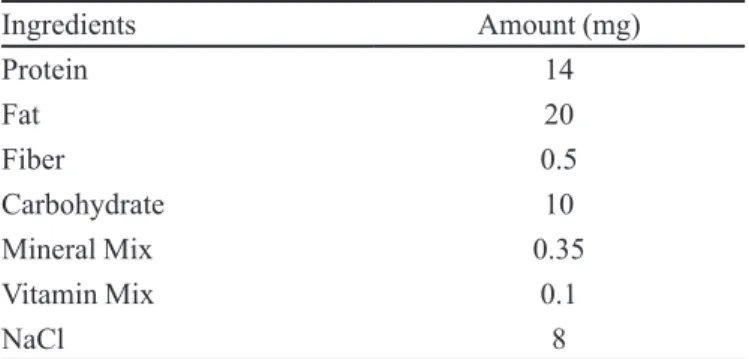
TABLE II - Diet Composition for mice for inducing Hypertension (Qianli et al., 2004)
Ingredients Amount (mg)
Protein 14
Fat 20
Fiber 0.5
Carbohydrate 10
Mineral Mix 0.35
Vitamin Mix 0.1
NaCl 8
TABLE III - Micromeritic properties along with encapsulation eficiency, %yield and assessment of mucoadhesivity of formulations
Sl. No. Formulation Code
% Encapsulation
eficiency (%E.E)
% Yield Particle Size (µm)
Mucoadhesivity [Detachment
Strength (N/Cm2)]
Assessment of Duration of Mucoadhesion
in (min)
% C.I Hausner’s Ratio
Angle of Repose 1 CRSAEU1 66.2753±0.0014 91.1529 317.5±15.08 0.021909 1.4±0.100 9.1±0.152 1.091±0.009 13.2±0.4612 2 CRSAEU2 78.759±0.001 88.6315 335.0±11.527 0.021582 1.5±0.05773 9.1±0.152 1.091±0.009 12.19±0.5411 3 CRSAEU3 82.598±0.0005 81.6 358.5±11.527 0.021909 1.4±0.100 15.96±0.647 1.16±0.0055 12.103±0.3874 4 CRSAEU4 98.8248±0.0012 86.206 291.5±12.527 0.022236 1.5±0.100 25±0.7637 1.25±0.009 15.13±0.39004 5 CRSAEU5 99.337±0.0100 91.636 264.5±7.527 0.023217 1.6±0.100 8.3±0.1301 1.083±0.0012 11.24±0.4652 6 CRHPSAI 65.84±0.080 85.71 358.5±11.527 0.022236 1.33±0.3055 14.3±0.7419 1.14±0.0100 12.0115±0.245
---of the polymer available for gelation by the crosslinking agent. These studies were done in triplicate.
As the drug is water insoluble, so most of the drug got entrapped in the polymer matrix resulting higher drug content and high percentage encapsulation efficiency. Moreover as the amount of both the polymers increased,
the entrapment eficiency also increased.
Micromeritic properties
As observed in Table III, the particle size of the different batches of microcapsules ranged from 264.5-358.5 µm as determined by optical microscopy method. The formulation CRSAEU5 showed the least particle size
of 264.5 µm±1.527 among the other formulations, showing
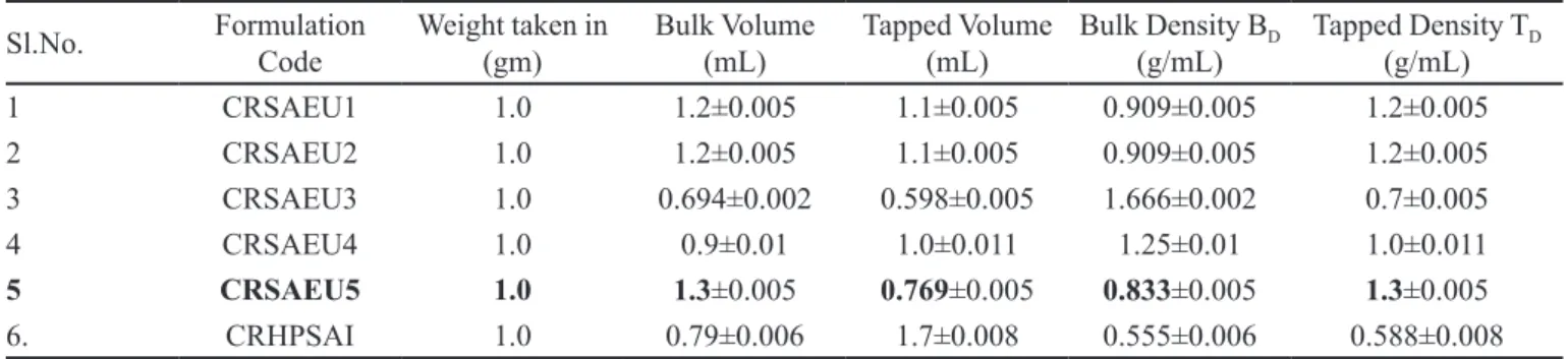
that is a better formulation. Moreover as seen from Table III, CRSAEU5 is the superior among all the other formulations as the hausner’s ratio, % compressibility index and angle of repose is smallest among all the other formulations (Ranjha, Khan, Nazeem, 2010). The bulk and tapped density data are given in Table IV.
Swelling studies
The results of swelling studies of all the formulations as compared to CRHPSAI as shown in Figure 1, indicates that with the increase in polymer concentration, the water
absorption capacity of the formulations also increase. These studies were done in triplicate. These studies are in
conirmation with earlier studies (Xiudong et al., 2004). The statistical studies of the optimized formulation CRSAEU5 is shown in Figure 2.
Mucoadhesion studies
As observed from the Table III, CRSAEU5 shows the highest mucoadhesive property. With the increase in both polymer concentrations, there is an increase in mucoadhesion. Also this is supported by results of assessment of duration of mucoadhesion as cited in Table III.As shown in Figure 3, microcapsules adhesioned to the rat intestine of 3 cm2 area, where length is 3cm and
breadth is 1 cm.
In vitro release studies
The in-vitro release studies were carried out in pH 6.8 phosphate buffer. It was observed that at alkaline pH, greater stress was caused to the polymers thereby causing release of drugs. The microcapsules swelled excessively followed by erosion in the buffered alkaline medium. Eudragit NE30D shows the gelling property in alkaline medium which is considered beneficial for sustaining drug release from the microcapsules. The microcapsules
TABLE IV - Bulk and tapped density of formulations (Mean ±S.D)
Sl.No. Formulation Code
Weight taken in (gm)
Bulk Volume (mL)
Tapped Volume (mL)
Bulk Density BD (g/mL)
Tapped Density TD (g/mL)
1 CRSAEU1 1.0 1.2±0.005 1.1±0.005 0.909±0.005 1.2±0.005
2 CRSAEU2 1.0 1.2±0.005 1.1±0.005 0.909±0.005 1.2±0.005
3 CRSAEU3 1.0 0.694±0.002 0.598±0.005 1.666±0.002 0.7±0.005
4 CRSAEU4 1.0 0.9±0.01 1.0±0.011 1.25±0.01 1.0±0.011
5 CRSAEU5 1.0 1.3±0.005 0.769±0.005 0.833±0.005 1.3±0.005
6. CRHPSAI 1.0 0.79±0.006 1.7±0.008 0.555±0.006 0.588±0.008
FIGURE 1 - Comparative % swelling studies of all formulations.
FIGURE 2 - % swelling studies of formulation CRSAEU5
TABLE V - Release kinetics of carvedilol from microcapsules
Sl. No. Formulation Code
Zero order Kinetics
First order Kinetics
Higuchi Kinetics
Hixson Crowell Kinetics
Korsmeyer-Peppas Kinetics
R2 k
0 R
2 k
1 R
2 k
H R
2 k
HC R
2 n
1 CRSAEU1 0.964 7.131 0.825 -0.153 0.980 28.69 0.945 -0.278 0.968 0.423
2 CRSAEU2 0.951 6.654 0.763 -0.138 0.980 26.94 0.934 -0.253 0.966 0.443
3 CRSAEU3 0.948 6.678 0.827 -0.135 0.976 27.02 0.926 -0.256 0.968 0.446
4 CRSAEU4 0.967 6.233 0.861 -0.079 0.943 24.56 0.931 -0.188 0.883 0.436
5 CRSAEU5 0.963 3.520 0.967 -0.022 0.966 14.06 0.963 -0.071 0.945 0.459
6 CRHPSAI 0.948 14.01 0.938 -0.250 0.984 40.71 0.991 -0.501 0.993 0.654
showed sustained release for a period of 13 hours due to the hindered diffusion of the drug from the gel matrix of Sodium alginate and Eudragit NE30D formed in situ
(Sharma, Sarangi, Pradhan2009) and thereby causing
retarded drug release. The release of drugs was retarded with the increase in the polymer ratio. From the Figure 3, it is clearly visible that formulation CRSAEU5 has the highest property of retarding the drug release. The results were compared with Formulation CRHPSAI (devoid of Eudragit NE30D) and it is seen that drug release from
CRHPSAI is 98% within 7 hours. Thus Eudragit NE30D
is highly effective in retarding the drug release. This study was done in triplicate.
Kinetics of drug release
Inorder to understand the mechanism and kinetics of invitro drug release, the data were analyzed with various kinetic equations like Zero order(% Cumulative drug release Vs time in hours), First order plot(log
FIGURE 3 – Comparative Drug Release Proile of microcapsules at pH 6.8; CRSAEU4(illed (Diamond,Ser.1),CRSAEU1(Filled S q u a re , S e r. 2) , C R S A E U 5 (F i l l e d t r i a n g l e , S e r i e s 3) , C R S A E U 2 (C r o s s , S e r. 4) , C R S A E U 3 (S t a r, S e r. 5) , CRHPSAI(Filled Circle,Ser.6), Mean % cumulative drug
release±S.D, n=3
of Cumulative % drug unreleased Vs Time), Higuchi model(% Cumulative Drug released Vs Square root of time) and Korsmeyer-Peppas Plot (log% Cumulative
drug released Vs log of time). Coeficient of Correlation
values were calculated for the linear curves obtained by regression analysis of the above plots.
As observed from Figure 3, it is clear that formulation CRSAEU5 retards the drug release for a longer period of time than in comparison to other formulations, thereby controlling the drug release. From Table V, it is clear that, CRSAEU5 follows first order release mechanism and
value of “n” is 0.459 which indicates that the mechanism
of the drug to diffusion controlled (Sharma, Sarangi,
Pradhan 2009; Mukherjee et al., 2005).
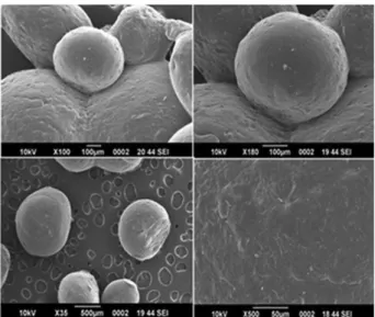
Surface topography
After considering all the parameters of evaluation
formulation CRSAEU5 is considered as most eficient.
Surface morphologies of the formulation CRSAEU5 as
conirmed by SEM studies shows that there are no cracks
or crevices present on the surface as shown in Figure 4.
Zeta potential measurement
The Zeta potential value of CRSAEU5 as seen from Table VI is found to be more towards the positive side than in comparison to the pure drug. The increase in zeta potential may be due to the contribution of HPBCD and Eudragit NE30D.This increase in zeta potential of the formulation also contributes to the increase in mucoadhesive property of the formulation as seen in
Table III. This result is in conirmation with earlier studies
(Dhawan, Singla, Sinha , 2004).
FTIR analysis
As observed in Figure 5, FTIR studies indicated weak interactions of carvedilol with HPBCD at 1:1 ratio prepared in the form of microcapsules with different
polymers as shown in Figure 7. In carvedilol spectra, absorption peaks were observed at 3349.7701 cm-1, 2933.55621 cm-1 due to hydroxyl and amine stretching respectively. Other peaks were at 1233.5979 cm-1 due to C-O group (epoxides),1094.86001 cm-1 due
to aryl alkyl ethers and alkyl ether C-O stretching respectively. Whereas in the PM, there is an exhibition
at 3339.7409 cm-1, 2913.4977 cm-1 due to hydroxyl
and amine stretching respectively which matches with
pure drug. Also there is an exhibition at 1273.715 cm-1, 1064.77225 cm-1 which matches with that of pure drug
showing still the presence of aryl alkyl ethers and alkyl ether C-O stretching respectively. In case of Formulation CRSAEU5, there is appearance of broad wave number
of 3261.17844 cm-1 indicating the presence of hydroxyl
stretching, and there is very less intensity of presence of
1064.77225 cm-1 indicating the entrapment of carbazol
moiety into host cavity during inclusion complex. The results are in confirmation with Hirlekar and Kadam
(2009).
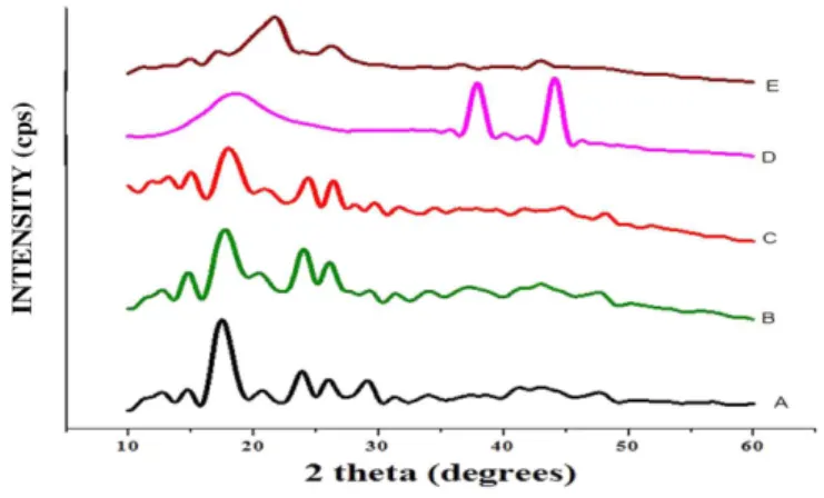
XRD analysis
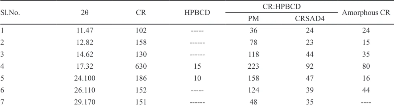
The XRD patterns of pure Carvedilol (CR), physical mixture (PM), pure HPBCD, formulation CRSAEU5 are illustrated in Figure 6andFigure 7. The diffraction pattern of the physical mixture is simply superimposition or summation of the drug and HPBCD with similar sharp peaks.Nevertheless some changes like peak locations,reduction in peak intensities were observed in the diffractograms of physical Mixture(Table IX) indicating the possibility of some kind of interactions between CR and HPBCD.The formulation also presented a diffraction pattern quite similar to that of physical mixture but with much lower intensities.Also there was disapperance of diffraction peaks of drug in the formulation CRSAEU5. Rajashree Hiralkar et al. found the similar results for
CR-MβCD complexes (Hirlekar, Kadam2009). These phenomena conirmed that an inclusion complex between
TABLE VI - Zeta potential of optimized formulation, polymers, pure drug
Sl.No. Formulation/Polymer/ Drug
Zeta potential (mV)
1 CRSAEU5 -20.2
2 sodium Alginate -67.3
3 Eudragit NE30D -17.2
4 HPBCD -19.4
5 carvedilol -28.2
FIGURE 5 - FTIR spectra of CRSAEU5, physical mixture (PM), HPBCD, carvedilol, sodium alginate, Eudragit NE30D.
FIGURE 6 - X-ray diffractograms of CRSAEU5,
the CR and HPBCD.The results in Table VII,showed the crystalline characteristics of the drug CR disapperared when complexed with HPBCD as seen in CRSAEU5. Moreover the peak intensities was found to be reduced in amorphous CR.
DSC analysis
DSC studies can be used for the recognition of inclusion complexes (Hirlekar, Kadam, 2009). When
the guest molecules were embedded in cyclodextrin cavity, their melting point , boiling or sublimation points generally shifted to a different temperature (Magnusdottir,
Masson, Loftsson, 2002). The thermograms of CR along with Physical mixtures, formulation CRSAEU5 and Pure HPBCD are shown in Figure 8. The thermogram of CR was typical of a highly crystalline compound characterized by a sharp endothermic peak at 118 oC which corresponded
to its melting point too (Miro et al., 2006). The DSC thermogram of HPBCD showed a broad endotherm which attained a maximum around 100 oC due to release
of water molecules. The PM also did not retain the drug
endothermic peak and showed a broad peak at 100.91 oC
which showed that the complex of the drug and HPBCD in solution state loses the crystalline properties of the drug and thus converts the drug amorphous. Similarly, formulation CRSAEU5 showed broad endothermic peak
at 95-105 oC. This may be due to the shift of characteristics peak of CR, which was observed at 117 oC, thereby
indicating weak interaction of drug CR with HPBCD. This indicates that there is formation of an amorphous drug, thereby indicating the formation of Inclusion Complex of CR in HPBCD.
TABLE VII - Peak intensities of CR in XRD patterns of CR-HPBCD systems
Sl.No. 2θ CR HPBCD CR:HPBCD Amorphous CR
PM CRSAD4
1 11.47 102 --- 36 24 24
2 12.82 158 --- 78 23 15
3 14.62 130 --- 118 44 35
4 17.32 630 15 223 92 80
5 24.100 186 10 158 47 16
6 26.110 152 --- 124 39 44
7 29.170 151 --- 48 35
----FIGURE 7 - X-ray diffractograms of Physical mixtures carvedilol with HPBCD in a ratio of 1:1.
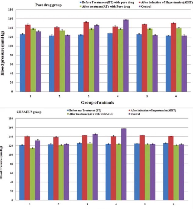
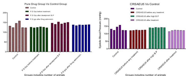
Invivo animal studies
The in vivo studies were carried out with the optimized formulation CRSAEU5 in Swiss albino mice to study the effect of microcapsules. For this purpose, the decrease in blood pressure was observed in hypertension induced mice (with diet mentioned in Table II), which were treated with CRSAEU5 formulation. The results were compared with group treated alone with pure drug CR and Control group. The effects were observed after 24 h from the day of treatment. The data of the blood pressure
are presented in Figure 9.
From the Figure 9, it is clear that, the microcapsule
formulation coded as CRSAEU5, was effective in decreasing the blood pressure as compared to pure drug group. This may be due to the increase in drug solubility
in the body luids by inclusion of complexing agent in the
formulation.
The in vivo statistical ANOVA studies are shown in Figure 10, which shows that CRSAEU5 is most effective in controlling the hypertension in comparison to the pure drug. As the pure drug is lipophilic and very less bioavailable, but in the formulation the drug is complexed with HPBCD, thus the hydrophilicity of the drug is enhanced and thus it becomes more bioavailable.
CONCLUSIONS
In this study, co-grinding technique has been applied to produce inclusion complex of CR with HPBCD and then incorporating this inclusion complex in the polymer matrix by using orifice gelation technique by use of a mixture of polymers Eudragit NE30D and sodium alginate which resulted in smooth, free flowing microcapsules having improved drug delivery. Higher drug loading
eficiency was observed for all the formulations, and also
the drug release was observed for a period of 13 h. Thus the polymer Eudragit NE30D, showed promising result in retarding drug release. Moreover Eudragit NE30D did not show any incompatibility with neither the inclusion complex nor with the other polymer, i.e. with sodium alginate. Morphological analysis by Scanning Electron Microscopy showed that the formulations were almost spherical in shape and size. All the analytical studies showed that the drug and HPBCD had weak interactions leading to inclusion of drug carvedilol in the cyclodextrin
cavity thereby improving the dissolution proile of the
drug. The in vivo animal studies showed that the optimized formulation was effective in controlling the hypertension for a period of 24 h.
ACKNOWLEGDEMENTS
Authors wish to thank Glenmark
Pharma-ceuticals,India, for providing Carvedilol as gift sample, thanks to Roquette ,Lestrem,France for providing gift sample of Hydroxy propyl beta cyclodextrin,and also heartiest thanks to Evonik Degussa rohm Pharma, India,
for providing gift sample of Eudragit NE 30D.Authors wish to thank CIF, BIT, Mesra, Ranchi for providing all the technical help in completing this study.
List of Abbreviations
CR:Carvedilol
HPBCD: Hydroxypropyl beta-cyclodextrin EU: Eudragit NE30D
SA: sodium alginate
SEM: Scanning electron microscopy
FTIR: Fourier Transform Infrared Spectroscopy XRD: X-ray diffraction
DSC: Differential scanning Calorimetry Ci: Compressibility Index
REFERENCES
ARCHONTAKI, H.A.; VERTZONI, M.V.; ATHANASSIOU, M.M.H. Study on the inclusion complexes of bromazepam with beta and beta-hydroxypropyl cyclodexrin. J. Pharm. Biomed. Anal., v.28, n.3-4, p.761-769, 2002.
BAL, T.; MURTHY, N.P.; PANDEY, A. Evaluation of
mucoadhesive carvedilol microcapsules prepared by oriice
gelation technique. J. Pharm. Res., v.5, n.1, p.519-525, 2012.
CARR, R.L. Evaluating low properties of solid. Chem. Eng.,
v.72, n.3, p.163-168, 1965.
CHOWDARY, K.P.R.; RAO, S.Y. Design and in vitro evaluation of mucoadhesive microcapsules of glipizide for oral controlled release. A technical note. AAPS Pharm. Sci. Tech., v.4, n.39, p.1-6, 2003.
DEASY, P.B. Microencapsulation and related drug processes.
United StatesNew York: Mercel Marcel Dekker, 1984. v.20, p.234-237.
DHAWAN, S.; SINGLA ,K.A.; SINHA, R.V. Evaluation of
mucoadhesive properties of chitosan microspheres prepared by different methods. AAPS Pharm. Sci. Tech., v.5, n.4,
p.1-7, 2004.
DHALIWAL, S.; JAIN, S.; SINGH, P.H.; TIWARY, A.K.
Mucoadhesive microspheres for gastroretentive delivery of acyclovir: in vitro and in vivo evaluation. AAPS Pharm. Sci. Tech., v.10, n.2, p.322-330, 2008.
GUPTA, A.; GARG. S.; KHAR, K.R. Measurement of
bioadhesive strength of mucoadhesive buccal tablets: design of an in vitro assembly. Indian Drugs, v.4, n.30, p.152-155,
1992.
HIGUCHI, T.; CONNORS, K.A. Phase solubility techniques.
Adv. Anal. Chem. Instr., v.4, p.117-212, 1965.
HIRLEKAR, R.; KADAM, V. Preparation and characterization
of inclusion complexes of carvedilol with
methyl-β-cyclodextrin. J. Incl. Phenom. Macrocycl. Chem., v.63,
n.3-4, p.219-224, 2009.
KUMAR, G.; GUPTA, G.A. Development and in vitro
evaluation of osmotically controlled drug delivery system of carvedilol. Int. J. Pharm. Sci. Drug Res., v.1, n.2,
p.80-82, 2009.
MAGNUSDOTTIR, A.; MASSON, M.; LOFTSSON, T.
Cyclodextrins. J. Incl. Phenom. Macroc. Chem., v.44, n.1-4, p.213-218, 2002.
MIRO, A.; QUAGLIA, F.; GIANNINI, L.; CAPPELLO, B.;
ROTONDA, M.I.L. Drug/ cyclodextrin solid systems in the design of hydrophilic matrices: a strategy to modulate drug delivery rate. Curr. Drug. Deliv., v.3, n.4, p.373-378, 2006.
MUKHERJEE, B.; MAHAPATRA, S.; GUPTA, R.; TIWARI, A.;
ARORA, P. A comparison between povidone-ethylcellulose and povidone-eudragit transdermal dexamethasone matrix patches based on in-vitro skin permeation. Eur. J. Pharm. Biopharm., v.59, n.3, p.475-483, 2005.
PATIL SANJAY, B.; SAWANT KRUTIKA, K. Development, o p t i m i z a t i o n a n d i n v i t ro e v a l u a t i o n o f a l g i n a t e mucoadhesive microspheres of carvedilol for nasal delivery. J. Microencapsul., v.26, n.5, p.432-443, 2009.
PATIL, D.A.; PATIL, G.B.; DESHMUKH, P.K.; BELGAMWAR,
V.S.; FARSULE, R.A. Chitosan coated mucoadhesive multiparticulate drug delivery system for glicazide. Asian J. Pharm. Clin. Res.,v.2, n.2, p.62-67, 2009.
PITHA, J.; MILECKI, J.; FALER, H.; PANNELL, L.; VEKAMA, K. Hydroxy propyl beta cyclodextrin: preparation and characterization; effects on the solubility of drugs. Int. J. Pharm., v.29, n.1, p.73-82, 1986.
QIANLI, Y.U.; DOUGLAS, F.L.; DENISE, S.; TAMARA, F.L.;
JEFFREY, H.B.; RONALD, R.W. Characterization of high salt and high fat diets on cardiac and vascular function in mice. Cardiovasc. Toxicol., v.4, n.1, p.37-46, 2004.
RANJHA, N.M.; KHAN, H.; NAZEEM, S. Encapsulation and
characterization of controlled release lubiprofen loaded
microspheres using beeswax as an encapsulating agent. J. Mater. Sci. Mater. Med., v.21, n.5, p.1621-1630, 2010.
SALSA, T.; VEIGA, F.; PINA, M.E. Oral controlled release
dosage forms. J. Cellulose ether polymers in hydrophilic matrices. Drug Dev. Ind. Pharm., v.23, n.9, p.929-938, 1997.
SCHAEFER, H.W.; POLITOWSKI, J.; HWANG, B.; DIXON JR., F.; GUTZAIT, L.G.A.; ANDERSON, K.; DEBROSSE, C.; BEAN, M.; RHODES ,R.G. Metabolism of Carvedilol
in dogs, rats and mice. Drug Metab. Dispos., v.26, n.10,
p.958-969, 1998.
SHEWALE, B.D.; SAPKAL, N.P.; RAVI, N.A.; GAIKWAD,
N.J.; FURSULE, A. Effects of hydroxyl propyl beta cyclodextrin on the solubility of carvedilol. Indian J. Pharm. Sci., v.70, n.2, p.225-227, 2008.
SHARMA, H.K.; SARANGI, B.; PRADHAN, S.P. Preparation
and in-vitro evaluation of mucoadhesive microbeads containing timolol maleate using mucoadhesive substances of Dillenia indica L. Arch. Pharm. Sci. Res., v.1, n.2,
TANWAR, S.Y.; CHAUHAN, S.C.; SHARMA, A. Development and evaluation of carvedilol transdermal patches. Acta Pharm., v.57, n.3-4, p.151-159, 2007.
VYAS, A.; SARAF, S.; SARAF, S. Cyclodextrin based novel drug delivery systems. J. Incl. Phenom. Macrocycl. Chem., v.62, n.1-2, p.23-42, 2008.
WANG, Z.; DENG, Y.; SUN, S.; ZHANG, X. Preparation of
hydrophobic drug cyclodextrin complexes by lyophilization monophase solution. Drug Dev. Ind. Pharm., v.32, n.1,
p.73-78, 2006.
XIUDONG, L.; WEIMING, X.; QUN, L.; WEITING, Y.; YINGLI, F.; XIAOJUN, M.A.; QUAN, Y. Swelling
behaviour of alginate-chitosan microcapsules prepared by external gelation or internal gelation technology. Carbohydr. Polym., v.56, n.4, p.459-464, 2004.
Received for publication on 03rd September 2012