Scanning Electron Microscopic Analysis
of the Effect of Carisolv
TM
Gel on Periodontally
Compromised Human Root Surfaces
Daniela Corrêa GRISI1
Letícia Helena THEODORO1
José Eduardo César SAMPAIO1
Márcio Fernando de Moraes GRISI2
Sérgio Luiz de Souza SALVADOR3
1Department of Diagnostics and Surgery, Faculty of Dentistry of Araraquara,
São Paulo State University, Araraquara, SP, Brazil
2Department of Buccomaxillofacial Surgery and Traumatology and Periodontology,
Faculty of Dentistry of Ribeirão Preto, University of São Paulo, Ribeirão Preto, SP, Brazil
3Department of Clinical Analysis, Faculty of Pharmaceutical Sciences of Ribeirão Preto,
University of São Paulo, Ribeirão Preto, SP, Brazil
The aim of this study was to analyze, under scanning electron microscopy (SEM), the morphologic characteristics of root surfaces after application of CarisolvTM gel in association with scaling and root planing (SRP). Sixty periodontally compromised extracted
human teeth were randomly assigned to 6 groups: 1) SRP alone; 2) passive topical application of CarisolvTM + SRP; 3) active topical
application of CarisolvTM + SRP; 4) multiple applications of CarisolvTM + SRP; 5) SRP + 24% EDTA; 6) topical application of
CarisolvTM + SRP + 24% EDTA. CarisolvTM gel was applied to root surfaces for 30 s, followed by scaling and root planing,
consisting of 50 strokes with Gracey curettes in an apical-coronal direction, parallel to the long axis of the tooth. The only exception was group 4, in which the roots were instrumented until a smooth, hard and glass-like surface was achieved. All specimens were further analyzed by SEM. The results showed that the treatment with CarisolvTM caused significant changes in root surface
morphology of periodontally compromised teeth only when the chemical agent was actively applied (burnishing technique). CarisolvTM failed to remove the smear layer completely, especially with a single application, independently of the method of
application. Multiple applications of CarisolvTM were necessary to achieve a smear layer reduction comparable to that obtained with
24% EDTA conditioning.
Key Words: periodontal disease, scaling and root planning, CarisolvTM, scanning electron microscopy.
Correspondence: Prof. Dr. José Eduardo César Sampaio, Diagnóstico e Cirurgia, Faculdade de Odontologia de Araraquara, UNESP, Rua Humaitá, 1680, 14801-903 Araraquara, SP, Brasil. Tel: +55-16-201-6369. Fax: +55-16-201-6369. e-mail: [email protected] INTRODUCTION
In periodontal disease, root surface is exposed to the subgingival environment and bacterial plaque. Expo-sure to crevicular fluid, as well as to enzymes and metabolites produced by subgingival plaque bacteria induces physical and chemical alterations on root ce-mentum. Periodontitis-affected root surfaces are hypermineralized (1-3), contaminated with bacterial plaque (4) and other cytotoxic substances (5).
Traditional scaling and root planing (SRP)
proce-dures have relied on the mechanical removal of plaque, calculus, root-bound toxins and contaminated cemen-tum. Although the effectiveness of scaling and root planing has been well documented, the efficacy of this treatment has been questioned. Additionally, smear layer that remains after instrumentation can impair periodontal healing (6,7).
(7,10,11), decalcifying of planed root surfaces, expo-sure of dentin or cementum collagen matrix, thus providing a biologically acceptable surface for regen-eration of a new connective tissue attachment (12).
Different etching solutions, such as citric acid, tetracycline and EDTA, have been used as adjunctive therapies to scaling and root planing in order to over-come the limitations of these procedures.
CarisolvTM gel (Mediteam, Sävadelen, Sweden), a chemomechanical caries removal system was devel-oped to aid carious dentin excavation. This system consists of sodium hypochlorite and three aminoacids (lysine, leucine and glutamic acid) that are able to remove carious tissue without affecting the healthy dentin structure (13-16).
The use of chemical agents in association with mechanical treatment represents a possibility of a less traumatic procedure, preventing the excessive loss of root substance. In the field of Periodontics, the possi-bility of chemically dissolving calculus and contami-nated root cementum in order to facilitate their mechani-cal removal is one of the most promising applications of CarisolvTM gel.
Therefore, the purpose of this study was to investigate, under scanning electron microscopy (SEM), the morphologic characteristics of periodontally com-promised human root surfaces after application of CarisolvTM gel in association with scaling and root planning.
MATERIAL AND METHODS
Sixty periodontally compromised extracted hu-man teeth with supra- and subgingival calculus were used. The teeth were extracted for periodontal reasons from patients treated at the Department of Diagnostics and Surgery of the Faculty of Dentistry at Araraquara, (Brazil), and were stored in saline until use. The research protocol was approved by the local Ethics in Research Committee.
Diseased tooth surfaces with adhered calculus were chosen as the treatment areas and delimited with a round bur, and the teeth were randomly assigned to 6 groups (n=10), as follows. Group 1: Scaling and root planing (SRP) alone. The root surfaces were instru-mented with Gracey curettes (Hu-Friedy, Chicago, IL, USA), using 50 strokes in an apical-coronal direction, parallel to the axis of the tooth. Group 2: Passive topical
application of CarisolvTM gel + SRP. Carisolv™ gel was applied to the delimited area in each root for 30 s. Root surfaces were instrumented with Gracey curettes in the same way as described as in group 1. Group 3: Active topical application of CarisolvTM gel + SRP: CarisolvTM gel was burnished onto the delimited areas for 30 s, using a disposable microbrush tip. Root surfaces were instrumented with Gracey curettes, in the same way as described as in groups 1 and 2. Group 4: Multiple applications of CarisolvTM gel + SRP. CarisolvTM gel was applied several times interposing with SRP procedures, which were performed with Gracey curettes in order to provide a smooth, hard, glass-like surface. The teeth were instrumented with hand instruments simulating the clinical situation. Group 5: SRP + EDTA. Root surfaces were instrumented and a microbrush tip soaked in 24% EDTA gel (PrefGel; Biora HelpMed-Medical Supplies, São Paulo, SP, Brazil) was subsequently brushed onto the delimited areas for 2 min, the microbrush being resoaked every 30 s. Group 6: CarisolvTM gel + SRP + EDTA: Prior to instrumentation, CarisolvTM gel was applied to the delimited areas for 30 s. Root surfaces were instrumented with Gracey curettes as previously described. The instrumented root sur-faces were conditioned with 24% EDTA gel.
The treated surfaces were rinsed in 20 mL saline and the crowns were removed at the cementoenamel junction. The teeth were then horizontally and vertically sectioned with a diamond circular saw, using the treated area as a reference. Each tooth section was rinsed in saline and placed in 2.5% glutaraldehyde in 0.1 M phosphate buffer (pH 7.4) for a minimum of 24 h.
The specimens were washed and dehydrated in a series of graded alcohol solutions (50, 70, 80, 95 and 100%) for 10 min each. After 2 additional 10-min washings in absolute alcohol, the specimens were dried overnight in a desiccator jar, mounted on SEM stubs and sputter-coated with gold. Specimens were examined using a scanning electron microscope (JSM-T330A; JEOL, Tokyo, Japan). Photographs of the central por-tion of each specimen were taken at X1000 magnifica-tion. SEM examination was performed by a single blinded examiner. The following parameters were evalu-ated: surface morphology (regular, irregular or flaky surface), presence or absence of smear layer and presence or absence of dentinal tubules.
treat-ment groups. Because MANOVA determined a statisti-cally significant main effect for the treatments, Newman-Keuls test was used to determine differences between individual groups at 5% significance level.
RESULTS
The results of SEM analysis are presented in Table 1.
Group 1: The scaled, root-planed specimens
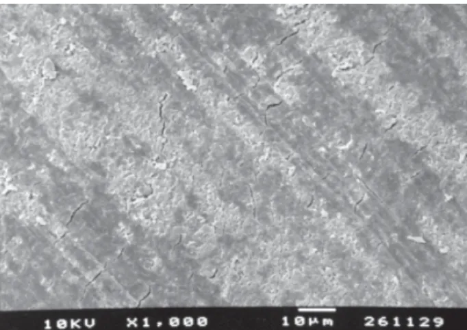
exhibited more irregular surfaces than the specimens in groups 3, 5 and 6 (p<0.05). However, there were no statistically significant differences in regular surfaces (p>0.05) between group 1 and the other groups. All specimens were covered with smear layer. There were no statistically significant differences between the SRP specimens and the other groups with respect to the presence of smear layer (p>0.05) (Fig. 1).
Group 2: Specimens treated with topical
applica-tion of CarisolvTM plus SRP had more irregular surfaces compared to group 6 (p<0.05). In comparison to the other groups, there were no statistically significant differences in regular (p>0.05) or flaky surfaces (p>0.05). All specimens were covered with smear layer, but there were no statistically significant differ-ences compared to the other treatments (p>0.05), except for group 4 (p<0.05) (Fig. 2).
Group 3: Specimens treated with active
applica-tion of CarisolvTM plus SRP had a regular surface, with no statistically significant differences compared to the other groups (p>0.05). In comparison to the SRP specimens, the active application of CarisolvTM gel resulted in fewer irregular surfaces (p<0.05). There were no statistically significant differences between the treatments with respect to the formation of flaky sur-faces (p>0.05). In
ad-dition, all specimens in this group were cov-ered with smear layer, with no statistically sig-nificant differences compared to the other treatments (p>0.05), except for group 4 (p<0.05) (Fig. 3).
Group 4:
Speci-mens exposed to mul-tiple applications of
CarisolvTM interposed with SRP procedures had more regular surfaces, with no significant difference com-pared to the other groups (p>0.05). No significant differences were observed between the treatments with respect to flaky surfaces (p>0.05). Smear layer rem-nants were found in 6 specimens. There was a statisti-cally significant reduction in the amount of smear layer when this group was compared to groups 2 and 3 (p<0.05) (Fig. 4).
Group 5: The scaled, planed and EDTA-treated
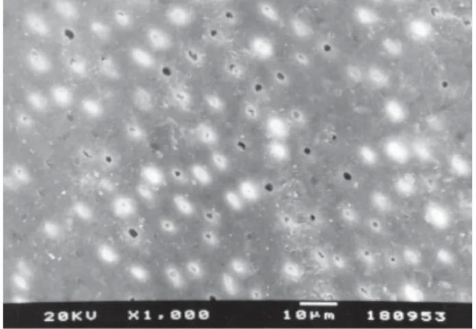
specimens showed more regular surfaces with no statistically significant differences (p>0.05). Compared to the SRP group, the specimens in this group had fewer irregular surfaces and more smear layer-free surfaces (p<0.05). Smear layer remnants were ob-served in 6 specimens. This group differed statistically from group 3 with respect to the presence of smear layer (p<0.05) (Fig. 5).
Group 6: Specimens exposed to topical
applica-tion of CarisolvTM followed by SRP and conditioning with EDTA gel had more regular surfaces, although not statistically different from the other groups (p>0.05). There were no significant differences between this group and the others with respect to flaky surfaces (p>0.05). In comparison to groups 1 and 2, the speci-mens in this group had fewer irregular surfaces (p<0.05). Conditioning with EDTA gel for 2 min after chemomechanical therapy removed more smear layer from the root surfaces compared to SRP alone or topical application of the CarisolvTM gel combined with SRP (p<0.05) (Fig. 6).
Although some specimens in groups 5 and 6 showed open dentinal tubules, no statistically significant differences (p<0.05) were observed among the pro-posed treatments with respect to tubule opening.
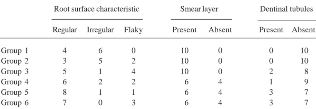
Table 1. Frequency of root surface characteristics, smear layer and dentinal tubules in each group.
Root surface characteristic Smear layer Dentinal tubules
Regular Irregular Flaky Present Absent Present Absent
Group 1 4 6 0 10 0 0 10
Group 2 3 5 2 10 0 0 10
Group 3 5 1 4 10 0 2 8
Group 4 6 2 2 6 4 1 9
Group 5 8 1 1 6 4 3 7
DISCUSSION
This study compared the morphological changes of periodontally compromised root surfaces submitted to either treatment with CarisolvTM gel combined with mechanical instrumentation or scaling and root planing alone.
When CarisolvTM gel was topically applied to root surfaces before scaling and root planing there were no significant changes in root surface morphology com-pared to mechanical treatment alone. Both treatments resulted in a higher frequency of rough surfaces and
were equally ineffective in removing smear layer. The irregular appearance of root surfaces treated with SRP or SRP + CarisolvTM were partially due to the presence of smear layer (Figs. 1 and 2).
Unlike passive application, active aplication (bur-nishing) of CarisolvTM gel produced more extensive morphological changes than SRP alone. This mode of application resulted in fewer irregular surfaces, prob-ably due to the mechanical abrading action of the microbrush soaked in CarisolvTM gel, which also al-lowed this agent to be in closer contact with the root surface (Fig. 3). These results are in agreement with
Figure 1. Group 1. SEM micrograph of diseased root surface after scaling and root planing. The surface is irregular and covered by smear layer remnants. Traces of instrumentation are evident.
Figure 2. Group 2. SEM micrograph of diseased root surface after topical application of CarisolvTM plus scaling and root
planing. The surface is irregular and covered by smear layer remnants.
Figure 4. Group 4. SEM micrograph of diseased root surface after multiple applications of CarisolvTM interposed with
scaling and root planing. The surface is regular and free of smear layer.
Figure 3. Group 3. SEM micrograph of diseased root surface after active application of CarisolvTM plus scaling and root
those of previous studies (17,18), which described considerable changes in surface morphology after bur-nishing with citric acid.
Although there was no statistically significant difference in root surface morphology after multiple applications of CarisolvTM compared to scaling and root planing alone, it is interesting to note that some speci-mens exhibited a mosaic-like structure resembling that of healthy cementum (Fig. 7). This structure seemed to be masked to the surface coating that is often formed on root surface, due to the hypermineralization process resulting from periodontal disease (2,3). In view of this,
it is reasonable to assume that if it is performed as usually done in clinical situations, the chemomechanical therapy would be able to remove the contaminated cementum layer and expose the healthy structure.
Regardless of the treatment, the surface appear-ance was extremely variable, ranging from irregular to regular, which may be explained by the variability in the anatomical structure of cementum and also by differ-ences in mineralization of the surface coating or even by a combination of both.
Flaky surfaces were more frequently observed in chemomechanically treated specimens (Fig. 8). These
Figure 5. Group 5. SEM micrograph of diseased root surface after scaling and root planing and conditioning with EDTA. The surface is regular and with patent dentinal tubules. There is no evidence of smear layer.
Figure 7. Group 4. SEM micrograph of diseased root surface after multiple applications of CarisolvTM plus scaling and root
planing, showing an amorphous appearance with small circular mounds, resembling healthy cementum.
Figure 8. Group 2. SEM micrograph of diseased root surface after topical application of CarisolvTM plus scaling and root
planing, showing a flaky appearance with remnants of smear layer.
Figure 6. Group 6. SEM micrograph of diseased root surface after topical applications of CarisolvTM plus scaling and root
morphological characteristics were similar to those observed by Bannerjee et al. (19). This could be due to the chemical effect of CarisolvTM gel on root surface of periodontally compromised teeth. The main constituent of CarisolvTM is sodium hypochlorite, which is mixed with three amino acids (lysine, leucine and glutamic acid). The resulting gel would be able to remove the organic components of root cementum or calculus, in the same way as it does with those of carious lesions. Additionally, this gel would be able to reduce smear layer formation because it is usually used simultaneously with mechanical instruments, thus acting as a lubricating gel. In the present study, remnants of smear layer covering the root surface were still evident after the combination of CarisolvTM gel and SRP procedures. This suggests that the chemomechanical treatment failed to reduce or eliminate the smear layer, especially with a single application of CarisolvTM, independently of the mode of application. Other investigators have also found that CarisolvTM failed to remove the smear layer from the dentin surface (15,20). This is not surprising because the use of CarisolvTM gel was proposed as an adjunctive therapy to scaling and root planing for removal of calculus and contaminated cementum. Hence, it should be applied before root scaling and not after this procedure as advised for EDTA. This could explain the limited effect of a single application of CarisolvTM gel on smear layer removal. Significant removal of smear layer was only achieved when root surfaces were etched with EDTA (Fig. 6), as observed in a previous study (15).
On the other hand, when multiple applications of CarisolvTM were performed, there was a significant decrease in smear layer compared to a single application of the gel either passively or actively. These findings are consistent with those of Banerjee et al. (19) who observed lack of smear layer after multiple applications of CarisolvTM gel for removal of carious dentin. It is important to note that several applications of CarisolvTM gel caused a smear layer reduction that could be com-pared to that of EDTA gel (Table 1) and was also able to remove the decayed cementum layer, exposing a healthy cementum layer underneath. However, topical application of EDTA gel was the only treatment capable of reducing smear layer compared to SRP procedures. The effectiveness of EDTA gel in removing smear layer and exposing dentin or cementum collagen matrix has been previously reported (11). It may be assumed that not only active but also multiple applications of CarisolvTM
gel would be required to alter root surface morphology and enhance the smear layer removal effect of CarisolvTM for treatment of periodontally compromised teeth.
Further studies are needed to investigate whether CarisolvTM could improve the removal of calculus and also establish whether the morphological alterations of root surfaces produced by chemomechanical therapy might provide a biologically acceptable environment for periodontal healing.
In conclusion, the chemomechanical therapy caused significant changes in root surface morphology of periodontally compromised teeth only when CarisolvTM was actively applied (burnishing technique). CarisolvTM failed to remove the smear layer completely, especially with a single application, independently of the method of application. Multiple applications of CarisolvTM were necessary to achieve a smear layer reduction compa-rable to that obtained with 24% EDTA conditioning.
RESUMO
A utilização do CarisolvTM tem sido proposta como um método
auxiliar à raspagem e ao alisamento radicular (RAR), a fim de facilitar a descontaminação da superfície da raiz. O objetivo deste estudo foi avaliar, através da microscopia eletrônica de varredura (MEV), as características das superfícies radiculares, após a aplicação do CarisolvTM em associação à RAR. Sessenta dentes
humanos extraídos devido à doença periodontal foram divididos em 6 grupos: 1) RAR ; 2) CarisolvTM (aplicação passiva) + RAR;
3) CarisolvTM (aplicação ativa) + RAR; 4) CarisolvTM (aplicações
múltiplas) + RAR; 5) RAR + EDTA a 24%; 6) CarisolvTM +
RAR + EDTA a 24%. CarisolvTM foi aplicado às superfícies
radiculares por 30 s, seguido de raspagem e alisamento radicular, que consistiu de 50 movimentos com curetas de Gracey no sentido corono-apical, co o instrumento paralelo ao longo eixo do dente. A única exceção foi o grupo 4, no qual as raízes foram instrumentadas até obter uma superfície lisa, dura e com aspecto vítreo. Os espécimens tratados foram preparados e examinados em MEV. Os resultados demonstraram que a associação do CarisolvTM aos procedimentos periodontais mecânicos
proporcionou modificações significativas na superfície radicular quando comparada à raspagem e ao alisamento radicular, apenas quando o CarisolvTM foi aplicado de forma ativa. A aplicação do
CarisolvTM uma única vez, apresentou um efeito limitado na
capacidade de remoção de smear layer, sendo que aplicações sucessivas apresentaram resultados comparáveis àqueles obtidos após a aplicação do EDTA.
ACKNOWLEDGEMENTS
REFERENCES
1 . Wirthlin MR, Pederson ED, Hancock EB, Lamberts BL, Leonard EP. The hypermineralization of diseases root sur-faces. J Periodontol 1979;50:125-127.
2 . Eide B, Lie T, Selvig KA. Surface coatings on dental cemen-tum incident to periodontal disease. I. A scanning electron microscopic study. J Clin Periodontol 1983;10:157-171. 3 . Eide B, Lie T, Selvig KA. Surface coatings on dental
cemen-tum incident to periodontal disease (II). A scanning electron microscopic confirmation of a mineralized cuticle. J Clin Periodontol 1984;11:565-575.
4 . Adriaens PA, Edward CA, De Boever JA, Loesche WJ. Ultra-structural observations on bacterium invasion in cementum and radicular dentin of periodontally diseased human teeth. J Periodontol 1988;8:493-503.
5 . Aleo JJ, DeRenzis FA, Farber PA, Varboncoeur AP. The presence and biological activity of cementum-bound endot-oxin. J Periodontol 1974;45:672-675.
6 . Polson AM, Caton J. Factors influencing periodontal repair and regeneration. J Periodontol 1982;53:617-625.
7 . Polson AM, Frederick GT, Ladenheim S, Hanes PJ. The production of a root smear layer by instrumentation and its removal by citric acid. J Periodontol 1984;55:443-446. 8 . Maynor GB, Wilder RS, Mitchell SC, Moriarty J.D.
Effective-ness of a calculus scaling gel. J Clin Periodontol 1994;21:365-368.
9 . Nagy RJ, Endow JP, Inouge AE, Otomo-Corgel J. The effects of a single course of a calculus-softening scaling and root planing gel. A ccanning electron microscopic study. J Periodontol 1998;69:806-811.
10. Sabirnoff JA, O’Leary TJ, Miller CH. The comparative effec-tiveness of various agents in detoxifying diseased root sur-faces. J Periodontol 1983;54:77-80.
11. Blomlöf J, Blomlöf L, Lindskog S. Effect of different
concen-trations of EDTA on smear removal and collagen exposure in periodontitis-affected root surfaces. J Clin Periodontol 1997;24:534-537.
12. Blomlöf J, Jannson L, Blomlöf L, Lindskog S. Root surface etching at neutral pH promotes periodontal healing. J Clin Periodontol 1996;23:50-55.
13. Ericson D, Zimmerman M, Raber H, Gotrick B, Bornstein R, Thorell J. Clinical evaluation of efficacy and safety of a new method for chemo-mechanical removal of caries. A multi-center study. Caries Res 1999;33:171-177.
14. Wennerberg A, Sawase T, Kultji C. The influence of CarisolvTM on enamel and dentin surface topography. Eur J Oral Sci 1999;107:297-306.
15. Cederlund A, Lindskog S, Blömlof J. Effect of chemo-me-chanical caries removal system (CarisolvTM) on dentin to-pography of non-carious dentin. Acta Odontol Scand 1999;57:185-189.
16. Avirdsson A, Liedberg B, Möller K, Lyvén B, Sellén A, Wennerberg A. Chemical and topographical analyses of dentin surfaces after CarisolvTM treatment. J Dent 2002,30:67-75.
1 7 Sterrett JD, Murphy HJ. Citric acid burnishing of dentinal root surfaces. A scanning electron microcopy report. J Clin Periodontol 1989;16:98-104.
18. Sterrett JD, Murphy HJ. Citric acid demineralization of ce-mentum and dentin: the effect of application pressure. J Clin Periodontol 1995;22:434-441.
19. Banerjee A, Kidd EA, Watson TF. Scanning electron micro-scopic observations of human dentin after mechanical caries excavation. J Dent 2000;28:179-186.
20. Yazici AR, Özgünaltay G, Dayangaç B. A scanning electron microscopic study of different caries removal techniques on human dentin. Oper Dent 2002;27:360-366.