INTRODUCTION
Epilepsy is a common condition that affects up to 1%-5% of the population.1 Sleep has long
been known to affect epilepsy and nearly 21% of patients had seizures solely during sleep.2
There is a reciprocal relationship between sleep and epilepsy.3 Sleep affects epilepsy and
epilepsy, in turn, affects sleep. Patients with epilepsy are particularly sensitive to the adverse effects of sleep disruption. Sleep disturbances are common in epilepsy causing impairment of daytime functioning and quality of life and impairment of seizures control.4,5 Furthermore,
primary sleep disorders such as obstructive sleep apnoea (OSA) may worsen epilepsy and
treatment of these sleep disorders has been shown to lead to improved seizure control. Both sleep disordered breathing (SDB) and epilepsy; however, are common, treatable conditions.
Sparse data are available regarding the prevalence of SDB from developing countries like India. Sleep-related breathing disorders are increasingly recognized as an important cause of morbidity. Attention to sleep in patient with epilepsy has important implications for diagnosis, seizure control, and quality of life.Sleep disorders in patients with epilepsy have rarely been investigated systematically. Even less is known regarding the epidemiology
Original Article:
Sleep disordered breathing in patients with epilepsy
B. Vengamma,1 J. Vijayabhaskara Rao, 1Alladi Mohan2
Departments of 1
Neurology, 2Medicine, Sri Venkateswara Institute of Medical Sciences, Tirupati
ABSTRACT
Background:Sleep has long been known to affect epilepsy. Little has been documented regarding the epidemiology of sleep disordered breathing (SDB) in patients with epilepsy from India.
Methods:Between April 2009 and September 2011, in the first stage of the study, 452 consecutive patients with epilepsy (cases) and 500 age- and gender-matched normal control subjects were screened using Epworth’s Sleepiness Scale (ESS). Of these, 98 (23%) had an ESS score of 10 or more, suggestive of excessive day time sleepiness (EDS). Of these, 30 patients (mean age was 35.8±13.1; 26 males) were taken up for the second stage of the study, underwent detailed evaluation and overnight in-hospital polysomnography (PSG) in the sleep laboratory.
Results: Their mean age was 35.8 ±13.1; 26 males); Of these, 13 (43.3%)(all males) had evidence of OSAS on PSG: 9 had mild OSAS [apnoea-hypopnoea index (AHI) 5-15]; 3 had moderate OSAS (AHI 15-30) and one patient had severe OSAS (AHI >30). On univariate analysis, a higher proportion of patients with nocturnal seizures had OSAS compared with those with diurnal seizures and combined periodicity (7/8 vs 6/16; p=0.009). Patients with uncontrolled seizures had a higher prevalence of OSAS (p=0.045).
Conclusions: Our observations suggest that EDS is an important symptom being six times more common in patients with epilepsy compared to healthy control subjects. Furthermore, PSG study indicated that 43.3% of epilepsy patients with EDS have OSAS. Thus, SDB appears to be an underdiagnosed disease in patients with epilepsy from India.
Key words:Epilepsy, Sleep disordered breathing, Obstructive sleep apnoea
Vengamma B,Vijayabhaskara Rao J, Mohan A. Sleep disordered breathing in patients with epilepsy. J ClinSci Res 2016;5:7-19. DOI: http://dx.doi.org/10.15380/2277-5706.JCSR.15.073A.
Corresponding author:Dr B. Vengamma, Former Director-cum Vice Chancellor, Senior Pr ofessor an d Head, Depar tmen t of Neurology, Sri Venkateswara Institute of Medical Sciences, Tirupati, India.e-mail:
Received:November 16, 2015: Accepted: December 22, 2015.
Online access
http://svimstpt.ap.nic.in/jcsr/jan-mar16_files/1oa16.pdf
of SDB in patients with epilepsy and the relationship between the two entities from India. Keeping these facts in mind, the present study was designed to assess the prevalence of SDB in patients with epilepsy at a tertiary care teaching centre in south India.
MATERIAL AND METHODS
During the period April 2009 to September 2010, consecutive patients diagnosed to have and treated for various forms of epilepsy at the neurology and medicine out-patients departments, epilepsy clinic, acute neurological care unit (ANCU), medical intensive care unit (MICU) and medicine and neurology wards of the Sri Venkateswara Institute of Medical Sciences, Tirupati were screened for inclusion in the study. Patients diagnosed to have various form of epilepsy6 who were willing to
participate in the study were included. Pregnant women, patients with life threatening co-morbid conditions and those not willing to participate in the study were excluded.
The study was approved by the Institutional Ethics Committee.
Written informed consent was obtained from all patients for participating in the study.The study was carried out in two stages.
First stage
In the first stage, 452 consecutive patients with epilepsy who had given consent to participate in the study; and age and gender matched 500 normal control subjects were screened for excessive day time sleepiness (EDS) using Epworth’s Sleepiness Scale (ESS).7Excessive
day time sleepiness (EDS) was defined as an ESS score greater than 10. Of the epilepsy patients in whom ESS was 10 or greater, 30 were randomly selected for undergoing detailed evaluation and PSG.
Second stage
Thirty subjects underwent a detailed evaluation that included clinical history focused on
sleep-related symptoms such as self-reported snoring, history of snoring from the relative, history of choking during sleep, associated conditions, and co-morbidities. Severity of snoring and choking was graded via one-on-one interview by an investigator blinded to the PSG data on a four-point frequency scale: never [0]; less than 1/week [1]; 1-2/week [2]; 3 to 4/week [3]; and 5 to 7/week [4]. All subjects were subjected to a detailed ear, nose and throat examination to rule out significant upper airway obstruction, such as, enlarged adenoids, pharyngeal tonsils and lingual tonsils significant deviated nasal septum with/without spur and macroglossia, blood pressure measurement and anthropo-metry.
Body weight (to the nearest 0.5 kg) and height (H, measured to the nearest 1 cm) was recorded in all subjects while wearing only light clothes and no shoes. Body-mass index (BMI) was calculated as body weight (in kilograms)/height (in metres)2. Neck circumference (in cm) was
measured at the level o f cricot hyro id membrane. Neck length (NL, cm) was measured from the occipital tubercle to the vertebra prominens. Percent predicted neck circumference (PPNC) was calculated from the measured neck circumference (NC) and the height (H, cm) using the following formula:8-11
PPNC=(1000 × NC) / (0.55 × H + 310). H to NL ratio was calculated by dividing the H (cm) by NL (cm).8-11Waist circumference (WC)
Central obesity was defined as WC 90 cm for men and 80cm for women.12 Abdominal
obesity was defined as a WHR >0.90 in men and >0.80 in women.13 Obesity was classified
as per the International Obesity Task Force Guidelines for Asians.14
In all the patients, the following laboratory investigations were carried out: complete haemogram, fasting and post-prandial blood glucose and glycosylat ed haemo globin (HbA1c) estimation, thyroid profile, serum chemistry including fasting lipid profile, chest radiograph and electrocardiography. Metabolic syndrome was diagnosed as per International Diabetes Federation definition.12
Electroencephalography (EEG) was done in all patients using GalileoNT Systems (EBNeuro, Firenze, Italy), 32 channel EEG machine. Computed to mography (CT) (Siemens Somatom Emotion Spiral CT Scanner) and/or magnetic resonance imaging (MRI) of the brain (Siemens Symphony Maestro Class, 1.5 Tesla MRI Scanner) was done in all patients.The lung volumes and their subdivisions were measured using a constant volume variable pressure body plethysmograph (P.K. Morgan Chatham, Kent, UK) as described previously.15,16
Polysomnography
Patients with epilepsy were called for sleep study to the sleep laboratory at 8 pm, on the day of their appointment. They were hooked to Sleepscan Vision (Biologic Systems Corp, Illinois, USA) by standard gold cups after cleansing the area of attachment by spirit followed by Omniprep. The patient was advised to sleep at around 21:00 hours. The recording sleep study was started after ensuring that the impedance of recording electrodes were set to zero. Various parameters that will be monitored included EEG, electro oculogram (EOG), electro cardiogram (ECG), chin and leg electromyogram (EMG), nasal airflow, tracheal breath sounds, thoracic wall and abdominal movements, transcutaneous oxygen saturation,
body position, and continuous positive air way pressure titration when required.
The data were recorded overnight until the subject woke up naturally from his sleep in the morning or till 7 am, which was the “lights on” time. The data stored in the system were retrieved later. The sleep data recorded and automatically scored by the computer were manually scored for sleep stages, apneas, and hypopnoeas. An experienced laboratory technician, who was blinded to any clinical data, scored all the polysomnograms. Apnea was defined as cessation of oronasal airflow for greater than 10 seconds. Obstructive sleep apnea was scored when airflow was absent but respiratory efforts were present. Hypopnoea was defined as a discernible reduction in respiratory airflow of 50% - 90% for greater than 10 seconds accompanied by a decrease of 4% or more in oxyhemoglobin saturation during sleep. AHI was calculated using the following formula17:
AHI = (total number of obstructive apneas + total no. of hypopnoeas) / total sleep time (hours).
As per the American Academy of Sleep Medicine17 obstructive sleep apnea syndrome
was defined as AHI greater than or equal to 5 accompanied by EDS. The severity of OSAS was classified as mild (AHI of 5 -15); moderate (AHI 16 - 30); and severe OSA (AHI > 30).
Statistical analysis
Variables following normal distribution were summarized by mean and standard deviation. Continuous variables that were normally distributed were compared using unpaired students’ t-test. Categorical variables were compared using chi-square test. Correlation between ESS, AHI and other anthropometric variables was studied using Pearson’s correlation coefficient.
Statistical softwaresPASW Statistics 18, Release 18.0.0, (IBM SPSS Statistics, Somers NY, USA); and MedCalc Version 11.3.0 for Windows 2000/XP/Vista/7 (MedCalc Software bvba, Belgium) were used for statistical analysis.
RESULTS
First stage of the study
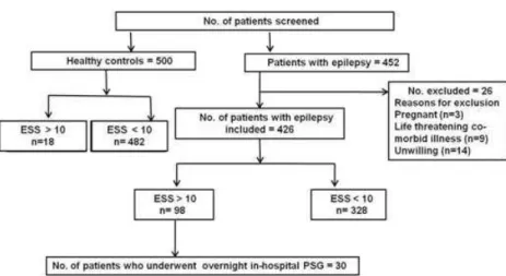
Between April 2009 and September 2011, 452 consecutive patients with epilepsy (cases) who had given consent to participate in the study and age and gender matched 500 normal control subjects were screened for EDS using ESS. Of these, 426 patients satisfied the inclusion criteria and screened with ESS for EDS (Figure 1). Their mean age was 37.5 ± 10.9 years; there were 295 males. Mean age of onset of seizures was 25.1 ± 11.5 years and the mean
duration of seizures was 136 ± 63.7 months. Majority of the patients had GTCS (49%), followed by CPS with (25%) and without (18%) secondary generalization.
Healthy co ntro l subjects (n=500) who consented to participate in the study were also screened with ESS for EDS (Figure 1). Their mean age was 37.3 ± 10.7 years; there were 369 males. The health control and the epilepsy group were age (p=0.389) and gender (2=2.1294) p=0.1445.
Of the 500 healthy control subjects who were screened, 18 (3.6%) were found to have EDS (ESS score 10). In comparison with healthy controls, patients with epilepsy had an almost 6-times higher prevalence of EDS (p=<0.001) (Table 1).
Second stage of the study
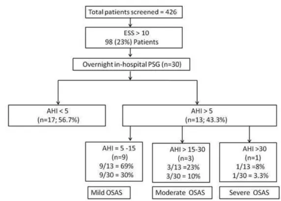
Of the 426 patients studied in the first stage of the study, 98 (23%) had an ESS score of 10 or more, suggestive of EDS. Their mean age was 36.4 ± 13.2 years; there were 84 males. Mean age of onset of seizures was 24.7 ± 12.7 years and the mean duration of seizures was 153.7 ± 93.3 months.
Majority of the patients had GTCS (52%), followed by CPS with (25%) and without
Figure 1: Study plan
Table 1: Prevalence of EDS
Patients with epilepsy (cases) Healthy control subjects Chi-square p-value
(n=426) (n=500)
No. (%) No. (%)
Patients with EDS 98 (23%) 18 (3.6%) 77.2817 p<0.0001
EDS=excessive daytime sleepiness
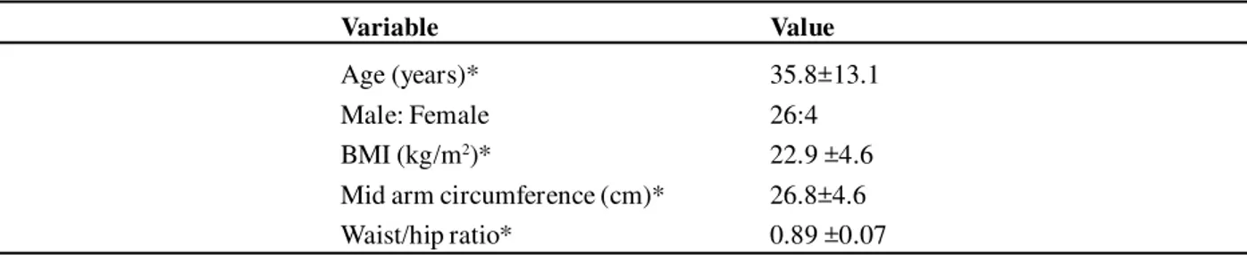
Table 2: Demographic characteristics of 30 patients with epilepsy who underwent polysomonography
Variable Value
Age (years)* 35.8±13.1
Male: Female 26:4
BMI (kg/m2)* 22.9 ±4.6
Mid arm circumference (cm)* 26.8±4.6 Waist/hip ratio* 0.89 ±0.07 * expressed as mean ± SD; BMI=body mass index
(14%) secondary generalization. Of these, 30 patients were taken up for the second stage of the study and underwent detailed evaluation and overnight in-hospital PSG in the sleep laboratory.
The baseline demographic characteristics of patients with epilepsy included in the study are shown in the Table 2. Majority of patients were in their third decade of life; males (n=26) outnumbered females (n=4). Their mean age was 35.8 ±13.1.
Median symptom duration of epilepsy was 72 [interquartile range (IQR) 72 to 180] months. All patients (n=30) manifested EDS (ESS score >10); 70% complained of snoring; (n=10) 33.3% had uncontrolled seizures. Median age of onset of seizures was 22 years (IQR 10.5 -37). GTCS was the most common seizure type (n=16; 53.3%) followed by CPS with (n=7; 23%) and without secondary generalization (n=4; 13%), among others.
Majority of patients had idiopathic epilepsy (n=22; 73%). Of the remaining 8 (27%) patients other causes included calcified granulonas and infarcts (3 patients each); head injury (n=1) and post surgery for craniopharyngioma (n=1).
There was equal distribution of nocturnal and diurnal periodicity of seizures (26.7% each). Seizures were well controlled in 20 (67%) patients; uncontrolled seizures occurred in 10 patients (33%). Most of the patients (n=24; 80%) required treatment with more than one AED.Six patients (20%) were receiving monotherapy with phenytoin, phenobarbitone and carbamazepine (2 patients each). Among patients receiving polytherapy, 18 were receiving 2 AEDs, while 6 patients were on triple drug regimen. Among patients receiving a 2-drug regimen, commonly used combination regimen was phenytoin plus carbamazepine (n=5) and phenobarbitone plus carbamazepine (n=4).
Table 3A: Anthropometric measurements
Variable Observations*
Body mass index (kg/m2) 22.9 ± 4.6
Neck length (cm) 15.6 ± 2.4
Neck circumference (cm) 34.3 ± 4.2
Percent predicted neck circumference 132 ± 14.5
Total height/neck length ratio 10.6 ± 1.20
Mid-arm circumference (cm) 26.8 ± 4.6
Waist circumference (cm) 84.0±14.2
Waist/hip ratio 0.89±0.07
* data are shown as mean ± SD Anthropometric measurements are shown in Table 3A. Only one patient had a neck circumference greater than 43 cm. As per the International Diabetes Federation (IDF) definition12central obesity observed in 12
(40%) patients (10 males). As per WHO criteria13 abdominal obesity was observed in 14
(47%) patients (11 males). As per t he International Obesity Task Force Guidelines for Asians,14 BMI was in the normal range in 13
(43.3%) patients; 5 (16.7%) patients were overweight; 6 (20%) were categorized as obese I; 3 patients (10%) each were categorized as obese II; and underweight.
None of patients had craniofacial deformities and otorhinolaryngological examination was normal in all patients. The EEG was abnormal in 8 patients; 4 patients had epileptiform discharges and 4 patients had predominant slow waves. The EEG was normal in the remaining 22 patients.Eight (26.7%) patients had abnormal imaging findings. These included calcified granulomas (n=3); left MCA infarct (n=2); right MCA infarct; left temporal lobe contusion; sequelae of surgery for craniopharyngio ma (n=1 patient each). Pulmonary function testing was normal in all the 30 patients.
The PSG data and prevalence of OSAS are presented in Tables 3B and 3C respectively. An
AHI of 5 or more was observed in 13 (43.3%) patients and all of them had evidence of EDS (ESS > 10). Thus, 43.3% of patients who underwent PSG had evidence of OSAS. Of these, 9 patients had mild OSAS; 3 had moderate OSAS and the remaining patient had severe OSAS (Table 3C; Figure 2). The patient with severe OSAS had no other risk factors except alcoholism.
Table 3C: Prevalence of obstructive sleep apnoea syndrome in 30 patients with epilepsy and excessive of daytime sleeping
Variable Observations
AHI [median (IQR)] 3.9 (1.4 - 11.5)
OSAS [No. (%)] 13/30 (43)
Mild OSAS (AHI > 5 <15) 09/30 (30) Moderate OSAS (AHI > 15 <30) 03/30 (10)
Severe OSAS (AHI > 30) 01/30 (03)
IQR = interquartile range
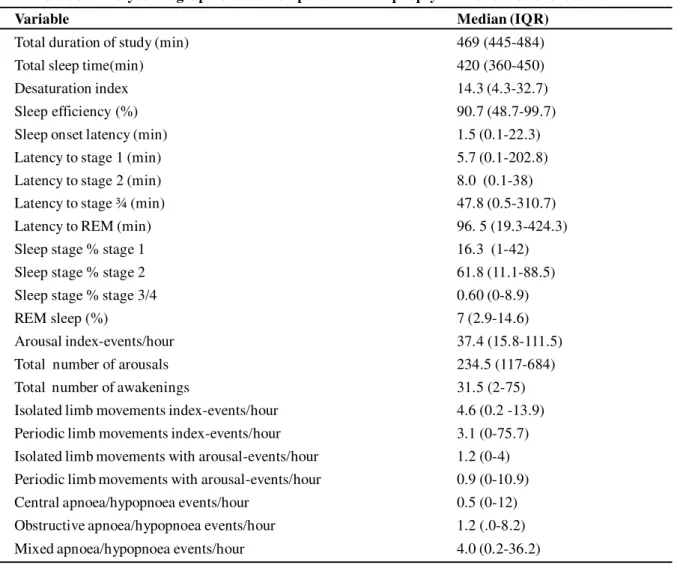
Table 3B: Polysomnographic data in 30 patients with epilepsy with evidence of excessive
Variable Median (IQR)
Total duration of study (min) 469 (445-484)
Total sleep time(min) 420 (360-450)
Desaturation index 14.3 (4.3-32.7)
Sleep efficiency (%) 90.7 (48.7-99.7)
Sleep onset latency (min) 1.5 (0.1-22.3)
Latency to stage 1 (min) 5.7 (0.1-202.8)
Latency to stage 2 (min) 8.0 (0.1-38)
Latency to stage ¾ (min) 47.8 (0.5-310.7)
Latency to REM (min) 96. 5 (19.3-424.3)
Sleep stage % stage 1 16.3 (1-42)
Sleep stage % stage 2 61.8 (11.1-88.5)
Sleep stage % stage 3/4 0.60 (0-8.9)
REM sleep (%) 7 (2.9-14.6)
Arousal index-events/hour 37.4 (15.8-111.5)
Total number of arousals 234.5 (117-684)
Total number of awakenings 31.5 (2-75)
Isolated limb movements index-events/hour 4.6 (0.2 -13.9)
Periodic limb movements index-events/hour 3.1 (0-75.7) Isolated limb movements with arousal-events/hour 1.2 (0-4) Periodic limb movements with arousal-events/hour 0.9 (0-10.9) Central apnoea/hypopnoea events/hour 0.5 (0-12) Obstructive apnoea/hypopnoea events/hour 1.2 (.0-8.2) Mixed apnoea/hypopnoea events/hour 4.0 (0.2-36.2)
IQR = interquartile range; REM = rapid eye movement sleep; AHI = apnoea-hypopnoea index; OSAS = obstructive sleep apnoea syndrome
between ESS AHI and other anthropometric variables is shown in Figure 3. A statistically significant positive correlation was observed between ESS and BMI (p<0.001); MAC (p=0.001); WC (p=0.001); HC (p=0.004); WHR (p=0.029). There was no statistically significant association between ESS and NL
(p=0.412); H/NL ratio (p=0.059); AHI (p=0.331); and oxygen desaturation index (0.368).
DISCUSSION
Table 3D: Comparison of demographic, clinical, anthropometric and laboratory variables between patients with and without OSAS
Variable No OSAS OSAS p-value
(n=17) (n=13)
Age at presentation (years) 32.5 ± 13.2 40.1 ± 12.1 0.18
Gender 0.113
Males 13 13
Females 04 00
Age at onset of seizures 26 ± 14.8 21.8 ± 15.2 0.462
Body mass index (kg/m2) 23.2 ± 3.9 22.6 ± 5.5 0.09
Neck length (cm) 15.5 ± 1.8 15.7 ± 3.1 0.29
Neck circumference (cm) 33.8 ± 3.8 35.1 ± 4.8 0.61
PPNC 134.5 ± 16.9 130.1 ± 12.6 0.42
Height to neck length ratio 10.47 ± 0.98 10.67 ± 1.47 0.39 Mid arm circumference (cm) 26.8 ± 3.9 26.8 ± 5.5 0.44 Waist circumference (cm) 83.5 ± 12.5 84.6 ± 16.7 0.46
Waist-hip ratio 0.88 ± 0.08 0.90 ± 0.05 0.46
Serum total cholesterol 180.7 ± 39.8 177.4 ± 41.1 0.84
Serum triglycerides 185.1 ± 58.0 171.6 ± 38.6 0.49
Serum HDL-cholesterol 36.1 ± 1.8 37.3 ± 2.6 0.14
OSAS=obstructive sleep apnoea syndrome; PPNC=percent predicted neck circumference; HDL=high density lipoproteins
Table 3E: Comparison of proportion of patients with a high BMI, high WC and high WHR between patients with and without OSAS
Variable No OSAS OSAS 2 p-value
(n=17) (n=13)
BMI (kg/m2)
>25 05 05
<25 12 08 0.271 0.446
High WC*
Yes 07 05
No 10 08 0.023 0.59
High WHR
Yes 09 05
No 08 08 0.621 0.339
* 90 cm for men, 80 cm for women
† > 0.90 cm for men, > 0.80 cm for women
BMI=body mass index; WC=waist circumference; WHR=waist-hip ratio; OSAS=obstructive sleep apnoea syndrome
deprivation may exacerbate seizures. Further, some seizures mainly occur during sleep. It is not uncommon for nocturnal seizures to be misdiagnosed as parasomnia and vice-versa. Recent observations indicate that primary sleep disorders such as OSA may aggravate epilepsy and treatment of these sleep disorders can
improve seizure control.18 The resulting
Figure 2: Burden of OSAS in patients with epilepsy
ESS=Epworth Sleepiness Scale; AHI=apnoea-hypopnoea index; OSAS=obstructive sleep apnoea syndrome
in patients with epilepsy is clinically important to ensure optimal treatment of both the conditions.
Some published questionnaire based studies19-22 had revealed a higher occurrence of
EDS in patients with epilepsy.In the present study, among 426 patients with epilepsy who were screened, EDS was almost six-times more co mmon compared to co ntro l subjects (p<0.0001; Table 2).
Several causes, such as, manifestation of seizure disorder, adverse effect of AEDs, uncontrolled seizures, increased sleep latency and number of awakenings during night, reduction or fragmentation of REM sleep, among others have been postulated to be responsible for increased EDS in these patients.23,24 The observations from the present
study indicate that the burden of EDS in Indian patients with epilepsy appears to be considerable and of similar magnitude to that observed in studies from the developed world even after allowing for the differences in the semiology of seizure disorders.
Since 1981, there have been anecdotal reports of epilepsy and OSA coexisting in the same subject.5,25,26 Subsequently, some published
studies from the west19,27-30had evaluated the
problem of SDB in adult patients with epilepsy utilizing PSG. In-hospital overnight PSG is the method of choice for the diagnosis of SDB. However, PSG facilities are not widely available in public sector hospitals of Andhra Pradesh state, except at our institute. Given this limitation and the cost factor, in-hospital overnight PSG could be done in 30 of the 98 patients with EDS. In the present study 43.3% of patients who underwent PSG had evidence of OSAS. In comparison, in community based st udies assessing the burden o f OSAS published from India employing PSG, the prevalence of OSAS ranged from 3.6% to 17%.8,9,31 These observations suggest that SDB
is an important underdiagnosed problem in patients with epilepsy.
1
6
Figure 3: Correlation matrix showing correlation between ESS AHI and other anthropometric variables All data are expressed as r (p-value)
ESS=Epworth Sleepiness Scale; BMI=body mass index; NC= neck circumference (cm); NL=neck length; PPNC=% predicted neck circumference; H/NL=height to neck length ratio; MAC=mid-arm circumference; WC=waist circumference; HC=hip circumference; WHR=waist-to hip-ratio; AHI=aponea-hyppopnoea index; DI=desaturation index
ep
d
is
o
rd
e
re
d
b
re
a
th
in
g
i
n
e
p
ile
p
sy
V
e
n
g
a
m
m
a
e
t
a
observed among women.10,32 All the 13 patients
who were diagnosed to have OSAS in the present study were men (Table 3D). Although it is not clear as to why OSA is more common in men than women, anatomical and functional properties of the upper airway, ventilatory response to the arousal from sleep, increased fat deposition around pharyngeal airway, hormonal differences, among others have been po stulated to be t he causes fo r male preponderance.32.
In the present study, majority of the patients (n=26) had never smoked and had never consumed alcoho l. However, alco hol consumption was the only documented risk factor in a patient with severe OSAS. In susceptible subjects, alcohol has been shown to relax upper airway dilator muscles, increase upper airway resistance and induce OSA. Therefore, alcohol intake has the potential to prolong apnea duration, suppress arousals, increase frequency of occlusive episodes and worsen the severity of hypoxemia.32,33 It is likely
that such factors could be operational in this patient.
Some studies28,34 have shown that patients with
epilepsy and OSAS were older, had a higher BMI and had a later onset of seizures than people with epilepsy without OSAS.28,34
However, there was no statistically significant difference in the demographic, anthropometric and laboratory variables listed above between patients with and without OSAS (Tables 3D and 3E) in the present study. These associations were not significant in other studies35,36 as well.
These issues need clarification in community based studies with a larger sample size. In some studies carried out in subjects with OSAS, the ESS score significantly correlated with the oxygen desaturation index (ODI), AHI, and BMI.37,38 In the present study, a statistically
significant positive correlation was observed between ESS and BMI (p=0.001); MAC
(p=0.001); WC (p=0.001); HC (p=0.004); WHR (p=0.029). There was no statistically significant association between ESS and NL (p=0.412); H/NL ratio (p=0.059); AHI (p=0.331); and desaturation index (0.368). As the sample size of patients with epilepsy who had undergone PSG in the present study is small (n=30), these correlations need to be further evaluated in studies with a large sample size. There was no statistically significant difference in the mean serum lipid profile among epilepsy patients with and without OSAS in the present study (Table 3D). Furthermore, the prevalence of metabolic syndrome was similar in epilepsy patients with and without OSAS in the present study (5/13 Vs 6/17; p=0.99). These findings are similar to the observations from a study9
conducted at New Delhi in which the authors reported that OSA had no independent association with lipid abnormalities, insulin resistance, serum leptin and adiponectin levels. In the present study, among the patients who have undergone PSG (n=30), a higher proportion of patients with nocturnal seizures had OSAS compared with those with diurnal seizures and combined periodicity (7/8 Vs 6/ 22; p=0.009). It has been reported that nocturnal seizures reduced the amount of stage II and IV sleep and increased the amount of stage I sleep with a consequent reduction in the sleep efficiency and increased drowsiness the day after.40 However, such an association has
not been documented in other studies.28
A higher proportion of patients in uncontrolled seizures had OSAS as compared with those with controlled seizures in the present study (7/10 Vs 6/20; p=0.045). SDB is likely to have contributed to poor control of seizures in these patients. OSA has been observed to be more frequent among patients with refractory epilepsy, and has been reported in one third of patients investigated for epilepsy surgery.34
benzodiazepines by increasing upper airway collapsibility and depressing arousal mechanisms may worsen OSAS.41 AEDs, such
as valproic acid and carbamazepine that can cause weight gain are also known to worsen OSAS.42 In the present study, there was no
statistically significant difference in the prevalence of OSAS among patients receiving a single AED or those receiving more than one AED for seizure control (3/7 Vs 10/23; p=0.66). The effect of the AED on SDB should be kept in mind when patients with epilepsy, especially those with EDS are initiated on antiepileptic treatment.
In-hospital, overnight PSG could not be carried out in all the 98 patients with EDS (ESS >10) but could be done only in 30 patients due to logistic reasons. So, meaningful multivariable analysis for defining the predictors of OSAS among patients with epilepsy could not be done.The present study also did not attempt to study the effect of CPAP treatment on seizure control due to logistic reasons.
Our observations suggest that SDB appears to be an underdiagnosed disease in patients with epilepsy from India. Increased awareness regarding the condition, screening of patients with EDS and overnight in-hopsital PSG can help in early recognition of this condition and may facilitate institution of CPAP treatment.
REFERENCES
1. Bur n eo JG, Tellez-Zen ten o J, Wiebe S. Understanding the burden of epilepsy in Latin America: a systematic review of its prevalence and incidence. Epilepsy Res 2005;66:63-74.
2. Passouant P. Historical aspects of sleep and epilepsy. Epilepsy Res Suppl 1991;2:19-22. 3. Chokroverty S. Sleep and its disorders. In: Bradley
WG, Daroff RB, Fenichel GM, Jankovic J, editors Neurology in clinical practice. 5th edition Philadelphia: Butterworth Heinemann Elsevier; 2008.p.1982.
4. Young T, Palta M, Dempsey J, Skatrud J, Weber S, Badr S. The occurrence of sleep-disordered
breathing among middle-aged adults. N Engl J Med 1993;328:1230-5.
5. Devin sky O, Eh r en berg B, Bar thlen GM, Abramson HS, Luciano D. Epilepsy and sleep apnea syndrome. Neurology 1994;44:2060-4. 6. Proposal for revised classification of epilepsies and
epileptic syndromes. Commission on Classification and Terminology of the International League Against Epilepsy. Epilepsia 1989;30:389-99. 7. Johns MW. A new method for measuring daytime
sleepiness: the Epworth sleepiness scale. Sleep 1991;14:540-5.
8. Sharma SK, Kumpawat S, Banga A, Goel A. Prevalence and risk factors of obstructive sleep apnea syndrome in a population of Delhi, India. Chest 2006;130:149-56.
9. Vijayan VK, Patial K. PRevalence of obstructive sleep apnea syndrome (OSAS) in delhi, India. Chest 2006;130:92S .
10. Sharma SK, Ahluwalia G. Epidemiology of adult obstructive sleep apnoea syndrome in India. Indian J Med Res 2010;131:171-5.
11. Davies RJ, Stradling JR. The relationship between neck circumference, radiographic pharyngeal anatomy, and the obstructive sleep apnoea syndrome. EurRespir J 1990;3:509-14.
12. The IDF consensus worldwide definition of the metabolic syndrome [Internet]. International Diabetes Federation. [cited 2016 Feb 26]. Available from: http://www.idf.org/front
13. Alberti KG, Zimmet PZ. Definition, diagnosis and classification of diabetes mellitus an d its complications. Part 1: diagnosis and classification of diabetes mellitus provisional report of a WHO consultation. Diabet Med J Br DiabetAssoc 1998;15:539-53.
14. The International Association for the Study of Obesity and the International Obesity Task Force. The Asia-Pacific perspective: redefining obesity and its treatment Australia: IASO and IOTF. 2000. 15. Mohan A, Kumar SN, Rao MH, Bollineni S, Manohar IC. Acute accidental exposure to chlorine gas: clinical presentation, pulmonary functions and outcomes. In dian J Ch est Dis Allied Sci 2010;52:149-52.
16. Mohan A, Premanand R, Reddy LN, Rao MH, Sharma SK, Kamity R, et al. Clinical presentation and predictors of outcome in patients with severe acute exacer bation of chr on ic obstructive pulmonary disease requiring admission to intensive care unit. BMC Pulm Med 2006;6:27. 17. Sleep-related breathing disorders in adults:
measurement techniques in clinical research. The Report of an Amer ican Academy of Sleep Medicine Task Force. Sleep 1999;22:667-89. 18. Eriksson SH. Epilepsy and sleep. CurrOpinNeurol
2011;24:171-6.
19. Malow BA, Bowes RJ, Lin X. Predictors of sleepin ess in epilepsy patien ts. Sleep 1997;20:1105-10.
20. Piperidou C, Karlovasitou A, Triantafyllou N, Terzoudi A, Constantinidis T, Vadikolias K, et al. Influence of sleep disturbance on quality of life of patients with epilepsy. Seizure 2008;17:588-94. 21. Khatami R, Zutter D, Siegel A, Mathis J, Donati
F, Bassetti CL. Sleep-wake habits and disorders in a series of 100 adult epilepsy patients—a prospective study. Seizure 2006;15:299-306. 22. de Weerd A, de Haas S, Otte A, Trenité DK-N,
van Erp G, Cohen A, et al. Subjective sleep disturbance in patients with partial epilepsy: a questionnaire-based study on prevalence and impact on quality of life. Epilepsia 2004;45:1397-404.
23. Hoeppner JB, Garron DC, Cartwright RD. Self-reported sleep disorder symptoms in epilepsy. Epilepsia 1984;25:434-7.
24. Car rion MJM, Nunes ML, Mar tin ez JVL, Portuguez MW, da Costa JC. Evaluation of sleep quality in patients with refractory seizures who undergo epilepsy surgery. Epilepsy Behav EB 2010;17:120-3.
25. Tartara A, Manni R. Epilepsy and sleep apnea syndrome: a two cases report. Boll Lega It Epilessia 1985;50:247-8.
26. Wyler AR, Weymuller EA. Epilepsy complicated by sleep apnea. Ann Neurol 1981;9:403-4. 27. Malow BA, Fromes GA, Aldrich MS. Usefulness
of polysomnogr aph y in epilepsy patients. Neurology 1997;48:1389-94.
28. Manni R, Terzaghi M, Arbasino C, Sartori I, Galimberti CA, Tartara A. Obstructive sleep apnea in a clinical series of adult epilepsy patients: frequency and features of the comorbidity. Epilepsia 2003;44:836-40.
29. Chihorek AM, Abou-Khalil B, Malow BA. Obstructive sleep apnea is associated with seizure occur r en ce in older adults with epilepsy. Neurology 2007;69:1823-7.
30. Weatherwax KJ, Lin X, Marzec ML, Malow BA. Obstructive sleep apnea in epilepsy patients: the Sleep Apn ea scale of the Sleep Disorder s Questionnaire (SA-SDQ) is a useful screening
instrument for obstructive sleep apnea in a disease-specific population. Sleep Med 2003;4:517-21. 31. Reddy EV, Kadhiravan T, Mishra HK, Sreenivas
V, Handa KK, Sinha S, et al. Prevalence and risk factors of obstructive sleep apnea among middle-aged urban Indians: a community-based study. Sleep Med 2009;10:913-8.
32. Lam JCM, Sharma SK, Lam B. Obstructive sleep apnoea: definitions, epidemiology & natural history. Indian J Med Res 2010;131:165-70. 33. Mitler MM, Dawson A, Henriksen SJ, Sobers M,
Bloom FE. Bedtime ethanol increases resistance of upper airways and produces sleep apneas in asymptomatic snorers. Alcohol ClinExp Res 1988;12:801-5.
34. Malow BA, Levy K, Maturen K, Bowes R. Obstructive sleep apnea is common in medically r efr actor y epilepsy patien ts. Neur ology 2000;55:1002-7.
35. Malow BA, Weatherwax KJ, Chervin RD, Hoban TF, Marzec ML, Martin C, et al. Identification and treatment of obstructive sleep apnea in adults and children with epilepsy: a prospective pilot study. Sleep Med 2003;4:509-15.
36. Malow BA, Foldvary-Schaefer N, Vaughn BV, Selwa LM, Chervin RD, Weatherwax KJ, et al. Treating obstructive sleep apnea in adults with epilepsy: a randomized pilot trial. Neurology 2008;71:572-7.
37. Thong JF, Pang KP. Clinical parameters in obstructive sleep apnea: are there any correlations? J Otolaryngol - Head Neck Surg J Oto-Rhino-LaryngolChirCervico-Faciale 2008;37:894-900. 38. Chen R, Xiong K, Lian Y, Huang J, Zhao M, Li J,
et al. Daytime sleepiness and its determining factors in Chinese obstructive sleep apnea patients. Sleep Breath Schlaf Atm 2011;15:129-35. 39. Sharma SK, Kumpawat S, Goel A, Banga A,
Ramakrishnan L, Chaturvedi P. Obesity, and not obstructive sleep apnea, is responsible for metabolic abnormalities in a cohort with sleep-disordered breathing. Sleep Med 2007;8:12-7. 40. Bazil CW, Castro LH, Walczak TS. Reduction of
rapid eye movement sleep by diurnal and nocturnal seizures in temporal lobe epilepsy. Arch Neurol 2000;57:363-8.
41. Legros B, Bazil CW. Effects of antiepileptic drugs on sleep architecture: a pilot study. Sleep Med 2003;4:51-5.