1Department of Neurosurgery, Brazilian Air Force Hospital, Rio de Janeiro, RJ, Brazil; 2M.D., Ph.D, Head of Department.
Received 4 October 2005, received in final form 16 January 2006. Accepted 17 February 2006
Dr. José A. Landeiro - Brazilian Air Force Hospital - Estrada do Galeão 4101 - Rio de Janeiro RJ - Brasil. E-mail: [email protected]
TECTAL PLATE TUMORS
Bruno C.R. Lázaro
1, José A. Landeiro
2A B S T R A C T - Tectal plate is a rare location for a tumor. Many papers have described diff e rent types of pathol-ogy arising in that location including tumors, vascular lesions, inflamatory and infectious processes. In this paper we describe our experience in treating seven patients with tectal plate lesions, with different ages and types of pathology: five patients presented with low grade gliomas, one with lung cancer metastasis and the last presenting with a tectal plate cavernoma. Open surg e ry was perf o rmed in three cases (due to tumor enlargement or need for the exact diagnosis). In the other cases, the treatment of non-comunicat-ing hydrocephalus was the only treatment employed. The prognosis is of course dependent on the under-lining pathology. In our series, except in the metastatic tumor case and the cavernoma, the other types of lesion consisted of low grade gliomas. These lesions represent a different type of brain stem tumor shar-ing a common good prognosis, with a benign behavior. We believe that tectal tumors must be managed case by case. When a patient presents with a benign lesions in the tectal region, treating the main symp-tom – hydrocephalus - should be the first attempt in management of these lesions.
KEY WORDS: brain stem, tectal tumors, brain stem tumors, third ventriculostomy.
Tumores da região tectal
RESUMO - Tu m o res na região do teto mesencefálico são raros. Vários tipos de lesões como tumores, lesões v a s c u l a res, inflamatórias e infecciosas localizam-se nesta região. Nós revimos o tratamento adotado em sete pacientes com diferentes tipos de lesões tectais: cinco pacientes apresentando gliomas de baixo grau, um paciente com lesão metastática proveniente de câncer de pulmão e um com cavernoma. O tratamen-to cirúrgico com abordagem direta da lesão foi realizado em três casos (devido ao aumentratamen-to do volume tumoral ou quando houve necessidade da confirmação diagnóstica). Nos demais casos o tratamento para a hidrocefalia não-comunicante foi o método empregado. O prognóstico dessas lesões é baseado no tipo de patologia em questão. Em nossa série, com exceção do caso de metástase e do paciente com cavern o m a , as demais lesões foram gliomas de baixo grau. Estas lesões re p resentam um subgrupo diferenciado de t u m o res de tronco encefálico, apresentando bom prognóstico e tendo comportamento benigno com sobre-vida elevada. Acreditamos que tumores da região tectal devam ser avaliados caso a caso. Na hipótese diag-nóstica de uma lesão benigna, o tratamento do principal complexo sindrômico – hidrocefalia não-comu-nicante – é provavelmente a melhor conduta a ser empregada.
PALAVRAS-CHAVE: tronco cerebral, tumores da região tectal, ventriculostomia.
long survival rate - almost more than 5 years. The t h i rd subgroup is re p resented by focal tectal gliomas1. On the other hand, brain stem tumors in adults -especially gliomas - are poorly understood in term s of low incidence and diff e rent pattern of behavior. Over all, it is actually observed that the prognosis of brain stem tumors is better in adults than in childre n . The peak incidence prevailed in the third and fourt h decade in adults and in the first decade in childre n2. Among tumors found in the tectal plate, the most common is the astrocytoma, but other types have been described, such as oligodendroglioma, ependy-Brain stem tumors are a wide described
moma, ganglioglioma, medulloblastoma3, primitive n e u ro e c t o d e rmaltumors, metastasis; as well as lipo-m a1 1, melanoma1 2, dysembryoplastic neuro e p i t e l i a l tumor (DNT)1 3, cavernomas, abscess and periaque-ductal gliosis.
We present seven cases of tumors arising in the tectal plate managed in our institution.
METHOD
We present seven cases of patients with mesencephal-ic tectal plate tumors treated in our institution, in a re t ro-spective analysis, between 1994 – 2005. There are 3 m ale and 4 female patients in this group with ages comprising 17 to 70 years-old.
Five patients presented Parinaud syndrome; all cases p resented with noncommunicating hydrocephalus. Image studies were perf o rmed in all cases involving computed tomography scan (CT) and magnetic ressonance image (MRI) demonstrating lesions with the aspect of a low grade glio-ma in five cases, one case of high grade tumor (lung can-cer metastasis), and the other presenting a vascular lesion (cavernoma).
The follow up period was over 8 years (1 to 8 years in average); open surgery was performed in 3 cases - 2 cases (see ilustrative cases below), cavernoma and the metasta-sis case - obtaining a definitive diagnometasta-sis; third ventricu-lostomy was perf o rmed in the other fi ve cases (Table); in one case two pro c e d u res were necessary (see ilustrative cases below). There was one death – the metastatic tumor due to progressive disease (Table).
Ilustrative cases
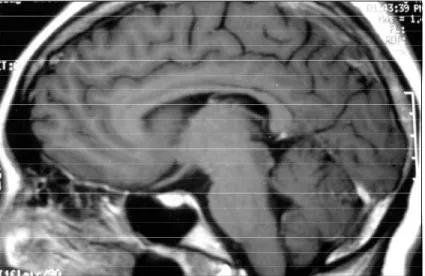
Case 1 –A 26-year-old woman presented to our hospi-tal with headache, nausea, papilledema and visual blur-ring. The CT scan revealed a noncommunicating hydro-cephalus and a hypodense lesion on the tectal region (Fig 1). MRI demonstrated a tectal plate lesion (Fig 2) with lack of contrast enhancement.
She initially underwent ventricular-peritoneal shunt placement achieving adequate control and impro v e m e n t of the symptoms, returning to normal activities and work afterwords.
One year later she returned to neurosurgical care pre-senting with worsening of the symptoms associated with incomplete Parinaud syndrome. A new MRI was perf o rm e d and showed hydrocephalus and an increase of the lesion size (Fig 3). The shunt appeared to be malfunctioned. The tumor was removed using the semi-sitting position achiev-ing a gross total removal of the lesion, as presented in MRI (Fig 4). She experienced an improvement of the symptoms, being discharged from the hospital one week later.
Histopatological analysis revealed a grade II astro c y-toma.
Case 2 – A 24-year-old man was admitted with a histo-ry of somnolence, disorientation, headache and Parinaud syndrome. He had undergone placement of a ventricular-peritoneal shunt 10 years before in another hospital for a diagnosis of “hydrocepha lus” (history collected from his mother). A CT scan was perf o rmed showing a noncomuni-cating hydrocephalus. MRI was perf o rmed corro b o r a t i n g the diagnosis showing a T1 hypointense lesion located at the tectal plate.
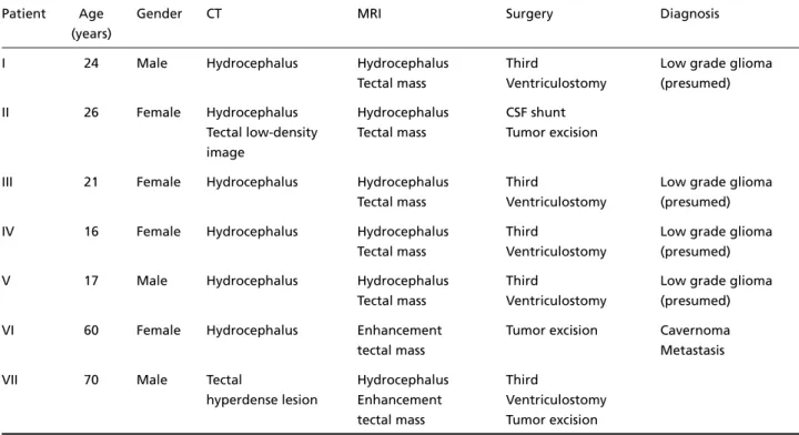
Table 1. tectal tumors case reports.
Patient Age Gender CT MRI Surgery Diagnosis
(years)
I 24 Male Hydrocephalus Hydrocephalus Third Low grade glioma
Tectal mass Ventriculostomy (presumed)
II 26 Female Hydrocephalus Hydrocephalus CSF shunt
Tectal low-density Tectal mass Tumor excision image
III 21 Female Hydrocephalus Hydrocephalus Third Low grade glioma
Tectal mass Ventriculostomy (presumed)
IV 16 Female Hydrocephalus Hydrocephalus Third Low grade glioma
Tectal mass Ventriculostomy (presumed)
V 17 Male Hydrocephalus Hydrocephalus Third Low grade glioma
Tectal mass Ventriculostomy (presumed)
VI 60 Female Hydrocephalus Enhancement Tumor excision Cavernoma
tectal mass Metastasis
VII 70 Male Tectal Hydrocephalus Third
Fig 2. MRI, T1 weighted sagital view, showing a non-enhanced tectal mass.
Fig 1. Brain CT scan in axial view presenting a noncommunicating hydrocephalus. Note the lowden -sity lesion on the tectal plate.
Fig 4. Postoperative MRI, T1 weighted sagital view, showing no gadolinium enhancement on the tec -tal plate.
Fig 3. MRI sagital view, T2 FLAIR, presenting a tectal mass leading to obliter -ation of the cerebral aqueduct.
He underwent an endoscopic third ventriculostomy with an imediate post opperative improving of the symptoms; the Parinaud syndrome was the last feature to disappear, occuring after one month. One year later he had re c c u rre n-ce of the symptoms. A new radiologic study was perf o rm e d showing once again a non-communicating hydro c e p h a l u s but the average size of the tectal tumor remained the same. A second third ventriculostomy was perf o rmed impro v i n g the symptomatology. He was discharged from the hospital 5 days after the procedure without symptoms (Fig 5).
DISCUSSION
In comparison to children, brain stem gliomas in adults is a less understood disease and present a much lower incidence (<2% of gliomas)1. The survival rate however can be very much longer with the peak inci-dence in the third and fourth decades2. The duration of symptoms is generally shorter in children while in adults it tends to be longer with tectal tumors, fourt h ventricle dorsally exophytic tumors and cerv i c o m e-d u l l a rygliomas having a more favorable pro g n o s i s when compared to difuse lesions.
Tectal plate gliomas have been re p o rted as a par-t i c u l a ry indolenpar-t lesion ofpar-ten remaining spar-table in size for many years. The majority of these lesions were described as low grade gliomas, presented with late-onset aqueductal stenosis often without associated brain stem signs8. The average age at the time of diagnosis is about 10 years in children, with the majo-rity of cases presenting with raised intracranial pre s-s u re s-secondary to obs-struction of the Sylvian aque-d u c t1 4, with some other cases presented with Pari-naud syndro m e1 5like the patients observed in our ilustrative cases, although this is also an uncommon f e a t u re. Even before the development of computed t o m o g r a p h y, tectal tumors were an unre c o g n i z e d because of late onset hydro c e p h a l u s6, once said that this can be the smallest lesion that can lead to death of the patient.
The radiologic investigation of these tumors have been up graded in the CT scan era with better visu-alization of the tectal region; but the majority of le-sions continue to appear as noncommunicating hy-d rocephalus alone, although calcification or a hypo-dense lesion on the tectal plate can be observed. MRI of these tumors reveals tectal distortion or thickning caused by a localized mass, leading to aqueductal c o m p ression and hydro c e p h a l u s1 6; characteristic T1 hypointensity and T2 hyperintensity1 7 - 2 1. MRI is an accurate and noninvasive method of diagnosis that can be indicated in all cases of late onset hydro c e p h a-lus and aqueductal obstruction, especially in adults.
Even when present, contrast enhancement after ga-dolinium injection was an independent factor of tumor grading17.
The characteristics of the lesion seen on the MRI is of course dependent on its pathologic basis. In our series we presented 5 cases of patients baring a tec-tal low grade glioma, with the typical MRI appeare n-ce just as described above. The other two cases – a brainstem metastasis and a tectal plate cavernoma -a p p e -a red -as -an irregul-ar enh-ancing lesion -and -a sm-all v a s c u l a r-like lesion presenting an impregnation of hemossiderin, respectivelly.
The prognosis of tectal plate lesion is a much de-bated issue in neuro s u rgical publications. It is re a s o-nable to expect that a malignant lesion, brain stem metastasis and rapidally infiltrating lesions pre s e n t a poor prognosis. But even in cases of tectal gliomas some controversies had arise.
Initially the pathology of intrinsic tectal lesions w e re considered to be similar to other brainstem tu-mors. In fact this was later discovered as not tru e . Many papers have oriented the presence of this tumor as a special site of low grade gliomas4-6,9,10,18. These tumors appear to present a better pro g n o s i s given to a slow growing rate and boundaries displa-cement instead of an infiltrative behaviour. However, the management of the patients remains contro v e r-sial. The majority of authors oriented the manage-ment including a mandatory histopatological analy-sis to acertain the low grade mark, and just after-w o rds the adequate treatment is traced. We think that the conjunction of history, age, neuro exam and MRI appearence of a tectal lesion can be very suspi-cious for a low grade tumor. In our cases, there is a tendency for treating the symptom – usually non-communicating hydrocephalus – as the first option, but it is not always possible when there is a high sus-picion of another type of tectal lesion, like the cav-ernoma and the lung cancer metastasis cases in our series.
S u rgical treatment consists on tumor resection or open biopsy; generally the surgical approach is that described for pineal region lesions, like the suboc-cipital-transtentorial or supracere b e l l a i n f r a t e n t o r-ial approach, prefered by this paper´s senior author and routinelly perf o rmed in this institution adopt-ing the semi-sittadopt-ing position.
e-phalus, a rigid endoscope can be utilized to perf o rm a third ventriculostomy, as we described, with no ma-jor technical complications; the visibility of the inner s u rface of the third ventricle, with all stru c t u res in-volved is very good22.
Tectal tumors may extrude from the tectum into the lumen of the cerebral aqueduct and subsequent-ly protrude to the third ventricle, pushing away the posterior commissure and enlarging the orifice. Some authors consider this lesion as intraventricular ones, treating each lesion individually23.
With the advent of the flexible neuroendoscope other types of therapy could be perf o rmed. Potential t reatment options include shunt placement2 4a n d aqueductal plasty2 4 , 2 5. The third ventriculostomy suc-cess and failure rate is similar to those cere b r a l - s p i n a l fluid (CSF) shunts – about 30%24.
In conclusion, tectal tumors re p resent a diff e re n-tiated category in neuro - o n c o l o g y. Despite a variety of lesion encountered in this region, most publictions indicate that low grade glioma is most pre v a-lent. However, the major difficulty is re g a rding the adequate treatment of these lesions. A number of a rticles orients an invasive approach consisting of sur-gical treatment, given the need for mass re d u c t i o n or just to obtain histopatological sample5,27,28; other authors decline to conservative treatment, justified by the benign behavior and slow growth of this tu-m o r s4 , 6 , 8 , 1 9 , 2 9 , 3 0. In our institution, the decision depends on individual analysis. We give pre f e rence in tre a t-ing the major symptom – hydrocephalus – with CSF shunts or preferably endoscopic third ventriculosto-m y. Our study deventriculosto-monstrate that surg e ry should be indicated when there is evidence of tumor enlarge-ment or in cases when the definitive diagnosis is imperative.
Acknowledgement – We thank Igor de Castro M .D
for editorial assistance and José Francisco Salomão M.D. for his support
REFERENCES
1. Guillamo JS, Monjour A, Taillandier L, et al. Brain stem gliomas in adults: prognostic factors and classification. Brain 2001;124:2528-2539. 2. Selvapandian S, Rajshekhar V, Chandy MJ. Brain stem glioma: com-parative study of clinico-radiological presentation, pathology and out-come in children and adults. Acta Neurochir 1999;141:721-727. 3. Pollack IF, Hoffman HJ, Humphreys RP, Becker L. The long-term
out-come after surgical treatment of dorsally exophytic brain stem gliomas. J Neurosurg 1993;78:859-863.
4. May PL, Blaser SI, Hoffman HL, et al. Benign intrinsic tectal “tumors” in children. J Neurosurg 1991;74:867-871.
5. Lapras CI,. Bognar L, Turjman E, et al. Tectal plate gliomas. Part I: micro-surgery of the tectal plate gliomas. Acta Neurochir 1994;126:76-83. 6. Yeh DD, Warnick RE, Ernst RJ. Management strategy for adult patients
with dorsal midbrain gliomas. Neurosurgery 2002;50:740.
7. Epstein FJ, Farmer JP. Brain stem glioma growth patterns. J Neuro s u rg 1993;78:408-412.
8. Pollack IF, Pang D, Albright AL. The long term outcome in childre n with late-onset aqueductal stenosis resulting from benign intrinsic tec-tal tumors. J Neurosurg 1994;80:681-688.
9. Vandertop WP, Hoffman HJ, Drake JM, et al. Focal midbrain tumors in children. Neurosurgery 1992;31:186-194.
10. Wang C, Zhang J, Liu A, et al. Surgical treatment of primary midbrain gliomas. Surg Neurol 2000;53:41-51.
11. Uchino A, Hasuo K, Matsumoto S, Masuda K. MRI of dorsal mesensephalic lipomas. Clin Imaging 1993;17:12-16.
12. Weiding SM, Press GA, HasselinkJR. Am J Neuroradiol 1988;9:214-215. 13. K u r t k a y a - Yapicier O, Elmaci I, Boran B, et al. Dysembrioplastic neu-roepithelial tumor of the midbrain tectum: a case report. Brain Tu m o r Pathol 2002;19:97-100.
14. Gomez-Gonsalvez FA, Menor F, Morant A, et al. Tectal tumors in pae-diatrics. A review of eight patients. Rev Neurol 2001;33:605-611. 15. Moffie D, Ongerboer de Visser BW, Stefanko SZ. Parinaud syndrome.
J Neurol Sci 1983;58:175-183.
16. Antunes NL, Tavora L, Souweidane M. Globular glioma of the tectum. Pediatr Neurol 1999;21:492-495.
17. Bognar L, Turjman F, Villanyi E, et al. Tectal plate gliomas Part II: CT scans and MRI imaging of tectal gliomas. Acta Neurochir 1994;127: 48-54.
18. Ramina R, Neto MC, Fernandes YB et al. Intrinsic tectal low grade astro-cytomas. Arq Neuropsiquiatr 2005;63:40-45.
19. Bowers DC, Georgiades C, A ronson LJ et al. Tectal gliomas: natural history of an indolent lesion in pediatric patients. Pediatr Neuro s u rg 2000; 32:265-271.
20. Hamilton MG, Lauryssen C, Hagen N. Focal midbrain glioma: long term survival in a cohort of 16 patients and the implications for man-agement. Can J Neurol Sci 1996;23:204-207.
21. Smith RR, Zimmerman RA, PackerRJ et al. Pediatric brain stem glioma; post-radiation clinical and radiographic follow-up. Neuro r a d i o l o g y 1990;32:265-271.
22. Oka K, Go Y, Kin Y, Tomonaga M. An observation of the third ventri-cle under flexible fiberoptic ventriculoscope: normal stru c t u re. Surg Neurol 1993;40:273-277.
23. Oka K, Kin Y, Go Y, et al. Neuroendoscopic approach to tectal tumors: a consecutive series. J Neurosurg 1999;91:964-970.
24. Bulsara KR, Villavicencio AT, Shah AJ, et al. Successful aqueductal plas-ty and stenting for tectal plate tumor after failed third ventricuostomy: a case report. Surg Neurol 2003;59:58-62.
25. Oka K, Yamamoto M, Ikeda K, Tomonaga M. Flexible endoneuro s u r-gical therapy for aqueductal stenosis. Neurosurgery 1993;33:236-242. 26. S c h roeder HW, Gaab MR. Endoscopic aqueductoplasty. Neuro s u rg e r y
1999;45:508-517.
27. Koziarsky A, Zielinski G, Podgorski JK, Wa rczynska A. One stage removal of periaqueductal glioma in adult via infratentorial suprac-e rsuprac-ebsuprac-ellar and transaqusuprac-eductal approachsuprac-es. Acta Nsuprac-eurochir 2004;146:169-173.
28. Robertson PL, Muraszko KM, Bru m b e rg JA, Axtell RA, Dauser RC, Turrisi AT. Pediatric midbrain tumors: a benign subgroup of brain stem gliomas. Pediatr Neurosurg 1995;22:65-73.
29. S q u i res LA, Allen JC, Abbot R, Epstein FJ. Focal tectac tumors: man-agement and prognosis. Neurology 1994;44:953-956.