RBCCV 44205-1535 DOI: 10.5935/1678-9741.20140052
Comparison of fractal dimension and Shannon
entropy in myocytes from rats treated with
tryptophan-glutamate and
histidine-tryptophan cetoglutarate
Comparação da dimensão fractal e entropia de Shannon em miócitos de ratos submetidos à cardioplegia
utilizando solução histidina-triptofano-cetoglutarato e histidina-triptofano com glutamato
Marcos Aurélio Barboza de Oliveira
1, MD; Antônio Carlos Brandi
2, MD; Carlos Alberto dos Santos
2,
MD; Paulo Henrique Husseni Botelho
2, MD; José Luís Lasso Cortez
3, MD; Moacir Fernandes de
Godoy
4, PhD; Domingo Marcolino Braile
5, MsC, PhD
1Faculdade de Medicina de São José do Rio Preto (FAMERP), São José
do Rio Preto, SP, Brazil; Centro Universitário de Votuporanga (UNIFEV), Santa Casa Votuporanga, Votuporanga, SP, Brasil.
2Hospital de Base São José do Rio Preto. São José do Rio Preto, SP, Brazil. 3Santa Casa Votuporanga, Votuporanga, SP, Brazil.
4Departamento de Cardiologia e Cirurgia Cardiovascular da Faculdade de
Medicina de São José do Rio Preto (FAMERP), São José do Rio Preto, Brazil. Transdisciplinary Unit for Chaos and Complexity Study (NUTECC--CNPq), São José do Rio Preto, Brazil.
5Faculdade de Medicina de São José do Rio Preto (FAMERP), São José do
Rio Preto, SP, Brazil.
Correspondence address:
Marcos Aurélio Barboza de Oliveira Avenida República do Líbano,2700 - casa 80
São José do Rio Preto. SP. Brazil - Zip code: 15092-440 E-mail: [email protected]
This study was carried out at S Faculdade de Medicina de São José do Rio Preto (FAMERP), São José do Rio Preto, SP, Brazil.
No inancial support.
Article received on November 19th, 2013
Article accepted on February 23rd, 2014
Abstract
Introduction: Solutions that cause elective cardiac arrest
are constantly evolving. but the ideal compound has not yet been found. The authors compare a new cardioplegic solution with histidine-tryptophan-glutamate (Group 2) and other one with histidine-tryptophan-cetoglutarate (Group 1) in a model of isolated rat heart.
Objective: To quantify the fractal dimension and Shannon
entropy in rat myocytes subjected to cardioplegia solution using histidine-tryptophan with glutamate in an experimental model. considering the caspase markers. IL-8 and KI-67.
Methods: Twenty male Wistar rats were anesthetized and
heparinized. The chest was opened. the heart was withdrawn and 40 ml/kg of cardioplegia (with histidine-tryptophan-cetoglutarate or histidine-tryptophan-glutamate solution) was infused. The hearts were kept for 2 hours at 4°C in the same solution. and
thereafter placed in the Langendorff apparatus for 30 min with Ringer-Locke solution. Analyzes were performed for immuno-histochemical caspase. IL-8 and KI-67.
Results: The fractal dimension and Shannon entropy were
not different between groups histidine-tryptophan-glutamate and histidine-tryptophan-acetoglutarate.
Conclusion: The amount of information measured by
Shan-non entropy and the distribution thereof (given by fractal dimen-sion) of the slices treated with histidine-tryptophan-cetoglutarate and histidine-tryptophan-glutamate were not different. showing that the histidine-tryptophan-glutamate solution is as good as histidine-tryptophan-acetoglutarate to preserve myocytes in isolated rat heart.
Descriptors: Heart Arrest. induced. Apoptosis. Myocardial
Cardioplegic solutions with low calcium concentration as the HTK can cause the so-called “calcium paradox”. de-stabilizing the cell membrane. which culminates in necrosis. leukocyte margination and apoptosis[10]. Histological chang-es induced by cardioplegic solutions could generate change in the amount and distribution of the information contained on the blade. It is well known that tissue structural changes can be quantiied by the fractal dimension and Shannon en -tropy[11.12].
The analysis of fractal dimension and Shannon entropy have been recently used in several areas of medicine such as cardiology. neurology. ophthalmology and radiology[11.13] and are useful in characterizing irregular and complex [11.13.14] structures. Using fractal analysis. Arruda et al.[11] and Doug-las et al.[12] correlated the degree of dedifferentiation and tu-mor invasiveness in prostate cancer and degree of rejection of cardiac tumors. respectively.
This study aims to assess if the study solution histi-dine-tryptophan-glutamate (HTG) is better than HTK (stan-dard solution) by fractal dimension and Shannon entropy in rat myocytes. considering the caspase markers. IL-8 and KI-67.
METHODS
After approval by the Research Ethics Committee on An-imal Experimentation of the Faculty of Medicine of São José do Rio Preto (Protocol number 015/2012). 20 male Wistar rats (10 in each group). were used. weighing 280 ± 29 grams. INTRODUCTION
During cardiac surgery it is usual the temporary arrest of the heart. allowing the surgeon to perform the surgery within the cardiac cavities environment free of blood and movement. Before there was any solution that produces safe cardiac arrest. it was by Gibbon in 1953 the merit of using a technique described by Senning in experimental atrial septal defect closure in dogs using ventricular ibrillation[1].
In 1955. Melrose et al.[2] introduced the concept of chem-ical stopping using solution containing 2.5% potassium ci-trate. which depolarizes the cell membrane and the conduct of the action potential. However. the high concentration of potassium caused focal myocardial necrosis and death in many patients. resulting in discontinuation of hyperkalemic cardioplegia as protective solution for nearly 20 years. In the mid 70s. alternative cardioplegia containing less potassium than Melrose’ solution were successfully introduced as the solution of St. Thomas[3]. low-volume blood cardioplegia[4.5] and solution of histidine-tryptophan-ketoglutarate (HTK)[6]. Despite these advances. the ideal cardioplegic solution has not yet been developed[7.8].
The substitution of glutamate ketoglutarate has impact still uncertain on the behavior of the myocardial muscle. but it will be an effective way to indirectly assess their incorpora-tion into the Krebs cycle. removing the mitochondrial pyru-vate. thus preventing acidosis which is notoriously damaging both to the cell as their enzymatic mechanisms[9].
Abbreviations. acronyms & symbols
HTG Histidina-triptofano-glutamato HTK Histidina-triptofano-cetoglutarato
IP Intraperitoneal
Resumo
Introdução: As soluções que provocam parada cardíaca
ele-tiva estão em constante evolução. porém. o composto ideal ainda não foi encontrado. Os autores comparam uma nova solução cardioplégica com histidina-triptofano-glutamato (Grupo 2) com histidina-triptofano-cetoglutarato (Grupo 1) em modelo de coração isolado de rato.
Objetivo: Quantiicar a dimensão fractal e entropia de
Shan-non em miócitos de rato submetidos à cardioplegia utilizando solução histidina-triptofano com glutamato em modelo experi-mental. considerando-se os marcadores caspase. IL-8 e Ki-67.
Métodos: Vinte ratos machos de raça Wistar foram
anestesia-dos e heparinizaanestesia-dos. O tórax foi aberto. realizado cardiectomia
e infundido 40 ml/Kg de solução cardioplégica apropriada. Os corações foram mantidos por 2 horas na mesma solução a 4oC e. após esse período. colocados em aparato de Langendorff por 30 minutos com solução de Ringer Locke. Foram feitas análises imunohistoquímicas para caspase. IL-8 e KI-67.
Resultados: A dimensão fractal e a entropia de Shannon dos
corações submetidos à parada cardíaca eletiva nos grupos 1 e 2 não foram diferentes.
Conclusão: A quantidade de informações avaliada pela
entropia de Shannon e a distribuição das mesmas (dada pela dimensão fractal) nas lâminas de coração de rato submetidas à cardioplegia com solução histidina-triptofano-acetoglutarato ou histidina-triptofano-glutamato não foram diferentes. o que mostra que a solução de histidina-triptofano-glutamato é tão boa quanto a histidina-triptofano-cetoglutarato na preservação dos miócitos em modelo de coração isolado de rato.
Descritores: Parada Cardíaca Induzida. Apoptose. Isquemia
All animals received care according to the recommenda-tions of the Committee on Care and Use of Laboratory An-imals - Institute of Laboratory Animal Resources (ILAR) - National Research Council. United States[15].
Experimental Protocol
The animals were anesthetized with an injection of 65 mg/kg intraperitoneal (IP) of sodium pentobarbital and re-ceived systemic heparin (500 IU/kg). After opening the chest. cardiectomy was performed. Hearts received Ringer’s lactate solution to “wash” the coronary tree and then cardioplegic solution according their group.
The hearts in this phase of the experiment were divided into two groups. Group 1 used HTK solution at 4°C and in Group 2. solution of histidine-tryptophan-glutamate (HTG) at 4°C. Table 1 shows the composition of each solution. In all cases the infusion of cardioplegia was taken as a single dose 40 ml/kg at the aortic root. followed by immersion of the organ in the same solution for 2 hours at 4°C.
After this time. the hearts were placed in a Langendorff system and perfused with Locke Ringer buffer oxygenated normothermic solution and a constant pressure of 100 cm H2O for gravitational method for 30 minutes. The drainage of the right ventricle was performed by opening the pulmonary artery. and preserved the right atrium in order to preserve the sinus node[16].
Three threads of epicardial pacemaker were inserted at equidistant points from the ventricles to the electrocardio-graphic documentation of cardiac events. The time of onset of ventricular ibrillation and the irst heartbeat counted from the start of infusion of Ringer Locke solution was noted.
After 30 minutes of infusion of Ringer Locke solution. the experiment was discontinued. The hearts were removed from the Langendorff system and fragments of the cardiac apex. which were stored in sterile Falcon tubes containing 10% formalin for subsequent histological and immunohisto-chemical preparation.
Histological and immunohistochemical technical preparation
Initially. the material was embedded in parafin. a proce -dure that provides resistance allowing for cutting thickness of 3 m and placed on silanized slides. The silanization of the blades consisted in preparing these with an adhesive ix -ing the fragment to the blades prevent-ing their detachment during the immunohistochemical procedure. For this. they were immersed in acetone PA (2 minutes). 4% silane solution diluted with acetone (2 minutes) and again in acetone PA (4 to 5 dips). The drying of the slides was performed in an oven at 60ºC.
The block was attached to the microtome. the slice thick-ness was set to 3 m and the cuts placed on silanized blade identiied and left in an oven at 60°C for 24 hours. The blade went through the process of deparafinization in xylene. fol -lowed by hydration in absolute alcohol I. II and III. inishing with six dives in tap water. incubated with 3% hydrogen per-oxide for 30 minutes to block endogenous peroxidase.
Antigen retrieval was performed in the steamer with spe-ciic buffer for each antibody for 30 minutes (Table 2). Then the slides were covered up with a solution containing fetal bovine serum (BSA) and incubated with the primary anti-body.
After this step. the slides were washed in PBS and incu-bated for 15 minutes with Starr Trek Universal HRP Detec-tion kit (Biocare Medical®). which consisted in biotinylated secondary antibody for 1 hour and streptavidin-peroxidase complex for 30 minutes. followed by washing with PBS for 15 minutes. The revelation was performed with substrate Table 1. Composition of solutions used
Substance Sodium chloride Potassium chloride Magnesium chloride Calcium chloride Potassium-hydrogen-2-ketoglutarate Glutamate Histidine
Histidine chloride. H2O Tryptophan
Mannitol
Water for injection
HTK (g/L) 0.8766 0.671 0.8132 0.0022 0.1842 ---27.9289 3.7733 0.4085 5.4651 a 1000 ml
HTG (g/L) 0.8766 0.671 0.8132 0.0022 ---0.1842 27.9289 3.7733 0.4085 5.4651 a 1000 ml
HTK: Histidine-tryptophan ketoglutarate; HTG: histidine-tryptophan-glutamate
Table 2. Relation of antibodies used
chromogen (DAB Betazoidchromogen) Starr Trek Universal HRP Detection kit (Biocare Medical®) for 2 to 5 minutes and counter-stained with Harrys hematoxylin for 40 seconds. The tissues were dehydrated in alcohol and bathed in ascending degree in xylene before mounting the slides in ERV-MOUNT amid (Erviegas®).
Negative control of reactions were obtained by omitting the primary antibody. Tonsil tissue was used for Ki-67 reac-tions and Caspase 3 and as positive control breast tissue for IL-8 reaction.
Slides were photographed and enzyme quantiied by Ax -ioVision software on X40 magniication Axioskop 2 Zeiss microscope. For each sample. three regions of cardiac tissue were selected.
Fractal dimension and Shannon entropy
The photographed slides were then binarized for reading of fractal dimension and Shannon entropy. They were esti-mated by the Box-counting method with the aid of the Im-ageJ software of the US National Institute of Health (NIH). widely used in the literature and available for free on the In-ternet (http://rsbweb.nih.gov/ij/).
This program considers the Box-counting in two dimen-sions. allowing the quantiication of the distribution of pixels in space. thus not considering the image texture. The inlu -ence of this is that two images with the same distribution of pixels. a binarized and one in gray levels. possess the same DF. With this. the DF calculated with ImageJ will be always between 0 and 2. not distinguishing different textures.
Statistical Analysis
The data were subjected to the Kolmogorov-Smirnov test and subsequently the parametric analysis by Student’s t test or nonparametric Mann-Whitney test and Fisher exact test for categorical data. Results were expressed as mean ± standard deviation or median (25.75 percentile). when neces-sary. P value <0.05 was considered signiicant. The program GraphPad Instat statistical calculations and Prism 6.0. both for Windows ® were used.
RESULTS
The average weight of the animals was 277.4±24.6 (Group 1) and 288±34.5 g (Group 2). respectively. with no signiicant difference between groups (P=0.4396). Regard-ing the average volume of RRegard-inger Locke collected from cor-onary sinus after 30 minutes (363.1±177.3 and 277.4±33.7 ml. respectively). there was no signiicant difference between groups (P = 0.1923).
Findings during perfusion with cardioplegic solution and Ringer Locke
All hearts showed adequate perfusion of cardioplegia and
Ringer Locke. demonstrated by clear staining in the ventric-ular wall. The average heart rate after 5 minutes of perfu-sion (233±36 and 188±53.4 beats per minute. respectively) showed a signiicant difference (P=0.0086). The time of on-set of ventricular ibrillation (49±28.2 and 45±17 seconds. respectively) and the time of irst heartbeat (153±78 and 117±96.8 seconds respectively) showed no signiicant differ -ence (P=0.5869 and P=0.187. respectively).
Analysis of fractal dimension and Shannon entropy
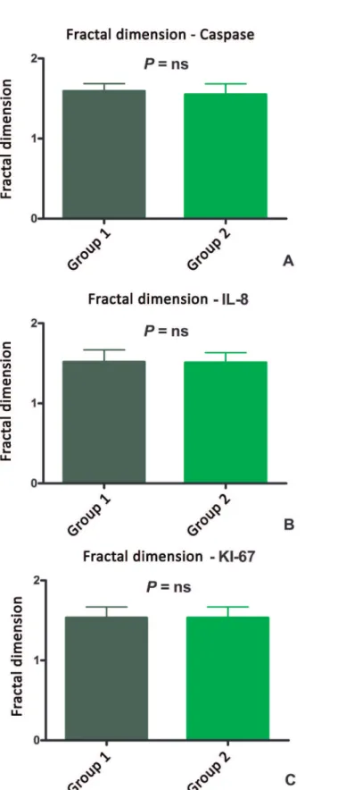
The fractal dimension using the caspase marker was 1.59 ± 0.09 (no unit) for group 1 and 1.55±0.13 for group 2. re-spectively (P=0.4400). KI-67 1.53±0.13 and 1.54±0.18. re-spectively (P=0.9595) and IL-8. 1.52 ± 0.15 and 1.51 ± 0.12. respectively (P=0.9164) (Figure 1).
The Shannon entropy with the caspase was 0.4±0.07 bits for group 1 and 0.38±0.08 bits for group 2 (P=0.5487). KI-67 0.36±0.1 bits and 0.37±0.13 bits. respectively (P = 0.9149). and IL-8 0.35±0.11 bits and 0.35±0.08 bits. respectively (P=0.9678) (Figure 2).
DISCUSSION
Although the effect of replacing ketoglutarate by glu-tamate in solution with histidine and tryptophan is still not known. glutamate has well-documented role when placed as a constituent of cardioplegic solution. The addition of glu-tamate in the perfusate maintains intracellular ATP and de-creases both lactate as pyruvate. which would contribute to acidosis. Exogenous glutamate and its transamination prod-ucts normally restore its contents decreased in the hypoxic myocardium. increase the concentration of succinate. which also leads to increased formation of ATP through anaerobic mitochondria pathway. thereby increasing the resistance to ischemia myocytes[9].
Fig. 2 - Histograms showing distribution of entropy in: (A) caspase. (B) IL-8 and (C) KI-67. Group 1: solution with ketoglutarate. Group 2: solution with histidine-tryptophan-glutamate.
Authors’ Roles & Responsibilities
MABO Main Author
ACB Elaboration of Graphics CAS Elaboration of Graphics PHHB Handling of animals JLLC Handling of animals
MFG Co-advisor and aid in inal writing
DMB Advisor and aid in inal writing
The fractal dimension is a useful parameter for the characterization of complex irregular structures. but when viewed mathematically. its analysis denotes regular igures with self-similarity features. or that is. resemble themselves when observed in different scales[24].
The fractal dimension of the object account the effec-tive number of degrees of freedom in the dynamical system. and therefore quantiies its complexity. Thus. it appears that images showing higher fractal dimensions are consequently more complex. However. we cannot quantify this complexity only by the visual aspect. The fractal dimension would then remedy this dificulty by adding a numeric value [12].
The size of the boxes for calculating the fractal dimension in the Box-counting method was standardized at 4. 8. 16. 32 and 64 pixels. It is known that the pixel size depends on the degree of resolution used. As commented by Tambasco et al. [24]. the size of the box used to bear a certain relationship with
the studied structure. because it can be so small that. in fact. were being evaluating the subcomponents of the structure or so great that. in fact. were being included in the measure-ment of components surrounding the structure of interest and not the structure itself. These values . however. are the de-fault values used in the literature and therefore probably not caused interference in the results[12].
In our study. there was no signiicant difference in fractal dimension between groups. Thus. we consider that the dis-tribution of the information contained in the slides of hearts treated with HTK solutions or HTG were not different.
An important contribution in Information Theory intro-duced by CE Shannon in 1948 was the concept of entropy as the amount of information in a system[25]. It is noteworthy that one should not confuse “entropy state” of Thermody-namics with “entropy concept” of the Information Theory[26]. According to Shannon. if X is the set of all messages of x. and p (x) is the probability (ranging from 0 to 1) of a message x. then the entropy of X will be[26]:
The Shannon entropy quantiies the degree of “uncer -tainty” or degree of “complexity” of information. With this formulation it is easy to understand that if the probability of occurrence of a particular event is 100%. or that is. if P=1 (no uncertainty). then the entropy contained in the message is zero (log 1 = zero)[26].
probability of each intensity of gray can be obtained easily by constructing a histogram of frequencies[26].
Zero entropy of an image is obtained when all the pix-els have the same color or the same amount of gray (100% probability. or that is 1; log 1=0). On the other hand. the maximum entropy may occur when the image contained the same amount of pixel for each intensities presented. Thus. we demonstrate that entropy is not related to the spatial layout of information. Two images may have the same number of pix-els with the same intensity and therefore the total entropy is the same. but spatially distributed [26] in a different manner.
In our study. there was no signiicant difference between the Shannon entropy between groups. Thus. we consider that the information contained in the slides of hearts treated with HTK or HTG solutions were not different.
The literature shows several studies in which there is a statistical difference between groups assessed with fractal dimension and Shannon entropy. drawing the reader’s atten-tion on its imaging discriminative character. but presenting no relevance when the results found did not reach statistical signiicance. as occurred in our study. The interpretation of the results of this study is that the HTG group did not alter the amount or distribution of the imaging information of rat hearts when compared to those treated with HTK.
Shannon entropy and the fractal dimension quantiies the distribution and the degree of complexity of the image. respectively. Thus. this technique is not comparable with Western-Blot or PCR. as these quantify the total amount of proteins studied and not their distribution or degree of com-plexity in the tissue.
CONCLUSION
The amount and distribution of the information assessed by the Shannon entropy and fractal dimension on blades of rat heart undergoing cardioplegia with HTK or HTG solu-tions were not different. which shows that the HTG solution is as good as HTK in preserving myocytes in isolated rat heart model.
REFERENCES
1. Miller BJ. Gibbon JH. Jr.. Greco VF. Smith BA. Cohn CH. Allbritten FF. Jr. The production and repair of interatrial septal defects under direct vision with the assistance of an extracorporeal pump-oxygenator circuit. J Thorac Surg. 1953;26(6):598-616.
2. Melrose DG. Dreyer B. Bentall HH. Baker JB. Elective cardiac arrest. Lancet. 1955;266(6879):21-2.
3. Fallouh HB. Kentish JC. Chambers DJ. Targeting for cardioplegia: arresting agents and their safety. Curr Opin Pharmacol. 2009;9(2):220-6.
4. Braile DM. Cardioplegia isotérmica anterógrada retrógrada de baixo volume. São José do Rio Preto. 1997. 27p.
5. Lima-Oliveira APM. Azevedo-Oliveira MTV. Taboga SR. Godoy MF. Braile DM. Cardioplegia utilizando baixo volume de agentes cardioplégicos: estudo morfológico em coração isolado de coelhos. Rev Bras Cir Cardiovasc. 2003;18(3):227-34.
6. Bretschneider HJ. Survival time and recuperative time of the heart in normothermia and hypotermia. Verh Dtsch Ges Kreislaufforsch. 1964;30:11-34.
7. Scrascia G. Guida P. Rotunno C. De Palo M. Mastro F. Pignatelli A. et al. Myocardial protection during aortic surgery: comparison between Bretschneider-HTK and cold blood cardioplegia. Perfusion. 2011;26(5):427-33.
8. Holper K. Meisner H. Hähnel C. Massoudy P. Technical reinements
in myocardial protection: infants-adults. Thorac Cardiovasc Surg. 1998;46 Suppl 2 292-5; discussion 6-7.
9. Pisarenko OI. Solomatina ES. Ivanov VE. Studneva IM. Kapelko VI. Smirnov VN. On the mechanism of enhanced ATP formation in hypoxic myocardium caused by glutamic acid. Basic Res Cardiol. 1985;80(2):126-34.
10. Rebeyka IM. Axford-Gatley RA. Bush BG. del Nido PJ. Mickle DA. Romaschin AD. et al. Calcium paradox in an in vivo model of multidose cardioplegia and moderate hypothermia. Prevention with diltiazem or trace calcium levels. J Thorac Cardiovasc Surg. 1990;99(3):475-83.
11. Arruda PFF. Gatti M. Facio Jr FN. Arruda JGF. Moreira RD. Murta
LO. et al. Quantiication of fractal dimension and Shannon’s
entropy in histological diagnosis of prostate cancer. BMC Clin Pathol. 2013;13:6.
12. Moreira RD. Moriel AR. Murta Junior LO. Neves LA. Godoy MF. Fractal dimension in quantifying the degree of myocardial cellular rejection after cardiac transplantation. Rev Bras Cir Cardiovasc. 2011;26(2):155-63.
13. Keipes M. Ries F. Dicato M. Of the British coastline and the interest of fractals in medicine. Biomed Pharmacother. 1993;47(9):409-15.
14. Karperien A. Jelinek HF. Leandro JJ. Soares JV. Cesar Jr RM. Luckie A. Automated detection of proliferative retinopathy in clinical practice. Clin Ophthalmol. 2008;2(1):109-22.
15. Committee on Care and Use of Laboratory Animals - Institute of Laboratory Animal Resources. Commission on Life Sciences. National Research Council. Guide for the care and use of laboratory animals. 8th ed. Washington: National Academies
Press; 2010. 211p.
16. Lahaye Sle D. Gratas-Delamarche A. Malardé L. Vincent S. Zguira MS. Morel SL. et al. Intense exercise training induces adaptation
in expression and responsiveness of cardiac β-adrenoceptors in
diabetic rats. Cardiovasc Diabetol. 2010;9:72.
17. Xu YJ. Saini HK. Zhang M. Elimban V. Dhalla NS. MAPK activation and apoptotic alterations in hearts subjected to calcium paradox are attenuated by taurine. Cardiovasc Res. 2006;72(1):163-74.
18. Fischer UM. Cox Jr CS. Laine GA. Mehlhorn U. Bloch W. Allen SJ. Induction of cardioplegic arrest immediately activates the myocardial apoptosis signal pathway. Am J Physiol Heart Circ Physiol. 2007;292(3):H1630-3.
19. Pirnia F. Schneider E. Betticher DC. Borner MM. Mitomycin C induces apoptosis and caspase-8 and -9 processing through a caspase-3 and Fas-independent pathway. Cell Death Differ. 2002;9(9):905-14.
20. Lee S. Huang CS. Kawamura T. Shigemura N. Stolz DB. Billiar TR. et al. Superior myocardial preservation with HTK solution over Celsior in rat hearts with prolonged cold ischemia. Surgery. 2010;148(2):463-73.
21. Anselmi A. Abbate A. Girola F. Nasso G. Biondi-Zoccai GG.
Possati G. et al. Myocardial ischemia. stunning. inlammation.
and apoptosis during cardiac surgery: a review of evidence. Eur J Cardiothorac Surg. 2004;25(3):304-11.
22. Lee Y. To proliferate or not to proliferate. Cardiovasc Res. 2010;86(3):347-8.
23. Walsh S. Pontén A. Fleischmann BK. Jovinge S. Cardiomyocyte cell cycle control and growth estimation in vivo: an analysis based on cardiomyocyte nuclei. Cardiovasc Res. 2010;86(3):365-73.
24. Tambasco M. Costello BM. Kouznetsov A. Yau A. Magliocco AM. Quantifying the architectural complexity of microscopic images of histology specimens. Micron. 2009;40(4):486-94.
2 5 . S h a n n o n C E . 1 9 4 8 . A M a t h e m a t i c a l T h e o r y o f Communication [Acesso 2/12/2013]. http://archive.org/details/ bellsystemtechni27amerrich
26. Arruda PFF. Quantiicação da dimensão fractal e entropia de