and Tripartite Efflux Pumps Share a Binding Motif for
TolC in Gram-Negative Bacteria
Minho Lee1., So-Young Jun2., Bo-Young Yoon2
, Saemee Song1, Kangseok Lee2*, Nam-Chul Ha1*
1Department of Life Science, Chung-Ang University, Seoul, Republic of Korea,2Department of Manufacturing Pharmacy, College of Pharmacy, Research Institute for Drug Development, Pusan National University, Busan, Republic of Korea
Abstract
The Hly translocator complex ofEscherichia colicatalyzes type I secretion of the toxin hemolysin A (HlyA). In this complex, HlyB is an inner membrane ABC (ATP Binding Cassette)-type transporter, TolC is an outer membrane channel protein, and HlyD is a periplasmic adaptor anchored in the inner membrane that bridges HlyB to TolC. This tripartite organization is reminiscent of that of drug efflux systems such as AcrA-AcrB-TolC and MacA-MacB-TolC ofE. coli. We have previously shown the crucial role of conserved residues located at the hairpin tip region of AcrA and MacA adaptors during assembly of their cognate systems. In this study, we investigated the role of the putative tip region of HlyD using HlyD mutants with single amino acid substitutions at the conserved positions.In vivoandin vitrodata show that all mutations abolished HlyD binding to TolC and resulted in the absence of HlyA secretion. Together, our results suggest that, similarly to AcrA and MacA, HlyD interacts with TolC in a tip-to-tip manner. A general model in which these conserved interactions induce opening of TolC during drug efflux and type I secretion is discussed.
Citation:Lee M, Jun S-Y, Yoon B-Y, Song S, Lee K, et al. (2012) Membrane Fusion Proteins of Type I Secretion System and Tripartite Efflux Pumps Share a Binding Motif for TolC in Gram-Negative Bacteria. PLoS ONE 7(7): e40460. doi:10.1371/journal.pone.0040460
Editor:Eric Cascales, Centre National de la Recherche Scientifique, France
ReceivedMarch 28, 2012;AcceptedJune 7, 2012;PublishedJuly 6, 2012
Copyright:ß2012 Lee et al. This is an open-access article distributed under the terms of the Creative Commons Attribution License, which permits unrestricted use, distribution, and reproduction in any medium, provided the original author and source are credited.
Funding:This work was supported by grants 2010-0015298 from the National Research Foundation of Korea, and the Next-Generation BioGreen 21 Program (SSAC, grant#: PJ009025), Rural Development Administration, Republic of Korea. The funders had no role in the study design, data collection and analysis, decision to publish, or preparation of the manuscript.
Competing Interests:The authors have declared that no competing interests exist.
* E-mail: kangseok@cau.ac.kr (KL); hnc@pusan.ac.kr (NH)
.These authors contributed equally to this work.
Introduction
Gram-negative bacteria have developed sophisticated secretion systems due to their characteristic double-layer membranes.
Secretion of the cytotoxina-hemolysin (HlyA) in many
uropatho-genic strains ofEscherichia coliis mediated by the protein complex Hly translocator, which belongs to the type I secretion system [1]. The 108-kDa HlyA can penetrate the membrane of host cells, creating a pore and causing them to lyse, which is an essential step for the bacteria to begin the infectious process [2].
The Hly translocator is comprised of HlyB, HlyD, and TolC [3]. The inner membrane protein HlyB is a member of the ATP-Binding Cassette (ABC) transporter family and transports HlyA from the cytosol by utilizing ATP hydrolysis [4,5]. TolC is the outer membrane channel protein and is often the final portal in the transport pathways of protein toxins or toxic molecules that have entered the cell [6,7,8]. HlyD is an adaptor protein that links HlyB to TolC, providing a continuous transmembrane duct for the export of HlyA. Previous studies have shown that HlyB and HlyD form a stable translocator complexin vivoin the absence of either HlyA or TolC, and TolC is then recruited into the translocator in the presence of the substrate HlyA [9]. A similar organization has been implicated in tripartite efflux pumps [10,11].
HlyD has a small N-terminal cytosolic domain connected to a large periplasmic domain by a single transmembrane helix [12]. It has been proposed that adaptor proteins share a similar overall
structural organization and conserved regions only in their C-terminal periplasmic domain [10]. The N-C-terminal cytosolic domain is unique in HlyD, and the transmembrane region is lacking or substituted with a lipid anchor in some of adaptor proteins [10]. Structural data for several functional homologues
such asE. coli AcrA, MacA and CusB andPseudomonas aeruginosa
MexA that contribute to drug or metal efflux systems have shown theC-terminal periplasmic domain [13,14,15,16]. The periplasmic domains of AcrA, MacA, CusB, and MexA commonly comprise a
membrane proximal (MP) domain, ab-barrel domain, a lipoyl
domain, and ana-helical domain that are linearly arranged in the tertiary structures [13,14,15,17,18,19]. Although all thea-helical domains are responsible for binding to their cognate outer membrane channel protein [20,21,22,23], they are structurally
variable. Thea-helical domains of AcrA and MacA consist of a
longa-hairpin of different lengths [13,14], while that of CusB is folded into a three-helix bundle structure [15,16]. Crystal structures, combined with genetic studies, have established that MacA and CusB exhibit a funnel-like hexameric assembly in their functional states [14,21,22,24].
AcrA and MacA are functional and structural homologues of
HlyD, and these are commonly associated with TolC inE. coli.
However, HlyD has a relatively low sequence similarity with AcrA and MacA compared with the homology between AcrA and
prediction programs unlike AcrA and MacA, suggesting that the TolC binding motif of HlyD might be different from that of the other adaptor proteins. Thus, the oligomeric state and TolC binding model of HlyD remains ambiguous.
Our research group has investigated the assembly of drug efflux pumps and proposed a tip-to-tip binding model between MacA (or AcrA) and TolC [14,20,21,22,23,25]. According to the model, the
a-helical tip region of the adaptor protein makes a
cogwheel-to-cogwheel interaction with the TolC a-barrel tip region. In this
study, we provide experimental evidence indicating that HlyD shares this mechanism of TolC binding.
Results
Sequence analysis of the putative HlyD a-helical region
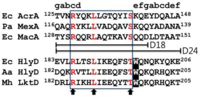
Since HlyD, MacA, and AcrA proteins are commonly associated with TolC, we hypothesized that HlyD shares a common structural motif with MacA and AcrA for binding to TolC. It should be noted that the conserved RxxxLxxxxxxS motif (x stands for any residue; we designate it the RLS motif), located at
thea-hairpin tip region of MacA, AcrA, and MexA, creates the
binding interface for the TolCa-barrel tip [21,22,23]. The RLS
motifs are universally located at the cogwheel region of the funnel-like hexamers of MacA, AcrA, and MexA [14,22,23,25]. Single mutations at the conserved arginine, leucine, and serine residues within the RLS motif affect the activity of efflux adaptors (i.e., direct binding to TolCin vitroand drug effluxin vivo) [20,21]. Here, it is worth noting that the S143A substitution in MacA retains some activity while the S143Y does not. This could be due to the bulkiness of tyrosine but also suggests that the serine residue of the RLS motif is more permissive to mutations (or less important for protein activity) [21].
Since HlyD does not show a significant sequence similarity with MacA or AcrA, we did a manual sequence alignment by considering the spacing between the conserved residues of the RLS motif. A putative RLS motif was identified in the region
encompassing amino acids 186 to 197 inE. coliHlyD. This motif
was also found in a subset of HlyD proteins from other species and homologous adaptor proteins engaged in other type I secretion systems. In this motif, serine was changed to threonine with conservation of a hydroxyl-bearing amino acid (Fig. 1). The motif has a substitution at the third serine site with a threonine residue, and thus we designated the motif as the RLT motif. This substitution at the third conserved residue seems reasonable because functional flexibility was observed at the residue in some adaptor proteins, as mentioned above. Further sequence similarity was present within the RLT motif, indicating that this motif is functionally important among HlyDs (Fig. 1).
Arg186, Leu190, and Thr197 of HlyD are important in the secretion of HlyA in vivo
To examine the functional importance of the three residues in the HlyD RLT motif, the residues were mutated in a cloned copy
of hlyDin the plasmid pLG815, which directs the expression of
hlyBand hlyD genes under the control of thecatgene promoter
[26] (Table 1). In addition, the start codon of the hlyDgene in
pLG815 was mutated to a stop codon, and the resulting plasmid
(pLG815-HlyD-null) was used as a negative control for hlyD
expression. The created pLG815-derived plasmids
(pLG815-HlyD-null, pLG815-HlyD-R186A, pLG815-HlyD-L190A,
pLG815-HlyD-T197A, and pLG815-HlyD-T197Y) were further modified by inserting a DNA segment encoding a c-Myc epitope (EQKLISEEDL) at the C-terminus of the HlyD coding region in pLG815, resulting in a pLG815-HlyD-c-Myc series of plasmids.
The pLG815-HlyD-c-Myc series of plasmids were transformed intoE. coli strain SE5000, harboring pLG813, which directs the
expression of hlyA and hlyC under the control of their intact
promoter (Kennyet al., 1992). The resulting transformants were
tested for their ability to hemolyze red blood cells. When HlyD mutants containing R186A, L190A, or T197Y were coexpressed with HlyA, HlyB, and HlyC inE. colistrain SE5000, the degree of hemolytic activity ofE. colicells on the M63-blood agar, as well as in liquid medium, were indistinguishable from those ofE. colicells expressing thehlyDnull mutant (Table 1 and Fig. 2A). Expression of HlyD containing the T197A mutation inE. colicells resulted in the formation of a clear zone on the M63-blood agar that was ten
times smaller in diameter than that formed by E. coli cells
expressing wild-type HlyD, indicating that this mutant renderedE.
coli cells partially hemolytic (Table 1). However, E. coli cells
expressing HlyD-T197A did not exhibit hemolytic activity that was detectable in the liquid medium, whereas those expressing wild-type HlyD showed strong hemolytic activity (Fig. 2A).
We further confirmed that the observed hemolytic activity ofE. colicells expressing HlyD mutants was related to the HlyA levels
secreted through the Hly translocator complex. E. coli strain
SE5000 coexpressing HlyA, HlyB, and HlyC together with wild-type or mutant derivatives of HlyD were grown in LB medium
with 10 mM CaCl2at 37uC for 4 hr to an OD600of 3.5. These
conditions induce maximal hemolysin secretion [26,27]. Culture supernatants and cell pellets were separated by centrifugation. Proteins were separated by SDS-PAGE and analyzed by Western blot with antibodies directed against HlyA. As shown in Fig. 2B (middle), the same amounts of HlyA were detected in cell extracts expressing wild-type or mutant HlyD proteins. The wild-type and mutant HlyD proteins tagged with the c-Myc epitope were normally expressed in the cell and inserted into the membrane fraction, according to Western blot with anti-c-Myc antibodies (Fig. 2B middle and bottom). As expected, HlyA was secreted into the medium when cells expressed wild-type HlyD. In contrast, HlyA was very poorly or not secreted from cells expressing HlyD proteins with mutation in the RLT motif (Fig. 2B top). These results demonstrate that Arg186, Leu190, and Thr197 play an important role in the function of HlyDin vivoand further suggest the functional importance of the HlyD RLT motif in the function of the Hly translocator.
Figure 1. Sequence comparison of thea-hairpins with the
anti-parallel coiled-coil structures.Sequences of the RLS (or RLT) motif from six adaptor proteins are aligned. The corresponding heptad positions are shown above the sequence. The three conserved residues in the RLS (or RLT) motif are indicated by arrows. The12 RLS (or RLT) motif residues are in a box, which was used in the MacA-HlyD12 hybrid. The 18 and 24 residues used in the construction of MacA-HlyD18 and MacA-HlyD24, respectively, are indicated (D18 and D24). The amino acid numbering is based on the protein precursors. In the sequence alignment, Ec, Pa, Aa, and Mh stand for E. coli, P. aeruginosa,
Actinobacillus actinomycetemcomitans, and Mannheimia haemolytica, respectively.
doi:10.1371/journal.pone.0040460.g001
Physical interaction between HlyD RLT motif and TolCin vivo
We have previously shown that the RLS motif of the tip region in MacA and AcrA is crucial for binding to the tip region of TolC. Therefore, we investigated the interaction between TolC and the RLT motif of HlyD usingin vivochemical cross-linking [9]. In this experiment, we used a pLG815-HlyD-c-Myc series of plasmids to express wild-type or mutant HlyD tagged with c-Myc at the C-terminus, and HlyB. pLG813 was used to express the substrate
HlyA and HlyC. Plasmid pTolC2 has a replication origin from pSC101 [28] and an ampicillin antibiotic marker, which are compatible with pLG813- and pLG815-derived plasmids. The
plasmid pTolC2 contained a cloned copy of thetolCgene, which
directs the synthesis of TolC with a hexahistidine-tag at the
C-terminus under the control of a lacUV5 promoter. The three
plasmids, pTolC2, pLG813, and one of the pLG815-HlyD-c-Myc series were transformed into an acrAB- and tolC-deleted E. coli
strain. Transient protein complexes were stabilized with the
Figure 2. HlyA activity ofE. coliSE5000 expressing wild-type or mutant HlyD.A. Hemolytic activity ofE. colicells expressing wild-type or mutant HlyD. The absorbance at 450 and 543 nm was detected using a spectrophotometer, and hemolytic activities were calculated according to the following formula: Percent Hemolysis = (1-ODs/ODt)6100, where ODs and ODt are the differences in optical density at 543 nm between the samples and 100% hemolyzed SRBC solution, and differences in optical density at 450 nm between nonhemolyzed (control) and 100% hemolyzed sheep red blood cell solution, respectively. Filled symbols and lines indicate hemolytic activity detected in culture supernatants (OD543/OD450). B. Effects of
expression of HlyD mutants (HlyD-R131A, HlyD-L186A, HlyD-T190A, HlyD-T197Y, HlyD-null) on HlyA secretion.E. coliSE5000 coexpressing HlyA, HlyB, HlyC, and wild-type or HlyD mutants (HlyD-R186A, HlyD-L190A, HlyD-T197A, HlyD-T197Y, or HlyD-Stop) were grown exponentially in LB with 10 mM CaCl2at 37uC, and proteins in the culture supernatant (equivalent to 3.5 OD600of cells) were TCA precipitated. The precipitated protein samples were
separated by SDS-PAGE and subjected to immunoblotting with HlyA antibody (upper panel). Total cell lysate was used for the western blot analysis of HlyA to detect its expression levels in the cell (lower panel).
chemical cross-linker DSP, which has a fixed 12-A˚ spacer arm that connects the primary amine groups of two adjacent proteins and contains a disulfide bond cleavable under reducing conditions. Cells were lysed and protein complexes were isolated by affinity chromatography via the His-tagged component (TolC-His). The eluted complexes were treated with sample buffer containing DTT and boiled to cleave the DSP molecule and release the individual components. These were resolved by SDS-PAGE and identified by immunoblotting using anti-His (TolC) and anti-c-Myc (HlyD variants) antibodies.
As shown in Fig. 3, the crosslinking of R186A and HlyD-T197A to TolC were 10% and 48% of that of wild-type HlyD to TolC, respectively, whereas crosslinking of HlyD-L190A and Hly-T197Y to TolC was not detected. These results indicated that an amino acid substitution in the RLT motif partially (R186A and T197A) or completely (L190A and T197Y) abolished the binding
of HlyD to TolCin vivo. These findings are consistent with the
pivotal role of the RLT motif in the secretion of HlyAin vivo, even with the flexibility at the third residue. Moreover, the wild-type HlyD mediated the access of HlyA to TolC, detected by using the crosslinker, whereas the mutant HlyD abolished or reduced the accessibility of HlyA to TolC (Fig. 3). Taken together, our results strongly suggest that the RLT motif of HlyD makes direct contact with TolC, as is the case with the RLS motif of MacA or AcrA.
The RLT motif plays an important role in binding to the TolCa-hairpin tip regionin vitro
To confirm whether the RLT motif of HlyD forms a binding
interface with the TolCa-barrel tip region, we carried out the
chimeric approach that was used in a previous study of the interactions between MacA and TolC [22]. In this study, three chimeric MacA proteins containing the RLT motif were
generated. The a-hairpin tip 24
(TYSRQQRLAQT-KAVSQQDLDTAAT; the underlined region indicates the RLS or RLT motif), 18 (TYSRQQRLAQTKAVSQQD), or 12
residues (RQQRLAQTKAVS) ofE. coliMacA were substituted
with 24 (DKTRLTSLIKEQFSTWQNQKYQKE), 18
(DKTRLTSLIKEQFSTWQNQKY), or 12 residues
(RLTSLI-KEQFST) ofE. coliHlyD, respectively, resulting in the chimeric
proteins MacA-HlyD24 hybrid, MacA-HlyD18 hybrid, and
MacA-HlyD12 hybrid. To examine whether the TolCa-barrel
tip region is involved in the HlyD binding, a TolC chimeric
protein that contains a TolC a-barrel tip of 24 amino acids
(MacA-TolCahybrid-dimer) was used, which was previously used
to show that the chimeric protein specifically binds to MacA and AcrA, and reflects the functional binding of TolC to the cognate adaptor proteins [22,23]. With the chimeric proteins, we
performed an in vitro binding assay on CNBr-activated agarose
resin. The TolC chimeric protein (MacA-TolCa hybrid dimer)
was coupled to the resin and then each MacA-HlyD hybrid protein was incubated to allow binding. As shown in Fig. 4A, only
Table 1.Degree of hemolysis byE. colicells expressing HlyD mutants.
HlyD mutation Degree of hemolysis (diameter of a clear zone1
) HlyD mutant Amino acid change Codon change
HlyD WT None None 2.560.25 cm
HlyD null M1RStop ATGRTAG None
HlyD R186A R186RA CGTRGCT None
HlyD L190A L190RA TTGRGCT None
HlyD T197A T197RA ACARGCT 0.260.02 cm
HlyD T197Y T197RY ACARTAT None
1
,104cells were spotted on M63-blood agar plate, incubated for 24 hrs, and the diameter of the clear zone was measured.
doi:10.1371/journal.pone.0040460.t001
Figure 3. Interaction between TolC and HlyDin vivo.Thein vivointeraction between HlyD and TolC was detected using a chemical cross-linking agent (DSP).E. coliBW25113DacrABDtolC210::Tn10cultures that coexpressed HlyA, HlyB, HlyC, hexa-His-tagged wild-type TolC, and c-Myc-tagged wild-type HlyD (WT) or one of the HlyD mutants (R186A, L190A, T197A and T197Y) are shown. All cultures were treated with (+) or without (2)
DSP. Affinity-purified TolC and cross-linked proteins (HlyA and HlyD) were separated by SDS-PAGE and immunoblotted using monoclonal antibodies to His-tag and c-Myc, and polyclonal antibodies to HlyA.
doi:10.1371/journal.pone.0040460.g003
the MacA-HlyD12 hybrid was bound to the TolC a-barrel tip region. This result indicates that the 12 residues of the RLT motif comprise the binding interface with the TolCa-barrel tip, and that the conformation of the RLT is similar to that of the RLS motif in MacA (see Discussion).
To assess the importance of the conserved residues in the RLT motif, we introduced mutations to the MacA-HlyD12 hybrid and subsequently measured the affinity to the TolC chimeric protein. The TolC chimeric protein and the HlyD chimeric proteins were applied to size-exclusion chromatography to measure the binding affinity. Unlike the wild-type MacA-HlyD12 hybrid protein, the
non-hemolyticE. coli HlyD mutant hybrid proteins containing a
single amino acid substitution (R186A, L190A, T197Y) did not interact with the TolC hybrid, while the HlyD T197A variant protein showed a partial affinity for the TolC hybrid (Fig. 4B). A similar result was produced by anotherin vitrobinding assay based on the TolC chimeric protein-coupled agarose resin, although the T142A variant protein showed as strong an affinity for the TolC hybrid as the wild-type HlyD hybrid protein (Fig. 4C). Combined within vivobinding assays, the results of thein vitrobinding assays
showed that the HlyD RLT motif is not only functionally related to, but also interacts with, the TolCa-barrel tip region.
Discussion
In this study, we identified the RLT motif in HlyD as a functional counterpart of the RLS motif in drug efflux pumps and demonstrated that the RLT motif of HlyD plays an important role in the action of the HlyBD-TolC translocator. A single substitution mutation in the RLT motif of HlyD abolished binding to TolC in the in vivo cross-linking assay and secretion of HlyA to the extracellular environmentin vivo. A chimeric protein containing
the intact RLT motif was able to bind to the TolCa
-barrel-tip-containing protein, indicating that the RLT motif comprises the binding interface to the TolCa-barrel tip. Therefore, our findings imply that HlyD and TolC share the same binding mode with MacA (or AcrA) and TolC.
What does the structure of the binding domain of HlyD resemble? Many crystal structures of adaptor proteins in tripartite efflux pumps have a long,a-hairpin domain that is responsible for
Figure 4. Interaction between the HlyD RLT motif and the TolCa-barrel tip region.A. The MacA-TolCahybrid (T) was coupled to the
the TolC binding. However, the sequence alignment does not clearly define the corresponding domain in HlyD except for the
RLS motif, suggesting that HlyD does not have the canonicala
-hairpin domain. Furthermore, the chimeric MacA proteins harboring the HlyD 24 or 18 residues did not function as MacA surrogates in binding to the TolC chimeric protein in our results, unlike the chimeric protein with the only HlyD RLT motif. This observation suggests that the adjacent residues of the RLT motif
might interfere to form the a-hairpin structure of MacA. The
trimeric model of HlyD was proposed based on chemical cross-linking results [9]. However, recent studies have also converged on the hexameric model of adaptor proteins in tripartite efflux pumps, although the debate continues [11,22,23,25,29]. There-fore, a hexameric model of HlyD should be considered. The hexameric HlyD model is geometrically reasonable, because the dimeric HlyB ABC transporter and the trimeric TolC are connected by HlyD with an oligomerization number of ‘6,’ the least common multiple between 2 and 3. As supportive evidence for the hexameric model, the RLT motif of HlyD loaded into the hexameric platform of MacA showed a strong affinity for the TolC
a-barrel in the binding assay using the chimeric HlyD proteins.
This result further suggests that the assembly of the Hly translocator complex is similar to those of the drug efflux pumps
with a modifieda-hairpin domain, such that HlyD makes a direct
contact with TolC through a structural motif similar to the adaptor proteins in the drug efflux pumps.
To date, the assembly of the type I secretion system remains unclear. This study reveals a crucial motif in the adaptor protein for TolC binding, and shows parallels with the assembly of tripartite efflux pumps. Although further structural and functional studies are required to envisage the fully assembled complex, this study provides insight into the assembly mechanism for a tunnel across the periplasm and through the outer membrane for toxin translocation.
Materials and Methods
Bacterial strains and expression plasmids for genetic studies
The non-pathogenic E. coli strain SE5000 (rpsL ara139
D[lacIPOZYA]U169 recA57 thi) was used for the in vivo study.
Cultures were grown at 37uC in LB medium, normally
supple-mented with 10 mM CaCl2, for the hemolytic assay. Plasmid
pLG815 carriedhlyBDgenes cloned from the wild-type pathogenic
strain LE2001 [30]. Plasmid pLG813 was a pACYC derivative
encoding the hlyCA genes [31]. Plasmids pLG815 HlyD-null,
HlyD-R186A, HlyD-L190A, HlyD-T197A, and HlyD-T197Y were constructed using the overlap extension PCR method. The
resulting PCR product was digested with AccI and ApaI, and
ligated into the same sites in pLG815 to produce the plasmids
described above. pLG815 HlyD C-myc, HlyD-R186A C-myc,
HlyD-L190A C-myc, HlyD-T197A C-myc, and HlyD-T197Y
C-myc plasmids were constructed by cloning the DNA fragment
encoding the C-terminal region of HlyD with a PCR-amplified
c-Myc-tag into theAccI andXbaI sites of pLG815. Plasmid pTolC2
was constructed by subcloning the NotI and XbaI fragments
containing the coding region of TolC from pTolC1 [20] into the same sites in pPM30. Primers used for plasmid generation are listed in Table S1.
Construction ofActinobacillus actinomycetemcomitans (Aa) MacA-TolCa hybrid-dimer
Construction of the Aa MacA-TolCa hybrid-dimer has been
previously described [22].
Construction and expression ofE. coliMacA-HlyD12 hybrid,E. coliMacA-HlyD18 hybrid, andE. coli MacA-HlyD24 hybrid
To construct the expression vector for theE. coliMacA-HlyD12
hybrid, the three DNA fragments encodingE. coliMacA residues
32–130, HlyD residues 186–197, and MacA residues 143–371, respectively, were joined using the overlapping PCR technique,
and then ligated into theNcoI andXhoI sites of pPROEX-HTA
(Invitrogen). A similar approach was utilized for construction of
the expression vector for the E. coli MacA-HlyD18 and E. coli
MacA-HlyD24 hybrids. For the MacA-HlyD18 hybrid, the DNA fragments encoding MacA residues 32–127, HlyD residues 183– 203, and MacA residues 149–371 were joined. For the MacA-HlyD24 hybrid, the DNA fragments encoding MacA residues 32– 127, HlyD residues 183–206, and MacA residues 152–371 were joined. The sequences of the primers used for the plasmids are described in Table S1. Expression and purification of the hybrid proteins were performed using the same procedure previously reported [22].
Size exclusion chromatography
Size exclusion chromatography was performed at a flow rate of 0.5 ml/min on Superdex S-200 HR 10/30 (GE-Healthcare) that was equilibrated with 20 mM Tris buffer (pH 8.0) containing 150 mM NaCl. Before injection onto the column, 1 mg/ml of
MacA-TolCa hybrid-dimer protein was incubated with the same
concentration of MacA-HlyD12 hybrid or its mutant proteins
(R186A, L190A, T197Y, and T197A) for 30 min at 4uC,
respectively. The fractions from the column were applied to SDS-PAGE, and the gel was stained with Coomassie blue.
In vitrobinding assay using CNBr-activated resin
The MacA-TolCahybrid-dimer protein protein (100ml; 1 mg/
ml) was coupled to CNBr-activated agarose resin (50mg;
GE-Healthcare) according to the manufacturer’s instructions. The same amount of resin was inactivated with 100 mM Tris buffer for control. Then, 1 mg/ml of MacA or MacA-HlyD hybrid protein (D24, D18, or D12), or MacA-HlyD12 variants (wild type, R186A, L190A, T197Y, and T197A) was incubated with the protein-coupled or inactivated resin. After incubation, the resin was thoroughly washed with PBS and then subjected to SDS-PAGE for analysis. The protein bands were visualized by staining with Coomassie Blue.
Detection of HlyA
Ten milliliters of culture was harvested by centrifugation at
6,0006g for 10 min, and then TCA (trichloroacetic acid) was
added to the supernatant to a final concentration of 10% (w/v) before incubation on ice for 1 hr. The supernatant protein was
pelleted by centrifugation at 16,0006 g for 10 min and
resuspended in 50ml of SDS sample buffer. Ten microliters of
saturated Tris solution was added to bring the sample back to neutral pH. Proteins were separated on a 10% SDS-PAGE gel and subjected to Western blot analysis using polyclonal antibodies to HlyA.
Measurement of hemolytic activity
The procedure for measuring the hemolytic activity of E. coli
cells has been previously described [32].
Separation ofE. colimembranes
The procedure for preparing membrane fraction ofE. colicells
has been previously described [33].
In vivocross-linking assay
The procedure for in vivo cross-linking using Dithiobis
[succinimidyl propionate] (DSP) has been previously described [9].E. coliBW25113DacrABDtolC210::Tn10cells carrying pLG815
HlyD-C-mycor its derivatives (HlyD-R186A-C-myc,
HlyD-L190A-C-myc, HlyD-T197A-C-myc, and HlyD-T197Y-C-myc) and
pTolC2 were grown in LB medium to OD600= 0.7 and used for
cross-linking experiments. Anti-His and anti-myc monoclonal
antibodies were used to detect TolC and HlyD with C-terminal hexahistidine (TolC) and c-Myc tags (HlyD), respectively.
Supporting Information
Figure S1 Elution profiles of MacA-HlyD12 hybrid proteins on a size exclusion chromatography.The elution volumes for the peaks are shown. The same size exclusion
chromatographic column was used, and the fractions for the wild type protein (wt) were analyzed by SDS-PAGE (See Fig. 4B). (DOCX)
Table S1 Primers used in this study. (DOCX)
Acknowledgments
We thank Dr. I. Barry Holland for providing antibodies to HlyA,E. coli strain SE5000, and plasmids pLG813 and pLG815 for our use.
Author Contributions
Conceived and designed the experiments: KL NH. Performed the experiments: ML SJ BY SS. Analyzed the data: ML SJ KL NH. Contributed reagents/materials/analysis tools: KL NH. Wrote the paper: ML SJ KL NH.
References
1. Holland IB (2011) ABC transporters, mechanisms and biology: an overview. Essays Biochem 50: 1–17.
2. Welch RA, Dellinger EP, Minshew B, Falkow S (1981) Haemolysin contributes to virulence of extra-intestinalE. coliinfections. Nature 294: 665–667. 3. Schulein R, Gentschev I, Schlor S, Gross R, Goebel W (1994) Identification and
characterization of two functional domains of the hemolysin translocator protein HlyD. Mol Gen Genet 245: 203–211.
4. Wang RC, Seror SJ, Blight M, Pratt JM, Broome-Smith JK, et al. (1991) Analysis of the membrane organization of anEscherichia coliprotein translocator, HlyB, a member of a large family of prokaryote and eukaryote surface transport proteins. J Mol Biol 217: 441–454.
5. Schmitt L, Benabdelhak H, Blight MA, Holland IB, Stubbs MT (2003) Crystal structure of the nucleotide-binding domain of the ABC-transporter haemolysin B: identification of a variable region within ABC helical domains. J Mol Biol 330: 333–342.
6. Wandersman C, Delepelaire P (1990) TolC, anEscherichia coliouter membrane protein required for hemolysin secretion. Proc Natl Acad Sci U S A 87: 4776– 4780.
7. Koronakis V, Sharff A, Koronakis E, Luisi B, Hughes C (2000) Crystal structure of the bacterial membrane protein TolC central to multidrug efflux and protein export. Nature 405: 914–919.
8. Fralick JA (1996) Evidence that TolC is required for functioning of the Mar/ AcrAB efflux pump ofEscherichia coli. J Bacteriol 178: 5803–5805.
9. Thanabalu T, Koronakis E, Hughes C, Koronakis V (1998) Substrate-induced assembly of a contiguous channel for protein export fromE.coli: reversible bridging of an inner-membrane translocase to an outer membrane exit pore. Embo J 17: 6487–6496.
10. Lewis K (2000) Translocases: a bacterial tunnel for drugs and proteins. Curr Biol 10: R678–681.
11. Su CC, Long F, Zimmermann MT, Rajashankar KR, Jernigan RL, et al. (2011) Crystal structure of the CusBA heavy-metal efflux complex ofEscherichia coli. Nature 470: 558–562.
12. Schulein R, Gentschev I, Mollenkopf HJ, Goebel W (1992) A topological model for the haemolysin translocator protein HlyD. Mol Gen Genet 234: 155–163. 13. Mikolosko J, Bobyk K, Zgurskaya HI, Ghosh P (2006) Conformational flexibility
in the multidrug efflux system protein AcrA. Structure 14: 577–587. 14. Yum S, Xu Y, Piao S, Sim SH, Kim HM, et al. (2009) Crystal structure of the
periplasmic component of a tripartite macrolide-specific efflux pump. J Mol Biol 387: 1286–1297.
15. Su CC, Yang F, Long F, Reyon D, Routh MD, et al. (2009) Crystal structure of the membrane fusion protein CusB fromEscherichia coli. J Mol Biol 393: 342– 355.
16. Su CC, Long F, Yu EW (2011) The Cus efflux system removes toxic ions via a methionine shuttle. Protein Sci 20: 6–18.
17. Symmons MF, Bokma E, Koronakis E, Hughes C, Koronakis V (2009) The assembled structure of a complete tripartite bacterial multidrug efflux pump. Proc Natl Acad Sci U S A 106: 7173–7178.
18. Higgins MK, Bokma E, Koronakis E, Hughes C, Koronakis V (2004) Structure of the periplasmic component of a bacterial drug efflux pump. Proc Natl Acad Sci U S A 101: 9994–9999.
19. Akama H, Matsuura T, Kashiwagi S, Yoneyama H, Narita S, et al. (2004) Crystal structure of the membrane fusion protein, MexA, of the multidrug transporter inPseudomonas aeruginosa. J Biol Chem 279: 25939–25942. 20. Kim HM, Xu Y, Lee M, Piao S, Sim SH, et al. (2010) Functional
interrelationships between the AcrA hairpin tip region and the TolC aperture tip region for the formation of bacterial tripartite efflux pump AcrAB-TolC. J Bacteriol 192: 4498–4503.
21. Xu Y, Sim SH, Song S, Piao S, Kim HM, et al. (2010) The tip region of the MacA alpha-hairpin is important for the binding to TolC to theEscherichia coli
MacAB-TolC pump. Biochem Biophys Res Commun 394: 962–965. 22. Xu Y, Song S, Moeller A, Kim N, Piao S, et al. (2011) Functional implications of
an intermeshing cogwheel-like interaction between TolC and MacA in the action of macrolide-specific efflux pump MacAB-TolC. J Biol Chem 286: 13541–13549.
23. Xu Y, Lee M, Moeller A, Song S, Yoon BY, et al. (2011) Funnel-like hexameric assembly of the periplasmic adapter protein in the tripartite multidrug efflux pump in gram-negative bacteria. J Biol Chem 286: 17910–17920.
24. Zgurskaya HI, Krishnamoorthy G, Ntreh A, Lu S (2011) Mechanism and function of the outer membrane channel TolC in multidrug resistance and physiology of enterobacteria. Front Microbiol 2: 189.
25. Xu Y, Moeller A, Jun SY, Lee M, Yoon BY, et al. (2012) Assembly and channel opening of the outer membrane protein in tripartite drug efflux pumps of Gram-negative bacteria. J Biol Chem 287: 11740–11750.
26. Pimenta AL, Racher K, Jamieson L, Blight MA, Holland IB (2005) Mutations in HlyD, part of the type 1 translocator for hemolysin secretion, affect the folding of the secreted toxin. J Bacteriol 187: 7471–7480.
27. Holland IB, Blight MA, Kenny B (1990) The mechanism of secretion of hemolysin and other polypeptides from gram-negative bacteria. J Bioenerg Biomembr 22: 473–491.
28. Cohen SN, Chang AC (1977) Revised interpretation of the origin of the pSC101 plasmid. J Bacteriol 132: 734–737.
29. Janganan TK, Bavro VN, Zhang L, Matak-Vinkovic D, Barrera NP, et al. (2011) Evidence for the assembly of a bacterial tripartite multidrug pump with a stoichiometry of 3:6:3. J Biol Chem 286: 26900–26912.
30. Mackman N, Holland IB (1984) Functional characterization of a cloned haemolysin determinant fromE. coliof human origin, encoding information for the secretion of a 107K polypeptide. Mol Gen Genet 196: 129–134. 31. Kenny B, Taylor S, Holland IB (1992) Identification of individual amino acids
required for secretion within the haemolysin (HlyA) C-terminal targeting region. Mol Microbiol 6: 1477–1489.
32. Bhakdi S, Muhly M, Fussle R (1984) Correlation between toxin binding and hemolytic activity in membrane damage by staphylococcal alpha-toxin. Infect Immun 46: 318–323.
33. Pimenta AL, Young J, Holland IB, Blight MA (1999) Antibody analysis of the localisation, expression and stability of HlyD, the MFP component of theE. coli