459
Braz J Med Biol Res 37(4) 2004
Leptospira interrogans genome features
Brazilian Journal of Medical and Biological Research (2004) 37: 459-478 ISSN 0100-879X
Genome features of
Leptospira
interrogans
serovar Copenhageni
1Centro de Biotecnologia, Instituto Butantan, São Paulo, SP, Brasil
2Instituto de Química, 3Instituto de Biociências and
4Instituto de Ciências Biomédicas, Universidade de São Paulo, São Paulo, SP, Brasil
5Escola Superior de Agricultura Luiz de Queiroz, Universidade de São Paulo,
Piracicaba, SP, Brasil
6Laboratório de Bioinformática, Instituto de Computação,
Universidade Estadual de Campinas, Campinas, SP, Brasil
7Koninklijk Instituut voor de Tropen/Royal Tropical Institute (KIT), KIT Biomedical
Research, Amsterdam, The Netherlands
8The David Geffen School of Medicine at University of California, Los Angeles,
CA, USA
9Division of Infectious Diseases, Veterans Affairs Greater Los Angeles Healthcare
System, Los Angeles, CA, USA
10Virginia Bioinformatics Institute and Department of Computer Science, Virginia
Polytechnic Institute and State University Bioinformatics 1, Blacksburg, VA, USA A.L.T.O. Nascimento1,
S. Verjovski-Almeida2, M.A. Van Sluys3, C.B. Monteiro-Vitorello5, L.E.A. Camargo5, L.A. Digiampietri6, R.A. Harstkeerl7, P.L. Ho1, M.V. Marques4, M.C. Oliveira3, J.C. Setubal6,10, D.A. Haake8,9 and E.A.L. Martins1
Abstract
We report novel features of the genome sequence of Leptospira interrogans serovar Copenhageni, a highly invasive spirochete. Lep-tospira species colonize a significant proportion of rodent populations worldwide and produce life-threatening infections in mammals. Ge-nomic sequence analysis reveals the presence of a competent transport system with 13 families of genes encoding for major transporters including a three-member component efflux system compatible with the long-term survival of this organism. The leptospiral genome contains a broad array of genes encoding regulatory system, signal transduction and methyl-accepting chemotaxis proteins, reflecting the organism’s ability to respond to diverse environmental stimuli. The identification of a complete set of genes encoding the enzymes for the cobalamin biosynthetic pathway and the novel coding genes related to lipopolysaccharide biosynthesis should bring new light to the study of Leptospira physiology. Genes related to toxins, lipoproteins and several surface-exposed proteins may facilitate a better understanding of the Leptospira pathogenesis and may serve as potential candidates for vaccine.
Correspondence
A.L.T.O. Nascimento Centro de Biotecnologia, Instituto Butantan, Av. Vital Brazil, 1500 05503-900 São Paulo, SP Brasil
Fax: +55-11-3726-1505 E-mail: [email protected]
Research supported by FAPESP and CNPq.
Received 20 February, 2004 Accepted March 8, 2004
Key words
•Leptospira interrogans •Outer membrane proteins •Lipopolysaccharides •Transport
•Regulatory systems
Introduction
Spirochetes are motile, helically shaped bacteria which include the genera Leptospira, Leptonema, Borrelia and Treponema. Bor-relia and Treponema are the causative agents of Lyme disease, relapsing fever and
Infection produces a wide spectrum of clini-cal manifestations. The early phase of illness is characterized by fever, chills, headache, and severe myalgias. The disease progresses in 5 to 15% of the clinical infections to produce severe multisystem complications such as jaundice, renal failure and hemor-rhagic manifestations (2). In developed coun-tries, leptospirosis is associated with recre-ational activities (1) while in developing countries it produces large urban epidemics with mortality mainly during the rainy sea-son (3). Leptospirosis also represents a ma-jor economic problem producing abortions, stillbirths, infertility, failure to thrive, re-duced milk production, and death in animals such as cows, pigs, sheep, goats, horses, and dogs (1). Environmental control measures are difficult to implement because of the long-term survival of pathogenic leptospires in soil and water and the abundance of wild and domestic animal reservoirs (1). Lep-tospira are classified according to serovar status - more than 200 pathogenic serovars have been identified. Structural heterogene-ity in lipopolysaccharide (LPS) moieties ap-pears to be the basis for the large degree of antigenic variation observed among sero-vars (1). The development of vaccines has been pursued as a strategy for the prevention of leptospirosis. At present, vaccines are based on inactivated whole cell or mem-brane preparations of pathogenic leptospires which induce immune responses against lep-tospiral LPS (1). However, these vaccines do not induce long-term protection against infection and do not provide cross-protec-tive immunity against heterologous leptospi-ral serovars. Protein antigens conserved among pathogenic serovars may contribute to overcoming the limitations of the cur-rently available vaccines.
The genome sequence of Leptospira interrogans serovar Lai was recently pub-lished (4) and comparative genome analysis with L. interrogans serovar Copenhageni has been performed. We report here new
features of the L. interrogans serovar Co-penhageni that should contribute to under-standing the molecular mechanisms of lep-tospiral physiology, pathogenesis and facili-tate the identification of candidates for broad-range vaccines.
Material and Methods
The sequenced strain, Fiocruz L1-130, was isolated as described by Nascimento et al. (5). The sequencing strategy adopted fol-lows the basic outline of the Xylella genome project (6). Library construction, sequenc-ing, assembly, and finishing were carried out by the Agronomical and Environmental Ge-nomes consortium [http://aeg.lbi.ic.unicamp. br] and by Instituto Butantan. The genome was assembled using phrap from shotgun reads, cosmid reads and PCR-product se-quences. Scaffolding was performed using domestic software. Finishing criteria are based on consensus base phred quality of at least 20 and consensus base covered by at least one read sequence of each DNA strand (6). The first base of the sequence was cho-sen based on our hypothesis for the origin of the replication locus, which was in turn based on the presence of certain genes and on GC-skew variation. Genome annotation and com-parative genomics were done as previously described (7). Detection of potentially sur-face-exposed integral membrane proteins was carried out as described by Nascimento et al. (5). Sequences from 16S rDNA were manu-ally assembled using ESEE 3.2. Phyloge-netic analyses were performed based on two matrices (34 taxa and 1255 positions; 24 taxa and 1375 positions) using the program PAUP 4.0b8 (8). Divergence time was esti-mated based on 1445 positions of the 16S rRNA sequences. A constant rate of 1 to 2% per 50 million years was assumed (9).
461
Braz J Med Biol Res 37(4) 2004
Leptospira interrogans genome features
Results and Discussion
Genome analysis
The Leptospira genome consists of two circular chromosomes with a total of 4,627,366 base pairs (bp), chromosome I with 4,277,185 bp and chromosome II with 350,181 bp (5). Circular representations of both chromosomes are depicted in Figure 1. The origin of replication of the large repli-con was identified between the dnaA and dnaN genes, as in other bacterium genomes (10). GC nucleotide skew (G - C/G + C) analysis (11) confirmed the origin of replica-tion of the large replicon and indicated two putative sites for the small replicon.
As previously described (12), rRNA genes in L. interrogans are not organized into op-erons, as in most other bacteria, but are scattered over the chromosome I (Figure 1). L. interrogans serovar Copenhageni has one rrf gene, two rrl genes and two rrs genes coding for 5S, 23S and 16S rRNA, respec-tively. As in other parasitic strains, L. inter-rogans serovar Copenhageni has only one rrf (5S) gene, which is located close to the origin replication region as described before for other strains of L. interrogans (12). Com-paring the complete rrs (16S) sequences for L. interrogans, serovars Copenhageni, Lai and Canicola the identity among the se-quences is of 99.9 to 100%. The rrf (5S) sequence identity comparing Lai and Co-penhageni is 100% and the rrl (23S) is 99%. Based on ribosomal genes, Copenhageni and Lai are closely related, as supported by the whole genome comparison (5).
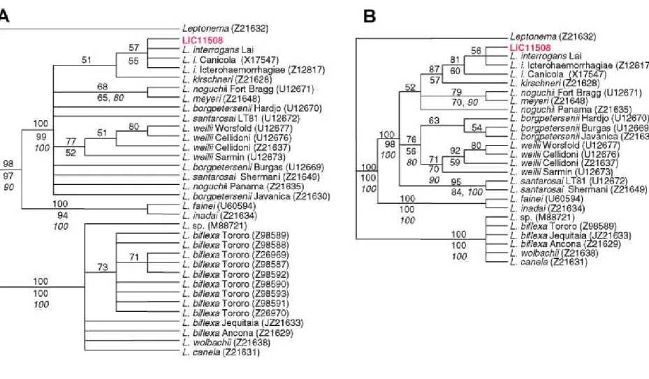
The Spirochaetes are divided into three major phylogenetic groups, or families: Spi-rochaetaceae, which includes Borrelia and Treponema among others; Brachyspiraceae and Leptospiraceae, which contain two gen-era, Leptospira and Leptonema (13). Phylo-genetic analyses based on 16S rDNA se-quences, using Leptonema as an outgroup, show that Leptospira are split into two
well-supported monophyletic groups (Figure 2), one of them formed by the pathogenic strains (e.g., L. interrogans) and the other formed by the non-pathogenic strains (e.g., L. biflexa). At the base of the clade of the pathogenic strains, L. inadai and L. fainei form a well-supported assemblage. Similar results were obtained by Postic et al. (14) based on 16S rDNA analyses. In these analyses L. interro-gans serovars formed a well-supported mono-phyletic cluster closely related to L. kirchneri (Figure 2B). Considering a constant diver-gence rate of 1 to 2% per 50 million years for the 16S rDNA (9), separation time between the two main assemblages (L. interrogans versus L. biflexa) was estimated at 590 to 295 million years.
A.L.T.O. Nascimento et al.
463
Braz J Med Biol Res 37(4) 2004
Leptospira interrogans
genome features
cobGKN genes, known to be involved in the cobalamin pathway (19) were not found. However, there are two predicted coding sequences inside this operon in chromosome II that could perform these steps. One has an oxidoreductase NAD-binding domain (LIC20133) and the other is a [2Fe-2S] ferre-doxin involved in electron transfer (LIC20135). In addition, other genes present in the genome coding for reductases such as LIC11145, LIC13354, LIC12391, and LIC10522 could also cope with these activi-ties. The presence of cysG in chromosome I, a gene that encodes a multifunctional pro-tein with methylase, oxidase and ferrochela-tase activities (20), may also be a cobalt-inserting enzyme in the B12 pathway. Other
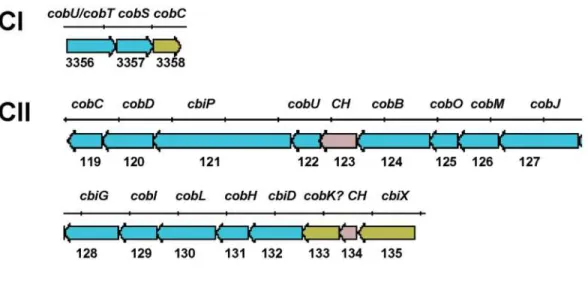
genes involved in this biosynthesis pathway were found in chromosome I (cysG/hemX/ cobA, cobT/cobU, cobS) (Figure 3). In fact, experimental evidence has recently shown that leptospires can grow in medium in the absence of B12 (Hartskeerl RA, unpublished results). This is contrary to the previous statement that L. interrogans is unable to synthesize B12, and that is why it is an essential component of the EMJH semi-syn-thetic medium (1,4). Thus, L. interrogans, unlike the spirochetes Borrelia burgdoferi and Treponema pallidum, have the complete repertoire of genes for de novo synthesis of protoheme and cobalamin. The functional link between the two replicons supports the view that the small replicon is in fact a second chromosome.
Transport
Among the 220 Leptospira transport pro-teins, we found 148 proteins comprising 108 different major primary and secondary trans-porters (Figure 4), as defined by Paulsen et al. (21). There are 34 major primary ATP-driven transporters including 30 ABC-trans-porters (53 proteins), the largest protein fam-ily in L. interrogans, as usually is the case for bacteria (22). The ABC superfamily
con-tains both uptake and efflux transport sys-tems, and ATP hydrolysis energizes the trans-port. The porters of the ABC superfamily consist of two integral membrane proteins (that determine specificity of the transported solute) and two cytoplasmic ATP-hydrolyz-ing proteins present as homodimers or heter-odimers. The uptake systems (but not the efflux systems) additionally possess extra-cytoplasmic solute-binding receptors (one or more per system), which in Gram-nega-tive bacteria are found in the periplasm. We found 21 ABC efflux systems, including drug and heavy metal export and detoxifica-tion, lipoprotein-releasing and hemolysin export systems. The remaining 9 are ABC uptake systems, including iron, sulfate, nickel, phosphate, dipeptide, amino acid, and carbohydrate uptake. There is one F-type ATP-synthase system (8 proteins), as mentioned above, and 3 P-type cation-trans-port ATPases (5 proteins).
465
Braz J Med Biol Res 37(4) 2004
Leptospira interrogans genome features
Figure 3. The cobalamin biosynthetic pathway of Leptospira interrogans. Genetic organization of the cobalamin biosynthetic locus in the two chromosomes (CI and CII) of L. interrogans serovar Copenhageni.
antiporter, a glycerol-3-P:Pi antiporter, two drug-sodium antiporters and two polysac-charide exporters. Secondary transporters also include 7 members of the TonB family of auxiliary proteins for energizing the outer membrane receptor-mediated active trans-port. Leptospira has many iron-transporting proteins in addition to the cobalamin/iron periplasmic binding protein component of the ABC-transport system (LIC13403) men-tioned above. Four members of the outer membrane receptor family have been identi-fied, which are probably involved in the transport of iron dicitrate (LIC10714), hemin (LIC10964), ferric hydroxamate (LIC11345) and ferrienterobactin (LIC12998). In addi-tion, an orthologue of the ferrous iron up-take (FeoB) protein of E. coli was found (LIC11402). FeoB has been characterized as an Fe2+ uptake system and possesses an
ATP/GTP binding motif at its N-terminal hydrophilic domain, therefore being prob-ably energized by ATP or GTP hydrolysis (23).
Oxygen defense
The Leptospira genome contains several genes encoding enzymes with peroxidase activities, such as catalase, glutathione per-oxidase and thiol perper-oxidase. However, su-peroxide dismutase orthologues and two important regulons, SoxRS and OxyR, nor-mally responsible for the defense against oxidative stress, are absent (5). It is possible that metalloporphyrins (24) could provide defense against oxidative damage, since L. interrogans is competent for porphyrin bio-synthesis and has metal uptake transport pro-teins. Genes coding for co-migratory bacte-rioferritin (LIC20093 and LIC10732), thiol peroxidase (LIC12765) and peroxiredoxin (LIC11219) with alkyl hydroperoxide re-ductase (AhpC) and thiol-specific antioxi-dant activities, respectively, were also iden-tified. Two predicted coding sequences for bacterioferritin (LIC11310 and LIC13209),
which may have functions analogous to ani-mal ferritin, are also present and may pro-vide both iron detoxification and storage, that would prevent free iron in Leptospira from driving oxidative reactions.
Regulatory systems and signal transduction
There are many genes encoding signal transduction proteins in Leptospira, indicat-ing that this organism has developed a vast array of regulatory systems that enable it to respond to environmental signals. This vari-ety of regulatory domains is found in non-obligate parasitic bacteria, indicating a much greater need to interpret the signals from the environment in order to respond properly, while obligate parasites have a much lower number and variety of domains in their sig-nal transduction proteins (25). There are 80 genes encoding components of the phospho-rylation-mediated signal transduction path-way: 29 histidine kinases (HK), 30 response regulators (RR) and 18 hybrid kinase/regu-lators (HK/RR). These members of the two-component systems present several domains organized into different arrangements (Fig-ure 5). Nineteen of the histidine kinases are located in the inner membrane, nine are cy-toplasmic and one is probably found in the periplasm, as predicted by the PSORT pro-gram [http://psort.nibb.ac.jp/].
macro-467
Braz J Med Biol Res 37(4) 2004
Leptospira interrogans genome features
phage phagocytosis (27), and probably this is also the case for Leptospira.
The response regulators are the cytoplas-mic effectors of the message, which become functional after being phosphorylated at an aspartate residue by the cognate histidine kinase. The RRs may possess a second effec-tor domain (Figure 5), which will perform its ultimate function, such as the DNA-binding helix-turn-helix domain that allows the RR to regulate transcription. Other noticeable domains found in L. interrogans RRs are the GGDEF and EAL motifs, which correspond to putative diguanylate cyclase and phos-phodiesterase domains, respectively, and a phosphatase domain similar to the mamma-lian phosphatase 2C that may be involved in the phosphorelay.
Cyclic nucleotides are likely to have a major regulatory role in Leptospira. There are 19 homologues of adenylate/guanylate cyclases, two specific phosphodiesterases and 7 cyclic nucleotide-binding proteins that probably have a regulatory function. There are 12 genes encoding proteins containing the GGDEF motif, seven of which are orga-nized in tandem in chromosome I (LIC11131 to LIC11125), and they also have a PAS/ PAC sensor domain in the amino-terminal region, suggesting that they are the product of gene duplication. The diguanylate cyclase activity of the GGDEF domain is required for a novel regulatory mechanism involving bis-(2',5')-cyclic diguanylic acid (c-di-GMP) as an allosteric activator (28). Two of the GGDEF-containing proteins also have the Cache domain, which is involved in chemo-taxis signal transduction (29), an important feature for Leptospira. There are eight puta-tive diguanylate phosphodiesterases contain-ing an EAL domain, with three of them becontain-ing associated with an RR domain (Figure 5). L. interrogans also presents in its genome three serine/threonine kinases and two hybrid HK/ RR with a GAF domain, which is a binding domain for cGMP (30).
Other interesting findings in the genome
include three related genes encoding puta-tive DNA-binding proteins (LIC20104, LIC20105 and LIC20178), which contain 6 transmembrane domains in the amino-termi-nus and one helix-turn-helix domain of the AraC type at the carboxyl-terminus. These genes are present in chromosome II, and two of them are clustered. Orthologues with a C-terminal DNA-2binding domain and a hy-drophobic N-terminal region were described in other bacteria, including Borrelia and Tre-ponema (31), but their function is unknown.
Motility and chemotaxis
burg-469
Braz J Med Biol Res 37(4) 2004
Leptospira interrogans genome features
dorferi, the majority of the structural and functional motility genes are positioned in potential operons. However, the operons probably underwent extensive rearrange-ments as they are generally smaller, often corresponding to only parts of the major Treponema and Borrelia operons. For ex-ample, the flgB operon in B. burgdorferi consisting of 26 genes (35) has been dis-rupted into 6 fragments dispersed at 5 posi-tions in the leptospiral genome. Differences in number of genes and operon organization might be associated with the high flexibility of pathogenic leptospires enabling them to survive and adapt to a variety of environ-ments and hosts.
Hemolysins
The primary lesion caused by Leptospira is the damage to the endothelium of small blood vessels, leading to hemorrhage and localized ischemia in multiple organs. As a consequence, renal tubular necrosis, hepato-cellular damage, meningitis, and myositis may occur in the infected host (1). Hemoly-sins may play a fundamental role in this toxic process. Several genes coding for predicted hemolysins were identified in the L. interro-gans genome. Some of them are structurally related to sphingomyelinases C (LIC10657, LIC12631, LIC12632, LIC11040, and LIC13198). Although generically called sphingomyelinases, it is possible that these genes code for hydrolases that act not only on sphingomyelins but also on other sphingolipids. Erythrocytes may represent a target for these enzymes since they are rich in glycosphingolipids like the antigenic de-terminants of the ABO system. Interestingly, LIC12631 and LIC12632 are organized as an operon, which may suggest a concerted expression and action.
The other class of genes coding for hemolysins, tlyABC, was identified. Although they were assigned to the same TlyABC class, they do not present structural
similar-ity. These putative hemolysins were first identified in the spirochete Brachyspira hyo-dysenteriae. Hemolytic and cytotoxic activi-ties of the recombinant TlyA, TlyB and TlyC proteins, expressed in E. coli, were detected (36). TlyB belongs to the family of Clp ATP-dependent proteases (37) and there are 3 genes coding for structurally related proteins (LIC10339, LIC11814 and LIC12017). Thus, 5 genes of the tlyABC class (LIC10284, LIC10339, LIC11814, LIC12017, and LIC13143) were identified in the L. interro-gans genome.
LIC12134 codes for an HlpA-related pro-tein which was characterized as a hemolysin in Prevotella intermedia, a common oral bacterium associated with periodontitis (38). Another identified gene (LIC10325) is re-lated to the hlyX gene which codes for a predicted hemolysin previously identified in L. borgpetersenii serovar Hardjo (Acces-sion number AAF09252, unpublished re-sults). LIC11352 should also be mentioned as the gene which codes for LipL32 or HAP-1, a ubiquitous lipoprotein of patho-genic Leptospira (see Lipoproteins section below) with hemolytic activity (39).
Surface-exposed proteins
the cytoplasmic membrane. The leptospiral genome also contains both the standard sig-nal peptidase and the lipoprotein sigsig-nal pep-tidase. The standard signal peptidase is in-volved in the hydrolysis of signal peptides of transmembrane outer membrane proteins and periplasmic proteins, releasing them from the cytoplasmic membrane. Lipoprotein sig-nal peptidase hydrolyzes the sigsig-nal peptides of lipoproteins prior to lipidation of the N-terminal cysteine.
Transmembrane outer membrane proteins
Analysis of the L. interrogans genome reveals 83 beta-sheet transmembrane outer membrane proteins (OMPs) and all except one (OmpL1, LIC10973) (40) were previ-ously unknown. An example of a newly identified leptospiral protein with a trans-membrane beta-sheet structure is LIC10642 which is predicted to be a member of the OMP superfamily.
Among these newly identified OMPs is a family of predicted coding sequences (LIC20202, LIC10995, LIC11465, and LIC11103) belonging to the superfamily of alpha/beta hydrolases, which includes bac-terial lipases. Another transmembrane OMP is LIC11623, a member of a family of highly conserved proteins of Gram-negative bacte-ria, including N. meningitides Omp85, which is thought to be a chaperonin involved in the assembly of OMPs in the outer membrane (41). OmpL1 belongs to the class of nonspe-cific porins allowing passage of small mol-ecules (<1000 Daltons) across the leptospi-ral outer membrane (40). Nonspecific porins allow both influx of nutrients and efflux of products of metabolism. Another example is LIC11458, which is predicted to be a mem-ber of the porin superfamily.
Specific channels
Survival in the mammalian host environ-ment by bacterial pathogens requires the
acquisition of certain trace nutrients. For example, iron is essential for leptospiral growth and is bound by a number of high-affinity binding proteins in mammals, which restrict its availability. Bacteria scavenge trace nutrients, including iron, heme, and vitamin B12, from their environment utiliz-ing the cytoplasmic membrane protein TonB and a series of “TonB-dependent” OMPs (Figure 6). After binding, transport of the nutrient across the outer membrane into the periplasm by this type of channel is an en-ergy-dependent step requiring interaction of the TonB-dependent OMP with TonB. The L. interrogans genome contains a TonB orthologue (LIC10889) and a large number of TonB-dependent OMPs: LIC20151, LIC20214, LIC10998, LIC10964, LIC10714, LIC12374, LIC12898, LIC12998, LIC11694, LIC11345, LIC10882, LIC10881, and LIC10896.
The leptospiral genome also contains OMPs involved in efflux pathways (Figure 6). The type I efflux system is represented by an orthologue of TolC (LIC13135), the outer membrane exporter of hemolysin and drugs, along with an orthologue of CusC (LIC11941), an outer membrane exporter of copper ion. A second type of efflux pathway found in the leptospiral genome belongs to the resistance-nodulation-cell division su-perfamily, a three-member complex that cata-lyzes substrate efflux via an H+ antiport
471
Braz J Med Biol Res 37(4) 2004
Leptospira interrogans genome features
two orthologues of the cation efflux system protein CzcD (LIC11714 and LIC13205), which are members of the cation diffusion facilitator family. CzcD of Bacillus subtilis has been shown to catalyze divalent cation (Zn2+ or Cd2+) efflux in exchange for the
uptake of two monovalent cations (K+ and
H+) in an electroneutral process energized
by the transmembrane pH gradient (42).
Lipoproteins
Experimental evidence for fatty acid modification of leptospiral lipoproteins has been described for the outer membrane lipo-proteins LipL32 (LIC11352) (43), LipL36 (LIC13060) (44), and LipL41 (LIC12966) (45). The cytoplasmic membrane also con-tains lipoproteins, as demonstrated for
LipL31 (LIC11456) and LipL71 (LIC11003) (46). A total of 184 predicted coding se-quences in the L. interrogans genome were found to have a lipoprotein signal peptidase cleavage site (5). All proposed lipoprotein-coding sequences conform to the rule of having an L, I, V, or F in the -3 and/or -4 position relative to cysteine and most of them have A, G, S, or N in the -1 position relative to cysteine (47).
Proteolytic functions can be assigned to some of the newly identified lipoproteins: four lipoproteins are thermolysin homologues (LIC10715, LIC13320, LIC13321, and LIC13322), and one is a predicted metallo-protease (LIC11834). Several lipoproteins may be involved in hemolysis: two lipopro-teins are sphingomyelinase homologues (LIC10657 and LIC12632), and one is a
Figure 6. Model of leptospiral membrane architecture. Leptospires have two membranes, an outer membrane (OM) and a cytoplasmic or inner membrane (IM). As in Gram-positive bacteria, the peptidoglycan (PG) cell wall is closely associated with the IM. The leptospiral surface is dominated by lipopolysaccharide (LPS) carbohydrate side chains. Subsurface proteins include the cytoplasmic protein, GroEL, and the periplasmic endoflagella (EF). The IM contains lipoproteins such as LipL31 and transmembrane proteins such as signal peptidase (SP) and ImpL63. The OM contains lipoproteins including LipL41 and LipL36, and transmembrane proteins including the porin, OmpL1. Genomic sequence analysis reveals several novel types of outer membrane proteins (OMPs), including TonB-dependent OMPs involved in nutrient acquisition. BtuB is an example of a TonB-TonB-dependent OMP involved in uptake of vitamin B12. The type I efflux system is represented by TolC, which forms a complex with ATP-binding
phospholipase D homologue (LIC11754). Lipoprotein LIC10972 is predicted to be located in the outer membrane and is a MauG homologue belonging to a family of cyto-chrome c peroxidases that may be involved in defense against hydrogen peroxide. Other lipoproteins with putative enzymatic func-tions are homologues of glucose dehydro-genase (LIC12294) and cholesterol oxidase (LIC13202).
S-layer and peripheral membrane proteins
S-layers are thought to provide structural integrity to the bacterial surface (48). Al-though an S-layer has not been observed in pathogenic leptospires, the genome contains at least two proteins with S-layer homology (LIC10868 and LIC12952). Experimental evidence is needed to determine whether these proteins are actually S-layer compo-nents.
Like S-layer proteins, peripheral mem-brane proteins are not integral memmem-brane proteins. LipL45 is processed to a peripheral membrane associated with the outer mem-brane, P31LipL45 (49). P31LipL45 expression is
dramatically up-regulated in stationary phase cultures, and for this reason may have a membrane-stabilizing function. At this time it is unclear whether P31LipL45 is
surface-exposed. Interestingly, the leptospiral ge-nome contains a number of predicted coding sequences with homology to LipL45 (LIC20102, LIC20114, LIC13414, and LIC10123).
Surface-maintenance proteins
Bacteria are likely to have a variety of proteins involved in maintaining the surface structural integrity. One such protein, glyc-erophosphoryl diester phosphodiesterase, is a protein belonging to this category which has been found in all spirochetal genomes studied to date. E. coli has two forms of this enzyme, a cytosolic form, ugpQ, and a
peri-plasmic form, glpQ. The leptospiral genome contains two homologues of this enzyme, LIC13182 and LIC10293. The former should be the cytosolic form because it lacks a signal peptide while the latter should be the exported form because it has an N-terminal signal peptide. In other spirochetes, GlpQ is associated with the outer membrane (50).
Cytoplasmic membrane proteins
The leptospiral genome contains a num-ber of proteins that belong to a large family of prokaryotic proteins with homology to the peptidoglycan-associated portion of E. coli OmpA (51). These proteins are predicted to be either cytoplasmic membrane (LIC20250, LIC10592, LIC13479, and LIC10050) or periplasmic proteins (LIC10537 and LIC10191), rather than OMPs, which is con-sistent with experimental evidence that the spirochetal cell wall is more closely associ-ated with the cytoplasmic membrane than the outer membrane. An interesting protein family is the mechanosensitive ion channel (LIC20069 and LIC12671). Two members of this protein family of M. jannaschii have been functionally characterized and both form mechanosensitive ion channels (52). Therefore, this family is likely to also en-code mechanosensitive channel proteins in L. interrogans, playing a physiological role in bacterial osmoregulation.
Lipopolysaccharides
Anti-473
Braz J Med Biol Res 37(4) 2004
Leptospira interrogans genome features
bodies raised against LPS from different Leptospira strains during infections are re-lated to polysaccharide structure in terms of its sugar composition, number, repetitive-ness, and ramification (1). In Leptospira, as in many other bacteria, at least part of the genes coding for enzymes of the polysaccha-ride biosynthesis pathway are found clus-tered in a region of chromosomes named O-antigen gene cluster (rfb locus) (53).
The complete genome analysis reveals a large portion of about 119 kb (genome posi-tion 2.538.470-2.554.255), in which all the 108 predicted coding sequences are tran-scribed in the same orientation (Figure 7). Interestingly, this region is dense in genes related to LPS biosynthesis (Table 1) and includes the O-antigen gene cluster previ-ously described in L. interrogans serovar Copenhageni (53,54). In the first 14 kb of this region the predicted genes seem not to be related to LPS biosynthesis, but to DNA replication, and some genes code for riboso-mal proteins. In the other 105 kb of this region there are 94 predicted genes 56 of which are clearly related to LPS biosynthe-sis. These predicted genes encode enzymes for nucleotide sugar biosynthesis, such as the enzymes for dTDP-rhamnose biosynthe-sis (54) for CMP-N-acetylneuraminic acid and for perosamine synthase. In addition, many genes coding for sugar transferases, sugar modifications and for the O-antigen processing, Wzx-flippase and Wzy-poly-merase, involved in oligosaccharide export-ing and assembly of the LPS, were identi-fied. Some genes encoding proteins of the lipid A biosynthesis (lpxD) and transport (msbA) are also found is this region.
The comparison with the corresponding region in the genome of L. interrogans serovar Lai (4) revealed only three distinct predicted coding sequences: two extra cop-ies of genes for aminotransferase (LIC12197 and LIC12198) and the absence of a gene encoding galactoside O-acetyltransferase (LA1622).
Seventy-seven other genes, probably re-lated to LPS biosynthesis, were identified along the genome. All the genes related to lipid A biosynthesis described in E. coli were identified in the Leptospira genome (lpxA, lpxC, lpxD, lpxB, lpxK). However, the predominant fatty acids in lipid A of L. interrogans are dodecanoic and hexade-canoic acid instead of hydroxymyrystoyl (14 carbons), which is the signature of Gram-negative bacteria (1). Comparative analysis between LpxA of E. coli and P. aeruginosa (55) showed that few structural differences in this enzyme determine changes in the fatty acid chain size incorporated during lipid A biosynthesis. The lower endotoxicity of lep-tospiral LPS as compared to LPS from Gram-negative bacteria has been reported (56). It was also reported that leptospiral LPS duces a TLR2-dependent cell activation, stead of LTR4, the receptor frequently in-volved in the LPS immune response (57). The lower endotoxicity and the difference in the mechanism of cell immune response ac-tivation are supposed to be related to differ-ences in the lipid A structure.
Genes encoding enzymes of Kdo biosyn-thesis (kdsA, kdsB) such as Kdo-transferase (waaA or kdtA), which catalyzes the binding of Kdo to lipid IVA, were identified. Al-though typical Kdo was found in Leptospira LPS, it was reported to be substituted at different carbon positions (1).
Four paralogues encoding MsbA, the pro-tein that transfers the flippase of lipid A in
Table 1. Number of genes at the lipopolysaccha-ride 119-kb locus.
Predicted protein function
Nucleotide sugar biosynthesis 22
Sugar transferase 17
Sugar epimerase 10
Lipid A and polysaccharide transport 4 Hypothetical 26 (17 conserved) Aminoacyl tRNA synthase 3
DNA metabolism 4
Ribosomal protein 3
A.L.T.O. Nascimento et al.
Figure 7. Lipopolysaccharide region of Leptospira interrogans. The 119-kb region of L. interrogans serovar Copenhageni genome with 108 predicted coding sequences transcribed in the same orientation. Orange arrows indicate predicted genes encoding polysaccharide biosynthesis-related proteins. Shaded regions indicate the homologous genes from L. borgpetersenii serovar Hardjobovis (53). The region containing the predicted coding sequences, indicated by the letters F, E, A, B, D, C, was first indicated as the rfb genes related to rhamnose biosynthesis in
475
Braz J Med Biol Res 37(4) 2004
Leptospira interrogans genome features
the inner membrane from the cytoplasmic side to the periplasmic side, were identified. One of the msbA genes is located in the 119-kb region. Another msbA gene is located upstream of lpxK, as reported in many other genomes (55). It will be interesting to compare the set of genes coding for enzymes related to LPS bio-synthesis identified in the Leptospira genome to the orthologues in other microorganisms, in order to correlate LPS structural differ-ences and endotoxic activity.
Comparative genomics and insertion sequences
A three-way comparison between the L. interrogans genome and the genomes of B. burgdorferi and T. pallidum yields the fol-lowing results: 1167 (31%) of the genes in L. interrogans Copenhageni are found in B. burgdorferi and/or in T. pallidum, 666 (41%) of the genes in B. burgdorferi are found in the Copenhageni genome, and 589 (57%) of the genes in T. pallidum are found in the L. interrogans genome. A total of 362 pre-dicted genes were found to be shared by all three spirochetes, 45 of which are hypotheti-cal (detailed list in our project website: http:/ /aeg.lbi.ic.unicamp.br/world/lic/). These re-sults show that a thorough analysis of the genome can significantly contribute to the understanding of spirochete biology.
Our analysis of the L. interrogans ge-nome revealed the presence of the
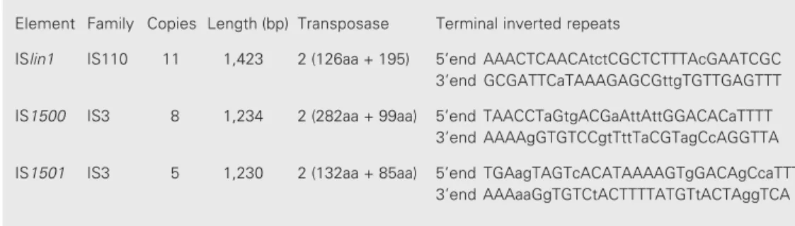
previ-ously described insertion sequence (IS) ele-ments IS1500, IS1501 (58), reminiscent in-sertions of IS1533 (59)and IS1502 (60)and an IS recently identified that we designated ISlin1 (5) (Table 2). IS1500, IS1501 and ISlin1 account for 24 insertions distributed throughout chromosome I. Together, IS ele-ments and transposases comprise 2% of the genome. These elements are related to major IS families such as IS110 and IS3 that are defined by their conserved amino acid motif (DDE) in the transposase. So far all the insertions were found in intergenic regions, having no mutagenic effects on L. interro-gans genes.
The L. interrogans genome provides new insights into biosynthesis pathway, transport families, environmental response, and patho-genesis. A broad array of regulatory system proteins enable this organism to respond to environmental signals. A new group of genes involved in LPS biosynthesis may contribute to the elucidation of serovar diversity among leptospires. Several categories of surface-exposed proteins required for colonization and survival in the mammalian host were identified. These proteins may have a role in mechanisms of leptospiral pathogenesis and protective immunity. Available vaccines are serovar-specific and have low efficacy (1). Surface-exposed proteins that are conserved among pathogenic serovars may be used for vaccine development for the prevention of leptospirosis.
Table 2. Insertion sequence (IS) elements present in Leptospira interrogans serovar Copenhageni.
Element Family Copies Length (bp) Transposase Terminal inverted repeats
ISlin1 IS110 11 1,423 2 (126aa + 195) 5’end AAACTCAACAtctCGCTCTTTAcGAATCGC 3’end GCGATTCaTAAAGAGCGttgTGTTGAGTTT
IS1500 IS3 8 1,234 2 (282aa + 99aa) 5’end TAACCTaGtgACGaAttAttGGACACaTTTT 3’end AAAAgGTGTCCgtTttTaCGTagCcAGGTTA
Acknowledgments
We are deeply indebted to Drs. I. Raw (Fundação Butantan, São Paulo, SP, Brazil) for use of laboratory facilities and valuable support. We thank Dr. Albert I. Ko (Fiocruz, Salvador, BA, Brazil) for the strain Fiocruz L1-130, the Agronomical and Environmental Genomes (AEG)
Consortium of the network Organization for Nucleotide Sequencing and Analysis (ONSA) for the genome sequenc-ing data, and Dr. L.C.C. Leite (Instituto Butantan) for a critical reading of this manuscript.
Linear representation of the Leptospira interrogans serovar Copenhageni chromosomes (CI and CII). Genes are colored according to their biological role. Arrows indicate the direction of transcription. The pie representation indicates the distribution of the number of genes according to their biological role. The numbers below protein-producing genes correspond to gene identification numbers (IDs).
Additional information is contained in the project website http://aeg.lbi.ic.unicamp.br/world/lic/
Insert InsertInsert
477
Braz J Med Biol Res 37(4) 2004
Leptospira interrogans genome features
References
1. Faine S, Adler B, Bolin C & Perolat P (1999). Leptospira and Lepto-spirosis. MediSci, Melbourne, Australia.
2. Bharti AR, Nally JE, Ricaldi JN et al. (2003). Leptospirosis: a zoonotic disease of global importance. Lancet Infectious Diseases, 3: 757-771.
3. Romero EC, Bernardo CC & Yasuda PH (2003). Human leptospiro-sis: a twenty-nine-year serological study in São Paulo, Brazil. Re-vista do Instituto de Medicina Tropical de São Paulo, 45: 245-248. 4. Ren SX, Fu G, Jiang XG et al. (2003). Unique physiological and
pathogenic features of Leptospira interrogans revealed by whole-genome sequencing. Nature, 422: 888-893.
5. Nascimento ALTO, Ko AI, Martins EAL et al. (2004). Comparative genomics of two Leptospira interrogans serovars reveals novel insights into physiology and pathogenesis. Journal of Bacteriology (in press).
6. Simpson AJ, Reinach FC, Arruda P et al. (2000). The genome se-quence of the plant pathogen Xylella fastidiosa. Nature, 406: 151-157.
7. Van Sluys MA, de Oliveira MC, Monteiro-Vitorello CB et al. (2003). Comparative analyses of the complete genome sequences of Pierce’s disease and citrus variegated chlorosis strains of Xylella fastidiosa. Journal of Bacteriology, 185: 1018-1026.
8. Swofford DL (2000). PAUP*-Phylogenetic Analysis Using Parsimony (*and other methods). Version 4. Sinauer Associates, Sunderland, MA, USA.
9. Ochman H, Elwyn S & Moran NA (1999). Calibrating bacterial evolu-tion. Proceedings of the National Academy of Sciences, USA, 96: 12638-12643.
10. Messer W (2002). The bacterial replication initiator DnaA. DnaA and oriC, the bacterial mode to initiate DNA replication. FEMS Microbiol-ogy Reviews, 26: 355-374.
11. Lobry JR (1996). Asymmetric substitution patterns in the two DNA strands of bacteria. Molecular Biology and Evolution, 13: 660-665. 12. Fukunaga M & Mifuchi I (1989). Unique organization of Leptospira
interrogans rRNA genes. Journal of Bacteriology, 171: 5763-5767. 13. Paster BJ & Dewhirst FE (2000). Phylogenetic foundation of
spiro-chetes. Journal of Molecular Microbiology and Biotechnology, 2: 341-344.
14. Postic D, Riquelme-Sertour N, Merien F, Perolat P & Baranton G (2000). Interest of partial 16S rDNA gene sequences to resolve heterogeneities between Leptospira collections: application to L. meyeri. Research in Microbiology, 151: 333-341.
15. Heidelberg JF, Eisen JA, Nelson WC et al. (2000). DNA sequence of both chromosomes of the cholera pathogen Vibrio cholerae. Na-ture, 406: 477-483.
16. Salanoubat M, Genin S, Artiguenave F et al. (2002). Genome se-quence of the plant pathogen Ralstonia solanacearum. Nature, 415: 497-502.
17. Bourhy P & Saint Girons I (2000). Localization of the Leptospira interrogans metF gene on the CII secondary chromosome. FEMS Microbiology Letters, 191: 259-263.
18. Guegan R, Camadro JM, Saint Girons I & Picardeau M (2003). Leptospira spp. possess a complete haem biosynthetic pathway and are able to use exogenous haem sources. Molecular Microbiol-ogy, 49: 745-754.
19. Rodionov DA, Vitreschak AG, Mironov AA & Gelfand MS (2003). Comparative genomics of the vitamin B12 metabolism and regula-tion in prokaryotes. Journal of Biological Chemistry, 278:
41148-41159.
20. Spencer JB, Stolowich NJ, Roessner CA & Scott AI (1993). The Escherichia coli cysG gene encodes the multifunctional protein, siroheme synthase. FEBS Letters, 335: 57-60.
21. Paulsen IT, Nguyen L, Sliwinski MK, Rabus R & Saier Jr MH (2000). Microbial genome analyses: comparative transport capabilities in eighteen prokaryotes. Journal of Molecular Biology, 301: 75-100. 22. Meidanis J, Braga MD & Verjovski-Almeida S (2002).
Whole-ge-nome analysis of transporters in the plant pathogen Xylella fasti-diosa. Microbiology and Molecular Biology Reviews, 66: 272-299. 23. Kammler M, Schon C & Hantke K (1993). Characterization of the
ferrous iron uptake system of Escherichia coli. Journal of Bacteriol-ogy, 175: 6212-6219.
24. Vujaskovic Z, Batinic-Haberle I, Rabbani ZN, Feng QF, Kang SK, Spasojevic I, Samulski TV, Fridovich I, Dewhirst MW & Anscher MS (2002). A small molecular weight catalytic metalloporphyrin antioxi-dant with superoxide dismutase (SOD) mimetic properties protects lungs from radiation-induced injury. Free Radical Biology and Medi-cine, 33: 857-863.
25. Galperin MY, Nikolskaya AN & Koonin EV (2001). Novel domains of the prokaryotic two-component signal transduction systems. FEMS Microbiology Letters, 203: 11-21.
26. Taylor BL & Zhulin IB (1999). PAS domains: internal sensors of oxygen, redox potential, and light. Microbiology and Molecular Biol-ogy Reviews, 63: 479-506.
27. Rickman L, Saldanha JW, Hunt DM, Hoar DN, Colston MJ, Millar JB & Buxton RS (2004). A two-component signal transduction system with a PAS domain-containing sensor is required for virulence of Mycobacterium tuberculosis in mice. Biochemical and Biophysical Research Communications, 314: 259-267.
28. Ausmees N, Mayer R, Weinhouse H, Volman G, Amikam D, Benziman M & Lindberg M (2001). Genetic data indicate that pro-teins containing the GGDEF domain possess diguanylate cyclase activity. FEMS Microbiology Letters, 204: 163-167.
29. Anantharaman V & Aravind L (2000). Cache - a signaling domain common to animal Ca(2+)-channel subunits and a class of prokary-otic chemotaxis receptors. Trends in Biochemical Sciences, 25: 535-537.
30. Ho YS, Burden LM & Hurley JH (2000). Structure of the GAF domain, a ubiquitous signaling motif and a new class of cyclic GMP receptor. EMBO Journal, 19: 5288-5299.
31. Subramanian G, Koonin EV & Aravind L (2000). Comparative ge-nome analysis of the pathogenic spirochetes Borrelia burgdorferi and Treponema pallidum. Infection and Immunity, 68: 1633-1648. 32. Bourret RB, Charon NW, Stock AM & West AH (2002). Bright lights,
abundant operons-fluorescence and genomic technologies advance studies of bacterial locomotion and signal transduction: review of the BLAST meeting, Cuernavaca, Mexico, 14 to 19 January 2001. Journal of Bacteriology, 184: 1-17.
33. Fraser CM, Casjens S, Huang WM et al. (1997). Genomic sequence of a Lyme disease spirochaete, Borrelia burgdorferi. Nature, 390: 580-586.
34. Fraser CM, Norris SJ, Weinstock GM et al. (1998). Complete ge-nome sequence of Treponema pallidum, the syphilis spirochete. Science, 281: 375-388.
Bacteriol-ogy, 179: 2289-2299.
36. ter Huurne AA, Muir S, van Houten M, van der Zeijst BA, Gaastra W & Kusters JG (1994). Characterization of three putative Serpulina hyodysenteriae hemolysins. Microbial Pathogenesis, 16: 269-282. 37. Squires C & Squires CL (1992). The Clp proteins: proteolysis
regula-tors or molecular chaperones? Journal of Bacteriology, 174: 1081-1085.
38. Beem JE, Nesbitt WE & Leung KP (1999). Cloning of Prevotella intermedia loci demonstrating multiple hemolytic domains. Oral Microbiology and Immunology, 14: 143-152.
39. Lee SH, Kim KA, Park YG, Seong IW, Kim MJ & Lee YJ (2000). Identification and partial characterization of a novel hemolysin from Leptospira interrogans serovar lai. Gene, 254: 19-28.
40. Shang ES, Exner MM, Summers TA, Martinich C, Champion CI, Hancock RE & Haake DA (1995). The rare outer membrane protein, OmpL1, of pathogenic Leptospira species is a heat-modifiable porin. Infection and Immunity, 63: 3174-3181.
41. Voulhoux R, Bos MP, Geurtsen J, Mols M & Tommassen J (2003). Role of a highly conserved bacterial protein in outer membrane protein assembly. Science, 299: 262-265.
42. Guffanti AA, Wei Y, Rood SV & Krulwich TA (2002). An antiport mechanism for a member of the cation diffusion facilitator family: divalent cations efflux in exchange for K+ and H+. Molecular Micro-biology, 45: 145-153.
43. Haake DA, Chao G, Zuerner RL, Barnett JK, Barnett D, Mazel M, Matsunaga J, Levett PN & Bolin CA (2000). The leptospiral major outer membrane protein LipL32 is a lipoprotein expressed during mammalian infection. Infection and Immunity, 68: 2276-2285. 44. Haake DA, Martinich C, Summers TA, Shang ES, Pruetz JD, McCoy
AM, Mazel MK & Bolin CA (1998). Characterization of leptospiral outer membrane lipoprotein LipL36: downregulation associated with late-log-phase growth and mammalian infection. Infection and Im-munity, 66: 1579-1587.
45. Shang ES, Summers TA & Haake DA (1996). Molecular cloning and sequence analysis of the gene encoding LipL41, a surface-exposed lipoprotein of pathogenic Leptospira species. Infection and Immuni-ty, 64: 2322-2330.
46. Haake DA & Matsunaga J (2002). Characterization of the leptospiral outer membrane and description of three novel leptospiral mem-brane proteins. Infection and Immunity, 70: 4936-4945.
47. Haake DA (2000). Spirochaetal lipoproteins and pathogenesis. Mi-crobiology, 146 (Part 7): 1491-1504.
48. Schuster B & Sleytr UB (2000). S-layer-supported lipid membranes. Journal of Biotechnology, 74: 233-254.
49. Matsunaga J, Young TA, Barnett JK, Barnett D, Bolin CA & Haake DA (2002). Novel 45-kilodalton leptospiral protein that is processed to a 31-kilodalton growth-phase-regulated peripheral membrane pro-tein. Infection and Immunity, 70: 323-334.
50. Cameron CE, Castro C, Lukehart SA & Van Voorhis WC (1998). Function and protective capacity of Treponema pallidum subsp. pallidum glycerophosphodiester phosphodiesterase. Infection and Immunity, 66: 5763-5770.
51. De Mot R & Vanderleyden J (1994). The C-terminal sequence con-servation between OmpA-related outer membrane proteins and MotB suggests a common function in both positive and gram-negative bacteria, possibly in the interaction of these domains with peptidoglycan. Molecular Microbiology, 12: 333-334.
52. Kloda A & Martinac B (2001). Structural and functional differences between two homologous mechanosensitive channels of Methano-coccus jannaschii. EMBO Journal, 20: 1888-1896.
53. de la Pena-Moctezuma A, Bulach DM, Kalambaheti T & Adler B (1999). Comparative analysis of the LPS biosynthetic loci of the genetic subtypes of serovar Hardjo: Leptospira interrogans subtype Hardjoprajitno and Leptospira borgpetersenii subtype Hardjobovis. FEMS Microbiology Letters, 177: 319-326.
54. Mitchison M, Bulach DM, Vinh T, Rajakumar K, Faine S & Adler B (1997). Identification and characterization of the dTDP-rhamnose biosynthesis and transfer genes of the lipopolysaccharide-related rfb locus in Leptospira interrogans serovar Copenhageni. Journal of Bacteriology, 179: 1262-1267.
55. Raetz CR & Whitfield C (2002). Lipopolysaccharide endotoxins. Annual Review of Biochemistry, 71: 635-700.
56. de Souza L & Koury MC (1992). Isolation and biological activities of endotoxin from Leptospira interrogans. Canadian Journal of Micro-biology, 38: 284-289.
57. Werts C, Tapping RI, Mathison JC et al. (2001). Leptospiral lipopoly-saccharide activates cells through a TLR2-dependent mechanism. Nature Immunology, 2: 346-352.
58. Boursaux-Eude C, Saint Girons I & Zuerner R (1995). IS1500, an IS3-like element from Leptospira interrogans. Microbiology, 141 (Part 9): 2165-2173.
59. Zuerner RL (1994). Nucleotide sequence analysis of IS1533 from Leptospira borgpetersenii: identification and expression of two IS-encoded proteins. Plasmid, 31: 1-11.