143
http://dx.doi.org/10.3345/kjp.2012.55.4.143 Korean J Pediatr 2012;55(4):143-146
eISSN 1738-1061 • pISSN 2092-7258
Case report
Two cases of female hydrocele of the canal of nuck
The processus vaginalis within the inguinal canal forms the canal of Nuck, which is a homolog of the processus vaginalis in women. Incomplete obliteration of the processus vaginalis causes indirect inguinal hernia or hydrocele of the canal of Nuck, a very rare condition in women. Here, we report 2 cases of hydrocele of the canal of Nuck that were diagnosed with ultrasonography in both cases and magnetic resonance imaging in 1 case to confirm the sonographic diagnosis. High ligation and hydrocelectomy were conducted in both patients. In 1 patient, 14 months later, the occurrence of contralateral inguinal hernia was suspected, but did not require surgery. The other patient had a history of surgery for left inguinal hernia 11 months before the oc currence of right hydrocele of the canal of Nuck. In both cases, the occurrence of an inguinal hernia on the contralateral side was noted.
Key words: Hydrocele, Canal of Nuck, Ultrasonography, Magnetic resonance imaging, Inguinal hernia
Yu Mi Choi, MD1, Gyu Min Lee, MD1, Jung
Bin Yi, MD1, Kyung Lim Yoon, MD1, Kye Shik
Shim, MD1, Chong Woo Bae, MD1, Sung Il
Choi, MD2, Hyun Cheol Kim, MD3
Departments of 1Pediatrics, 2Surgery, and 3Radiology, Kyung Hee University Hospital at Gangdong, Seoul, Korea
Received: 22 June 2011, Revised: 19 August, 2011 Accepted: 24 September 2011
Corresponding author: Kyung Lim Yoon, MD
Department of Pediatrics, Kyung Hee University Hospital at Gandong, 892 Dongnam-ro, Gangdong-gu, Seoul 134-727, Korea
Tel: +82-2-440-6132, Fax : +82-2-440-7175 E-mail: [email protected]
Copyright © 2012 by The Korean Pediatric Society
his is an open-access article distributed under the terms of the Creative Commons Attribution Non-Commercial License (http://creativecommons.org/licenses/by-nc/3.0/) which permits unrestricted non-commercial use, distribution, and reproduction in any medium, provided the original work is properly cited.
although there have been no reports about the recurrence of this anomaly.
Case reports
1. Case 1
A 4-year-old girl presented with a palpable mass in her right inguinal region. he mass was grape sized, tense, but not painful. he swelling was translucent and irreducible. She was in good physical health without any symptoms. There was no heating sensation nor erythematous change in the lesion. The differential diagnosis included hernia or hydrocele.
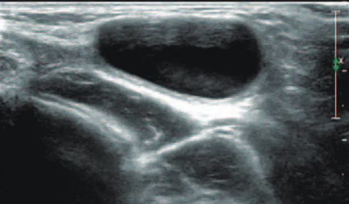
Inguinal ultrasonography with a 5 to 12 MHz linear array transducer revealed an ovoid shaped cystic mass of about 1.8 cm × 2.5 cm in the right inguinal region (Fig. 1). No bowel loop or omental fat was observed during the ultrasonographic examination. But, charicterization was difficult because it seemed a non-specific cystic
Introduction
144 YM Choi, et al. •Female hydrocele of the canal of nuck
lesion. MR imaging was requested to conirm the ultrasound inding and identify the relations with the surrounding structures. MR imaging showed a cystic mass in the right inguinal region (Fig. 2). he mass was well-deined cystic lesion without abnormal septation and solid portion.
High ligation and hydrocelectomy were performed. The round ligament with the cystic tense mass was excised. The pathologic examination revealed findings compatible with a hydrocele of the Canal of Nuck. Fourteen months later, she revisited the hospital because of a left inguinal mass that was considered to be a left inguinal hernia. However, the mass was reduced spontaneously and did not protrude for the next 4 years.
2. Case 2
An 18 month-old female patient presented with a small, right inguinal mass with tenderness. Ultrasound examination revealed a small sized, well-defined cystic mass about 0.74 cm×0.63 cm (Fig. 3). She had a history of herniorrhaphy of a left inguinal hernia which was diagnosed with ultrasound examination at the age of 7 months. High ligation of the hernial sac and hydrocelectomy was done, which
was conirmed by the pathology report. here was no occurrence of contralateral hydrocele or hernia for the next 3 years.
Discussion
The processus vaginalis, which is a homologue to the canal of Nuck, may be obliterated at any point between the internal inguinal ring and the scrotum, or it may happen incompletely. These variations account for the diverse classification of hernias (the extra-abdominal passage of viscus) and hydroceles (the extra-extra-abdominal passage of peritoneal fluid), including complete or scrotal hernias, communicating or non-communicating hydroceles, hydroceles of the spermatic cord in males, and the canal of Nuck in girls5).
Hydroceles are common in children, especially in boys. But hydrocele of the canal of Nuck is an uncommon clinical presentation6).
Although the exact process is unclear, it is generally agreed that obliteration of the processus vaginalis occurs only after the seventh month of gestation7), thus accounting for the higher incidence of inguinal hernias in the premature infant8). Although not clearly defined, there is a known heredity factor associated with the occurrence of hernias that is more frequent in twin gestations and in infants who have a family history of hernia9).
Swelling of the inguinal region in a female may result from a number of conditions, including inguinal hernia, tumor (lipoma, leiomyoma, sarcoma), cyst, abscess, lymphadenopathy, or hydrocele of the canal of Nuck1).
In most cases, the canal of Nuck should be distinguished from hernia. Bowel sounds over the swelling are strongly suggestive of a hernia.
Hydrocele of the canal of Nuck typically presents as a painless, translucent swelling in the inguinolabial region6). here is no nausea or vomiting. If possible, the child should be examined in the supine
Fig. 1. Inguinal sonography. Ultrasonography of the right inguinal area shows a cystic lesion without internal echoes. During the ultrasonography, no bowel or omental fat herniation was noted.
A B C
Korean J Pediatr 2012;55(4):143-146 • http://dx.doi.org/10.3345/kjp.2012.55.4.143 145
and standing positions6). In older children, Valsalva's maneuver should be performed to rule out hernia. If the bulge is apparent in the standing position and disappears while supine, a hernia is more plausible10).
Infection is an uncommon complication of hydroceles in children, and until now only 5 cases have been reported in the English language literature, all in boys. Infected hydrocele of the canal of Nuck is rare11).
In the past, a palpable inguinal mass was removed via surgery without an imaging study. Currently, sonography is the imaging modality of choice for evaluation of this type of mass12).
Because sonography is a relatively easy, accessible and accurate procedure4), we recommend that sonography be performed routinely in those females with an inguinal hernia containing an asymptomatic movable mass. But, we cannot identify whether there is a communication between cystic mass and peritoneal cavity with sonography. MR imaging can give more precise images including septation and a communication between cystic lesion and peritoneal cavity, and information on the anatomical relation with adjacent structures4,13). Therefore, MR imaging can help to diagnose in patients with inguinal cystic mass. The differential diagnosis of a cystic mass in the female groin region includes round ligament cysts, varicosities of the round ligament, inguinal herniation of the ovary, cystic lymphangiomas, epidermal inclusion cysts, abscesses, and pseudoaneurysms14).
The final diagnosis is made during surgery and confirmed by pathological examination6). Surgical resection of the hydrocele and ligation of the neck of the processus vaginalis should be considered as standard therapy.
Han et al.15) reported that in 2.6% of the patients who had been operated of unilateral hydrocele, contralateral hernia occurred. hey also reported that the occurrence of the contralateral hydrocele after the operation of unilateral hydrocele was 2.0% in males. Flum et al.16) reported that the recurrence rate in inguinal hernia is about 10% but has not been reported for a hydrocele of the canal of Nuck yet. In our cases, the occurrence of inguinal hernia was on the contralateral side from the hydrocele and operation was needed in one case. herefore, one should be aware that the ocurrence rate of the contralateral hernia is not low in patients with hydrocele of the canal of Nuck. So, we should consider the hydrocele of the canal of Nuck in patients who have a inguinal cystic mass and have a history of inguinal hernia on the other side.
In these cases, we suggest that MR imaging can help to identify an inguinal cystic mass when the diagnosis is vague by its sonographic indings in female. Because the inguinal hernia on the other side was detected either before or after the operation of hydrocele in both cases, we should consider the hydrocele of the canal of Nuck in patients who have an inguinal cystic mass and have a history of an inguinal hernia on the other side. Also, we should consider the risk of occurrence of contralateral inguinal hernia in patients with hydrocele of canal of Nuck.
References
1. Anderson CC, Broadie TA, Mackey JE, Kopecky KK. Hydrocele of the canal of Nuck: ultrasound appearance. Am Surg 1995;61:959-61.
2. Schwartz A, Peyser MR. Nuck's hydrocele (hydrocele muliebris). Int Surg 1975;60:91-2.
3. Kim SH, Seo IY, Cho HJ, Ku1 YM, Kim KH, Ahn CH, et al. Hydrocele of the Canal of Nuck. J Korean Surg Soc 2008;74:396-8.
4. Park SJ, Lee HK, Hong HS, Kim HC, Kim DH, Park JS, et al. Hydrocele of the canal of Nuck in a girl: ultrasound and MR appearance. Br J Radiol 2004;77:243-4.
5. Haynes JH. Inguinal and scrotal disorders. Surg Clin North Am 2006; 86:371-81, ix.
6. De Meulder F, Wojciechowski M, Hubens G, Ramet J. Female hydrocele of the canal of Nuck: a case report. Eur J Pediatr 2006;165:193-4.
7. Skandalakis JE, Gray SW, Ricketts RR, Skandalakis LJ. Anterior body wall. In: Skandalakis JE, Gray SW, editors. Embryology for surgeons: the embryological basis for the treatment of congenital anomalies. 2nd ed. Baltimore: Williams & Wilkins, 1994;578-80.
8. Rescorla FJ, Grosfeld JL. Inguinal hernia repair in the perinatal period and early infancy: clinical considerations. J Pediatr Surg 1984;19:832-7.
9. Czeizel A, Gárdonyi J. A family study of congenital inguinal hernia. Am J Med Genet 1979;4:247-54.
10. Graham SD Jr, Keane TE, Glenn JF. Glenn’s urologic surgery. 7th ed. Philadelphia: Lippincott Williams & Wilkins, 2009:735-49.
11. Ameh EA, Garba ES. Infected hydrocoele of the canal of Nuck in a
146 YM Choi, et al. •Female hydrocele of the canal of nuck
neonate. Urol Int 2003;71:226-7.
12. Huang CS, Luo CC, Chao HC, Chu SM, Yu YJ, Yen JB. The pre-sentation of asymptomatic palpable movable mass in female inguinal hernia. Eur J Pediatr 2003;162:493-5.
13. Safak AA, Erdogmus B, Yazici B, Gokgoz AT. Hydrocele of the canal of Nuck: sonographic and MRI appearances. J Clin Ultrasound 2007; 35:531-2.
14. Oh SN, Jung SE, Rha SE, Lim GY, Ku YM, Byun JY, et al. Sonography
of various cystic masses of the female groin. J Ultrasound Med 2007; 26:1735-42.
15. Han YJ, Nam SH, Kim DY, Kim SC, Kim IK. Contralateral incidence of pediatric inguinal hernia and hydrocele after unilateral operation. J Korean Assoc Pediatr Surg 2008;14:48-57.