Cop
yright
© ABE&M t
odos os dir
eit
os r
eser
vados
.
TSH reference range in older
adults: a Brazilian study
Valores de referência do TSH em indivíduos idosos: um estudo brasileiro
Pedro Weslley Rosario1,2, Maria Regina Calsolari2
ABSTRACT
Objective: To establish serum TSH reference values for a population of Brazilian elderly, and to compare them to those found in the adult population. Subjects and methods: Healthy volun-teers aged 70 to 85 years, without known thyroid disease or risk factors for thyroid dysfunc-tion, who did not use any medication that could potentially interfere with TSH, were selected. Subjects with goiter, palpable thyroid nodules, anti-thyroperoxidase antibodies, or altered free T4 were excluded. The sample consisted of 360 older adults (180 per sex). Results: TSH values corresponding to the 2.5th and 97.5th percentile of the sample were 0.2 and 4.62 mIU/L, respecti-vely. TSH > 2.5 mIU/L was seen in 25.26% of the volunteers, > 3 mIU/L in 15.26%, and > 4 mIU/L in 6.1% of them. TSH values were slightly higher than those previously reported for adults (18-60 years). Conclusion: This study suggests an upper limit for normal TSH of approximately 4.6 mIU/L for the Brazilian elderly population. Arq Bras Endocrinol Metab. 2014;58(4):389-93
Keywords
TSH; upper limit; older adults
RESUMO
Objetivo: Estabelecer os valores de referência do TSH sérico em uma população idosa brasilei-ra e compará-los ao encontbrasilei-rado na população adulta. Sujeitos e métodos: Foram selecionados voluntários saudáveis; com idade entre 70 e 85 anos; sem doença tireoidiana conhecida ou fato-res de risco para disfunção tireoidiana; sem uso de medicamentos potencialmente interferentes no TSH. Indivíduos com bócio, nódulo tireoidiano palpável, anticorpos antitireoperoxidase ou T4 livre alterado foram excluídos. A amostra foi composta de 360 participantes (180 de cada sexo). Resultados: Os valores correspondentes aos percentis 2,5 e 97,5 da amostra foram 0,2 e 4,62 mIU/L, respectivamente. TSH > 2,5 mIU/L foi visto em 25,26% dos indivíduos, > 3 mIU/L em 15,26% e > 4 mIU/L em 6,1%. Os valores de TSH foram discretamente maiores que os encontra-dos previamente em indivíduos adultos (18-60 anos). Conclusão: Nosso estudo sugere para a população idosa brasileira o limite superior normal do TSH de aproximadamente 4,6 mIU/L. Arq Bras Endocrinol Metab. 2014;58(4):389-93
Descritores
TSH; limite superior; idosos
1 Graduate Studies Program,
Santa Casa de Belo Horizonte, Belo Horizonte, MG, Brazil
2 Endocrinology Service, Santa
Casa de Belo Horizonte, Belo Horizonte, MG, Brazil
Correspondence to:
Pedro Weslley Rosario Instituto de Ensino e Pesquisa, Santa Casa de Belo Horizonte Rua Domingos Vieira, 590 30150-240 – Belo Horizonte, MG, Brazil
Received on Oct/15/2013 Accepted on Nov/15/2013
DOI: 10.1590/0004-2730000003065
INTRODUCTION
I
n addition to the discussion about reducing the upper limit for normal TSH, different (higher) values have been reported for older adults (1). In the NHANES III including 13,344 subjects older than 12 years (pregnant women were excluded) without known thyroid disease or other diseases demonstrated by laboratory tests, and without anti-thyroidantibo-dies, the 97.5th percentile of TSH was 4.12 mIU/L for
the entire sample, but 5.9 and 7.49 mIU/L for the age groups of 70-79 years and ≥ 80 years, respectively (1).
Cop
yright
© ABE&M t
odos os dir
eit
os r
eser
vados
.
the stimulus (hypothyroxinemia). A reduction, rather than an increase, in TSH was always considered to be a physiological alteration related to aging (4-8). Finally, other studies did not report higher TSH levels in older adults (4,9-15), but showed a trend of an inverse rela-tionship between age and TSH (4,9,13).
These data have to be considered together with the limitations of the NHANES III (1). The presence of thyroid disease, goiter, or use of thyroid or other medi-cations was self-reported and a detailed history was not obtained at the time of examination (16). On the basis of the same data, Spencer and cols. (17) concluded that the higher TSH levels seen in older adults did not ap-pear to relect the inluence of age
per se
, but rather a higher prevalence of occult thyroid disease. Unfor-tunately, Surks and Hollowell (1) did not determine the nature of the relationship between T4 and TSH in the populations studied (18), which could provide evidence of whether the increase in TSH relects oc-cult thyroid dysfunction. Regardless of this, all patients were from the United States and the indings may not be applicable to other populations (1).It is clear that further studies employing adequate sample selection, and involving other populations are needed to evaluate TSH levels in older adults. In this study, we evaluated the normal reference range of TSH in a population of Brazilian older adults (70-85 years) using rigorous sample selection (personal interview, clinical examination, and measurement of autoantibo-dies and free T4). In addition, we compared the results with those obtained for adults (≤ 60 years) of the same population in a previous study (19).
SUBJECTS AND METHODS
The origin of the subjects, selection criteria, protocol, method for the deinition of normal reference range, and immunoassays were the same as those used in a previous study (19). The two studies differed in the age of the participants: 18 to 60 years (19) and 70 to 85 years (present study).
The population studied was from the metropolitan region of Belo Horizonte (Minas Gerais, Brazil). Sub-jects were selected from clinical routine practice. Appar-ently healthy volunteers of both genders were initially interviewed and examined. Subjects who met the clini-cal criteria shown in table 1 were selected. Next, serum samples were obtained from the selected subjects in the morning (at about 8 a.m.) after an 8- to 10-h fast,
and anti-thyroperoxidase antibodies (TPOAb), free T4 and TSH were measured. Subjects presenting positive TPOAb and/or altered free T4 were excluded. Ultra-sonography (US) was not performed in all subjects be-cause of the following reasons: US is not required by the National Academy of Clinical Biochemistry (NACB) to establish reference intervals for thyrotropin (TSH) and thyroid hormones (20); previous studies have shown no change in the normal reference range for TSH when subjects with ultrasonographic anomalies were excluded from the initial sample (9,21-23), a inding also dem-onstrated in a previous study from our group (24); no consensus exists regarding the ultrasonographic indings associated with Hashimoto thyroiditis (9,21-23); and most subjects with chronic thyroiditis without circulat-ing TPOAb are identiied based on a history of autoim-mune disease, family history of thyroid disease, or pres-ence of goiter, which were exclusion criteria in this study. However, US was performed in subjects with TSH ≥ 4 mIU/L to evaluate the presence of occult chronic thy-roiditis. A total of 730 volunteers were recruited; from these, 716 (98%) agreed to be interviewed and exami-ned, and 356 were excluded because they did not fulill the selection criteria (Table 1). The sample was conside-red to be complete after the inclusion of 180 subjects of each gender [pre-deined and larger than that required by the NACB (20)]. The median age of the 360 volun-teers was 77 years and 32 (8.9%) subjects were smokers. The age distribution of the sample was: 70 to 80 years (n = 239) and ≥ 80 years (n = 121). The study was ap-proved by the local Research Ethics Committee.
Table 1. Inclusion criteria
Clinical criteria
Age 70 to 85 years
Absence of thyroid disease, no current or previous treatment with antithyroid drugs or L-T4, no history of 131I therapy or thyroidectomy
No use of potentially interfering medications, such as dopaminergic agonists or antagonists, neuroleptics, corticosteroids, estrogen, amiodarone, interferon, lithium, anticonvulsants, metformin, octreotide; or recent (in the past 8 weeks) exposure to iodinated contrast agents
No history of neck external radiotherapy
Absence of type 1 diabetes or other autoimmune diseases
No family history of thyroid disease
No acute disease in the last 3 months
Absence of goiter or any palpable thyroid anomaly
Complementary criteria
TPOAb negative
Normal free T4
Cop
yright
© ABE&M t
odos os dir
eit
os r
eser
vados
.
TSH was measured with a chemiluminescent as-say (Immulite 2000, Diagnostic Products Corpora-tion, Los Angeles, CA), with reference values of 0.4-4 mIU/L, functional sensitivity of 0.02 mIU/L, and intra- and interassay coeficients of variation < 7% for values ranging from 0.1 to 40 mIU/L. TPOAb and free T4 were also measured with a chemiluminescent assay (Immulite 2000, Diagnostic Products Corpora-tion, Los Angeles, CA), with reference values of up to 35 IU/mL and 0.8 to 2.0 ng/dL, respectively.
Ultrasonography was performed with a linear mul-tifrequency 12-MHz transducer for morphological analysis. The images were analyzed by experienced professionals who were unaware of the thyroid status of the patient. Diffuse hypoechogenicity was used as a criterion for the deinition of autoimmune thyroiditis.
The normal reference range for TSH was deined as follows: 1) 2.5th and 97.5th percentiles of the values
ob-tained for the sample; 2) logarithmic transformation of the values obtained, calculation of the mean ± 1.96 SD of these values, and exponentiation to obtain the limits corresponding to the original scale (20). The Spearman correlation test was used to analyze the correlation be-tween TSH and age. The nonparametric Kruskal-Wallis test and the parametric Shapiro-Wilk test were used to compare TSH values between groups. Fisher’s exact test or the X2 test was used to detect differences in the
proportion of cases. P value < 0.05 was considered to be statistically signiicant.
RESULTS
The distribution of TSH values in the sample is shown in igure 1. Mean, median, minimum, maximum, and 2.5th and 97.5th percentiles of the values obtained were
1.85, 1.8, 0.2, 5.84, 0.31 and 4.62 mIU/L, respec-tively. TSH > 2.5 mIU/L was seen in 25.26% of the participants, > 3 mIU/L in 15.26%, and > 4 mIU/L in 6.1% of them. There was no difference in TSH levels between men versus women (p = 0.3), smokers versus non-smokers (p = 0.43), or subjects aged 70-79 years versus ≥ 80 years (p = 0.64); TSH levels were also not correlated with age (p = 0.4).
The upper reference limit (mean + 1.96 SD) was es-timated for the log-transformed data, which were expo-nentiated back to the original scale. The resulting value (4.65 mIU/L) was very close to the 97.5th percentile of
TSH values (4.62 mIU/L).
< 0.5 25
20
15
10
5
0
Percent
0.5-1 1-1.5 1.5-2 2-2.5 2.5-3 3-3.5 3.5-4 4-4.5 > 4.5 TSH (mlU/L)
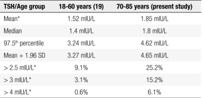
Table 2. Comparison of TSH values between adults [18-60 years (19)] and older people (70-85 years)
TSH/Age group 18-60 years (19) 70-85 years (present study)
Mean* 1.52 mIU/L 1.85 mIU/L
Median 1.4 mIU/L 1.8 mIU/L
97.5th percentile 3.24 mIU/L 4.62 mIU/L
Mean + 1.96 SD 3.27 mIU/L 4.65 mIU/L
> 2.5 mIU/L* 9.1% 25.2%
> 3 mIU/L* 3.1% 15.2%
> 4 mIU/L* 0.6% 6.1%
* p < 0.05.
Figure 1. TSH distribution in the entire cohort (n = 360).
It should be noted that none of the 22 subjects with TSH ≥ 4 mIU/L and submitted to US presented al-terations suggestive of chronic thyroiditis.
The comparison of TSH values between adults aged 18 to 60 years (19) and older people (70-85 years) is shown in table 2.
DISCUSSION
To our knowledge, this is the irst study evaluating the normal reference range for TSH in a population of Bra-zilian older adults. In addition, the selection criteria, number of participants (20), and adequate sample se-lection should be highlighted.
Cop
yright
© ABE&M t
odos os dir
eit
os r
eser
vados
.
have family history of thyroid disease, personal history of autoimmune disease or goiter, and these conditions were exclusion criteria in the present study. Second, if cases of occult thyroiditis exist, it would be expected that they are more frequent among subjects with el-evated TSH. However, US did not suggest this diag-nosis in subjects with TSH ≥ 4 mIU/L. In addition, previous studies found no difference in the upper limit of TSH after excluding patients with altered US (9,21-23), suggesting that these cases of inapparent thyroid-itis (negative TPOAb) have no signiicant impact.
The hypotheses to explain the physiological eleva-tion of TSH in older adults would, in fact, be a re-duction in pituitary sensitivity to thyroid hormones (altered setpoint), higher production of isoforms with lower biological activity, and a decline in the functional capacity of the thyroid with age.
Although conirming higher TSH concentrations in older adults, the values found in this study are lower than those reported in the NHANES III (1). This dis-crepancy can be readily explained by population dif-ferences (genetic background, ethnicity, iodine sufi-ciency, among others). However, we believe that that NHANES values might be overestimated due to the less rigorous selection of the reference population. The presence of thyroid disease, goiter, or use of thyroid or other medications was self-reported, and a detailed history was not obtained at the time of examination (16). Supporting the failure in the selection to exclude occult thyroid diseases, 93 subjects included in the ref-erence population had TSH > 10 mIU/L, range 10.4 to 83 mIU/L (26). Another recent study (from Scot-land) that was larger than the NHANES III and did not include TPOAb as a selection criterion, a fact that may have overestimated TSH values, reported an upper limit for normal TSH of 4.96 mIU/L in the age group of 70-80 years (25), very similar to the limit observed here (4.62 mIU/L).
Serum concentrations of TSH < 4.5 mIU/L are acceptable even in adults (< 70 years). Similarly, TSH levels > 10 mIU/L are considered to be altered, ir-respective of age (26). Therefore, the adoption of a different limit for the elderly population would have an impact on subjects with intermediate TSH levels, who may be erroneously classiied as having subclini-cal hypothyroidism (SCH) or as inadequately treated if undergoing L-T4 replacement therapy; however, the representativeness of these cases is variable (1,25,27). Fortunately, even if SCH is erroneously diagnosed in
older adults due to the lack of a speciic TSH limit for age, it is possible that only few patients will be treated. In this age group, the repercussions of SCH seem to be less consistent (28,29), and treatment of patients with TSH < 10 mIU/L apparently has no beneits (29,30).
This study suggests an upper limit for normal TSH of approximately 4.6 mIU/L for the Brazilian elderly population (70-85 years).
Disclosure: no potential conlict of interest relevant to this article was reported.
REFERENCES
1. Surks MI, Hollowell JG. Age-speciic distribution of serum thy-rotropin and antithyroid antibodies in the U.S population: impli-cations for the prevalence of subclinical hypothyroidism. J Clin Endocrinol Metab. 2007;92:4575-82.
2. Carle A, Laurberg P, Pedersen IB, Perrild H, Ovesen L, Rasmussen LB, et al. Age modiies the pituitary TSH response to thyroid fail-ure. Thyroid. 2007;17:139-44.
3. Over R, Mannan S, Nsouli-Maktabi H, Burman KD, Jonklaas J. Age and the thyrotropin response to hypothyroxinemia. J Clin Endocrinol Metab. 2010;95:3675-83.
4. Mariotti S, Franceschi C, Cossarizza A, Pinchera A. The aging thy-roid. Endocr Rev. 1995;16:686-715.
5. Stan M, Morris JC. Thyrotropin-axis adaptation in aging and chronic disease. Endocrinol Metab Clin N Am. 2005;34:973-92. 6. Adler SM, Burman KD. Abnormalities in thyroid function
param-eters and subclinical thyroid disease in the elderly: a brief review. 2007. Available at: www.hotthyroidology.com
7. Peeters RP. Thyroid hormones and aging. Hormones (Athens). 2008;7:28-35.
8. Mitrou P, Raptis SA, Dimitriadis G. Thyroid disease in older peo-ple. Maturitas. 2011;70:5-9.
9. Volzke H, Alte D, Kohlmann T, Ludemann J, Nauck M, John U, et al. Reference intervals of serum thyroid function tests in a previ-ously iodine-deicient area. Thyroid. 2005;15:279-85.
10. Hershman JM, Pekary AE, Berg L, Solomon DH, Sawin CT. Serum thyrotropin and thyroid hormone levels in elderly and middle-aged euthyroid persons. J Am Geriatr Soc. 1993;41:823-8. 11. Davey R. Thyroxine, thyrotropin, and age in a euthyroid hospital
patient population. Clin Chem. 1997;43:2143-8.
12. Huang CC, Chen YC, Chen LK, Hwang SJ, Lin HD. Relationship between age and serum thyrotropin among asymptomatic older people in Taiwan. Arch Gerontol Geriatr. 2010;51:117-20.
13. Hoogendoorn EH, Hermus AR, de Vegt F, Ross HA, Verbeek AL, Kiemeney LA, et al. Thyroid function and prevalence of anti-thy-roperoxidase antibodies in a population with borderline suficient iodine intake: inluences of age and sex. Clin Chem. 2006;52:104-11. 14. Marwaha RK, Tandon N, Ganie MA, Mehan N, Sastry A, Garg
MK, et al. Reference range of thyroid function (FT3, FT4 and TSH) among Indian adults. Clin Biochem. 2013;46:341-5.
15. Tunbridge WM, Evered DC, Hall R, Appleton D, Brewis M, Clark F, et al. The spectrum of thyroid disease in a community: the Whick-ham survey. Clin Endocrinol (Oxf). 1977;7:481-93.
Cop
yright
© ABE&M t
odos os dir
eit
os r
eser
vados
.
17. Spencer CA, Hollowell JG, Kazarosyan M, Braverman LE. Nation-al HeNation-alth and Nutrition Examination Survey III thyroid-stimulat-ing hormone (TSH)-thyroperoxidase antibody relationships dem-onstrate that TSH upper reference limits may be skewed by occult thyroid dysfunction. J Clin Endocrinol Metab. 2007;92:4236-40. 18. Fatourechi V. Upper limit of normal serum thyroid-stimulating
hormone: a moving and now an aging target? J Clin Endocrinol Metab. 2007;92:4560-2.
19. Rosario PW, Xavier AC, Calsolari MR. TSH reference values for adult Brazilian population. Arq Bras Endocrinol Metabol. 2010;54:603-6.
20. Baloch Z, Carayon P, Conte-Devolx B, Demers LM, Feldt-Rasmus-sen U, Henry JF, et al. Laboratory medicine practice guidelines. Laboratory support for the diagnosis and monitoring of thyroid disease. Thyroid. 2003;13:3-126.
21. Zöphel K, Wunderlich G, Grüning T, Koch R, Döge H, Kotzerke J. Where does subclinical hypothyroidism start? Implications for the deinition of the upper reference limit for thyroid stimulating hormone (TSH). Nuklearmedizin. 2005;44:56-61.
22. Hamilton TE, Davis S, Onstad L, Kopecky KJ. Thyrotropin levels in a population with no clinical, autoantibody, or ultrasonographic evidence of thyroid disease: implications for the diagnosis of sub-clinical hypothyroidism. J Clin Endocrinol Metab. 2008;93:1224-30. 23. Kratsch J, Fiedler GM, Leichtle A, Brugel M, Buchbinder S, Otto
L, et al. New reference intervals for thyrotropin and thyroid hormones based on National Academy of Clinical Biochemistry criteria and regular ultrasonography of the thyroid. Clin Chem. 2005;51:1480-6.
24. Borges MAR, Rosario PW, Purisch S. Normal serum TSH for our adult population. Congresso Mineiro de Endocrinologia 2007 (Abstract).
25. Vadiveloo T, Donnan PT, Murphy MJ, Leese GP. Age- and gender-speciic TSH reference intervals in people with no obvious thy-roid disease in Tayside, Scotland: The Thythy-roid Epidemiology, Audit, and Research Study (TEARS). J Clin Endocrinol Metab. 2013;98:1147-53.
26. Boucai L, Hollowell JG, Surks MI. An approach for development of age-, gender-, and ethnicity-speciic thyrotropin reference lim-its. Thyroid. 2011;21:5-11.
27. Kahapola-Arachchige KM, Hadlow N, Wardrop R, Lim EM, Walsh JP. Age-speciic TSH reference ranges have minimal impact on the diagnosis of thyroid dysfunction.Clin Endocrinol (Oxf). 2012;77:773-9.
28. Ochs N, Auer R, Bauer DC, Nanchen D, Gussekloo J, Cornuz J, et al. Meta-analysis: subclinical thyroid dysfunction and the risk for coronary heart disease and mortality. Ann Intern Med. 2008;148:832-45.
29. Sgarbi JA, Teixeira PFS, Maciel LMZ, Mazeto GMFS, Vaisman M, Montenegro Jr. RM, et al. The Brazilian consensus for the clini-cal treatment of subcliniclini-cal hypothyroidism in adults: recom-mendations of the Thyroid Department of the Brazilian Society of Endocrinology and Metabolism. Arq Bras Endocrinol Metabol. 2013;57:166-83.