Case 9539
Endometriosis in the canal of Nuck
Monteiro V, Cunha TM
Genital (Female) Imaging
Section:
2011, Sep. 27
Published:
26 year(s), female
Patient:
Authors' Institution
V Monteiro 1 TM Cunha 2
Unidade Local de Saúde do Baixo Alentejo, EPE - Beja 1
Instituto Português de Oncologia de Lisboa Franscico Gentil 2
Email:[email protected]
Clinical History
A 26-year-old premenopausal nulliparous woman presented with a palpable right inguinal lump that had been present for 3 months. The patient reported pain and enlargement of the lump during menses. Her medical history was unremarkable.
Physical examination revealed a 3 cm subcutaneous right inguinal mass that was painful on palpation.
Imaging Findings
MRI revealed a mass with irregular contours extending through the right inguinal canal into the right lateral pubic area that was hypointense on T2-weighted images (Fig. 2). On fat-suppressed T1-weighted images (Fig. 3), the mass was hypointense with hyperintense foci, which indicates hemorrhagic areas. After intravenous gadolinium administration the mass showed only slight enhancement (Fig. 4). Another finding was hypointense irregular spiculated thickening of the torus uterinus and the uterosacral ligaments on T2-weighted images with hyperintense foci on
fat-suppressed T1-weighted images, consistent with deep pelvic endometriosis (Fig. 5).
The clinical presentation, anatomic location of the mass and MRI imaging findings, were suggestive of endometriosis in the canal of Nuck.
Surgical wide excision was performed and histological findings confirmed this diagnosis.
Discussion
Endometriosis is a common gynecologic disease affecting 5-10% of women during their
childbearing age [1]. It is characterized by implantation of functional endometrial tissue outside the uterine cavity, typically within the ovaries and peritoneum. However, endometriosis can also occur in extra-pelvic locations with a reported incidence of 0.8% [2], mainly involving extraperitoneal structures, like the round ligament, hernia sac and skin.
Endometriosis in the canal of Nuck is a rare disease, with an incidence of 0.4% [3] that was first described by Cullen in 1896. The canal of Nuck is an embryologic remnant of the processus
vaginalis peritonei.It is a "glovefinger-like" evagination of parietal peritoneum that accompanies the round ligament and extends through the inguinal canal into the labium majus and that normally undergoes obliteration during the first year of life. Occasionally, the canal of Nuck can remain patent, providing a communication between the peritoneal cavity and the inguinal canal and in such cases can be a site for endometriosis seeding.
The pathogenesis of endometriosis is still controversial, but the most widely accepted theory is that endometrial tissue is spread by retrograde menstruation.Most common symptoms are
dysmenorrhea, pelvic pain and infertility, although they are not specific and unusual symptoms are frequent when endometriotic implants occur in atypical locations.When endometriosis affects the canal of Nuck the most common symptom is a groin inguinal lump (96%), predominantly found on the right side (87%) [4], as in the presented case, and the reasons for this seems to be that
endometrial cells will remain in the right side for a longer time due to gravity and the left round ligament may be protected by the sigmoid colon.The groin lump may be painful and may enlarge during menses, as it was reported by the patient.
Pelvic MR imaging is an excellent method for identifying hemorrhagic content that characterizes endometriomas and its large field of view and multiplanar capabilities allows a correct mapping of multiple deeply infiltrating endometrial implants, facilitating the surgical planning [5]. On MR images, the lesions generally appear low to intermediate signal intensity on T1-weighted images, hypointense on T2-weighted images, and minimally enhanced after the injection of
gadolinium-based contrast material. Hyperintense foci may be seen on T1-weighted images, representing ectatic endometrial glands with hemorrhagic content [6].
confirmation. Laparoscopy is also indicated to evaluate for concomitant pelvic and intra-abdominal endometriosis.
Final Diagnosis
Endometriosis in the canal of Nuck
Differential Diagnosis List
Desmoid tumour, Inguinal hernia
Figures
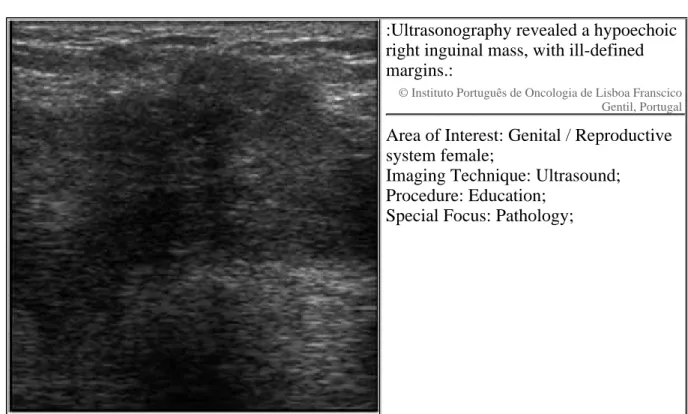
Figure 1 Ultrasound
:Ultrasonography revealed a hypoechoic
right inguinal mass, with ill-defined
margins.:
© Instituto Português de Oncologia de Lisboa Franscico Gentil, Portugal
Area of Interest: Genital / Reproductive
system female;
Imaging Technique: Ultrasound;
Procedure: Education;
Special Focus: Pathology;
Figure 2 Axial and Sagittal T2-weighted images
:T2-weighted images revealed an
irregular lesion (arrow) with low
signal intensity, extending through the
right inguinal canal into the right
pubic area.:
© Instituto Português de Oncologia de Lisboa Francisco Gentil, Portugal
Figure 3 Axial T1 and Fat-supressed T1-weighted images
:(A) The mass was mainly
hypointense on T1-weighted images.
(B) Small hyperintense foci were also
seen within the lesion, more clearly
revealed on fat-suppressed
T1-weighted images (arrow).:
© Instituto Português de Oncologia de Lisboa Francisco Gentil, Portugal
Area of Interest: Genital /
Reproductive system female;
Imaging Technique: MR;
Procedure: Imaging sequences;
Special Focus: Pathology;
Figure 4 T1-weighted images after intravenous contrast administration
:After the injection of gadolinium
contrast material the mass showed
minimal enhancement.:
© Instituto Português de Oncologia de Lisboa Franscico Gentil, Portugal
Area of Interest: Genital /
Reproductive system female;
Imaging Technique: MR;
Procedure: Imaging sequences;
Special Focus: Pathology;
Figure 5 Deep pelvic endometriosis
:T2-weighted images (A) showed a
hypointense spiculated lesion
involving the utero-sacral ligaments
(arrows) with small hyperintense foci
(*) on fat-suppressed T1-weighted
images (B), indicating hemorrhage.:
© Instituot Português de Oncologia de Lisboa Francisco Gentil, Portugal
MeSH
[C13.371.163]
Endometriosis
A condition in which functional endometrial tissue is present outside the UTERUS. It is often confined to the PELVIS involving the OVARY, the ligaments, cul-de-sac, and the uterovesical peritoneum.
References
[1] Woodward PJ, Sohaey R, Mezzetti TP Jr (2001) Endometriosis:radiologic-pathologic RadioGraphics 21:193-216
correlation
[2] Markham SM, Carpenter SE, Rock JA (1989) Extrapelvic endometriosis Obstet Gynecol Clin North Am 16:193-219
[3] Strasser EJ, Davis RM (1977) Extraperitoneal inguinal endometriosis Am Surg 43:421-422
[4] A Kirkpatrick, CM Reed, LT Bui-Mansfield, MJ Russell, W Whitford (2006) Endometriosis of AJR 186:56-57
the Canal of Nuck
[5] Chamié LP, Blasbalg R, Pereira RM, Warmbrand G, Serafini PC. (2011) Findings of pelvic Radiographics
endometriosis at transvaginal US, MR imaging, and laparoscopy Jul-Aug;31(4):E77-100
[6] Del Frate C, Girometti R, Pittino M, Del Frate G, Bazzocchi M, Zuiani C. (2006) Deep retroperitoneal pelvic endometriosis: MR imaging appearance with laparoscopic correlation Radiographics Nov-Dec;26(6):1705-18.
Citation
Monteiro V, Cunha TM (2011, Sep. 27)
Endometriosis in the canal of Nuck {Online}