Serum testosterone as a biomarker for second prostatic
biopsy in men with negative first biopsy for prostatic cancer
and PSA>4ng/mL, or with PIN biopsy result
_______________________________________________
Alexandros Fiamegos
1, John Varkarakis
1, Michael Kontraros
1, Andreas Karagiannis
1, Michael
Chrisofos
1, Dimitrios Barbalias
1, Charalampos Deliveliotis
11 2nd Department of Urology, University of Athens, Sismanoglio General Hospital, Athens, Greece
ABSTRACT
ARTICLE
INFO
______________________________________________________________ ______________________
Introduction: Data from animal, clinical and prevention studies support the role of androgens in prostate cancer growth, proliferation and progression. Results of serum based epidemiologic studies in humans, however, have been inconclusive. The present study aims to define whether serum testosterone can be used as a predictor of a posi-tive second biopsy in males considered for re-biopsy.
Material and Methods: The study included 320 men who underwent a prostatic biopsy in our department from October 2011 until June 2012. Total testosterone, free testos-terone, bioavailable testosterone and prostate pathology were evaluated in all cases. Patients undergoing a second biopsy were identified and biopsy results were statisti-cally analyzed.
Results: Forty men (12.5%) were assessed with a second biopsy. The diagnosis of the second biopsy was High Grade Intraepithelial Neoplasia in 14 patients (35%) and Pros-tate Cancer in 12 patients (30%). The comparison of prostatic volume, total testoste-rone, sex hormone binding globulin, free testostetestoste-rone, bioavailable testosterone and albumin showed that patients with cancer of the prostate had significantly greater levels of free testosterone (p=0.043) and bioavailable T (p=0.049).
Conclusion: In our study, higher free testosterone and bioavailable testosterone levels were associated with a cancer diagnosis at re-biopsy. Our results indicate a possible role for free and bioavailable testosterone in predicting the presence of prostate cancer in patients considered for re-biopsy.
Keywords:
Testosterone; Prostate; Neoplasms; Biopsy
Int Braz J Urol. 2016; 42: 925-31
_____________________
Submitted for publication: March 25, 2015
_____________________
Accepted after revision: November 29, 2015
INTRODUCTION
Prostate cancer is the second most com-mon cancer in men worldwide (1) and the only established risk factors for prostate cancer are age, race and family history (2). Androgens seem to play a key role in the natural history of prosta-te cancer. In fact, androgens are required for the normal growth and development of the prostate gland and high levels of androgens have long
been considered to be possible risk factors for prostate cancer (3, 4). Large doses of androgens can induce prostate cancer in rodents (5) and they also stimulate in vitro human prostate cancer cell proliferation (6). Prostate cancer presents only ra-rely among castrated men, while surgical or me-dical castration in prostate cancer patients causes tumor regression (7).
diagnosis or prognosis. In fact, many observational studies failed to demonstrate any clear association between testosterone levels and risk of prostate cancer diagnosis, and the exact mechanism of action of testosterone on prostatic tissue has not been to date fully elucidated. Prostate cancer diagnosis is currently based mainly upon digital rectal examination (DRE) and/or PSA measurement leading to a transrectal prostatic ultrasound guided biopsy (TRUS-b). A problem in this regard is assessing patients after a negative biopsy, as rates of prostate cancer diagnosis fall substantially thereafter.
In the present study we aim to determine whether serum testosterone can be used as a pre-dictor of a positive second biopsy in males con-sidered for re-biopsy, either after a negative first biopsy, or with a high grade intra prostatic neo-plasia (HGPIN) diagnosis.
MATERIAL AND METHODS
The study included 320 consecutive men who underwent a prostatic biopsy in our depart-ment from October 2011 until June 2012. All 320 patients were informed about participating in the study and have signed a consent form with full information about the procedure, the aims of the study and the possible outcomes. The study was approved by the Medical School of University of Athens board and the ethics committee of Sis-manoglion General Hospital of Athens. Decision for biopsy was based upon high serum PSA levels and/or suspicious DRE findings according to cur-rent practice as pointed out by the EAU guide-lines. The serum PSA level which led a patient to a biopsy was above 4ng/mL. Only 17 out of the 320 patients were led to biopsy with clinical suspicion from the digital rectal examination and had serum PSA level lower than 4ng/mL. The biopsy was per-formed transrectally under ultrasound guidance using a spring-driven biopsy gun, between 07:00 and 09:00 am. Before the TRUS biopsy procedure was undertaken, a complete history was acquired, including race and ethnicity and detailed dietary habits history, as well as peripheral vein blood samples for free and total PSA (if not already per-formed), free testosterone (fTe), total testosterone
(tTe), albumin (ALB) and sex hormone binding globulin (SHBG) levels measurement. During the TRUS biopsy, the prostatic size was measured and recorded. The biopsy was performed by one phy-sician. The sextant cores pattern was used with 12 core biopsy. This pattern was changed based on the TRUS findings concerning the size of the prostate and possibly suspicious regions and var-ied from 8 cores from a small prostate to 28 cores for large prostatic glands, but most of the patients underwent a 12 cores biopsy and the number of the cores taken proved to be of no importance for the diagnosis of prostate cancer or not. Tissue samples were examined by the pathologic labora-tory of our facility, by one pathologist.
The decision for a second biopsy was based on three criteria. The first one was high clinical suspicion due to digital rectal examination. The second one was repeated high PSA levels (>4ng/ mL), despite a negative result from the first biopsy. The third one is HGPIN diagnosis in the initial bi-opsy. The second biopsy was performed within a year from the first one and the meantime between the two biopsies varied due to the different criteria for second biopsy and due to patient preferences and awareness.
Total Testosterone (tTe) and sex hormone binding globulin (SHBG) were measured using electrochemiluminescence immunoassay “el-lia” for use in immunological analysis [elecsys 1010/2010 and modular analytics E170 (subunit elecsys) of Roche]. Albumin (ALB) was measured using a colorimetric assay endpoint method, and PSA with heterogeneous direct chemiluminescence (sandwich type, using two monoclonal antibod-ies). The free testosterone (fTe) and bioavailable testosterone (BioTe) levels were calculated imple-menting accepted published equations using tTe, SHBG and ALB (8, 9). We measured fTe and BioTe, as these are considered to be the actual amount of testosterone available at a cellular level (8).
Statistical analysis
between the two study groups chi-square and Fisher’s exact tests were used. Student’s t-tests were computed for the comparison of mean va-lues. All p values reported are two-tailed. Sta-tistical significance was set at 0.05 and analyses were conducted using SPSS statistical software (version 18.0).
RESULTS
Sample characteristics are presented in Table-1. All the patients were Caucasians, 318 Greeks, 1 Hungarian and 1 Italian concerning the race and the ethnicity. The analysis of the detailed nutrition habits history has shown no statistical important difference between the positive and the negative for cancer group. In total, a sample of 320 patients was selected and 40 of them (12.5%) had a re-biopsy. The decision for second biopsy was based on high clinical suspicion for prostate cancer but negative first biopsy. The patients of the second biopsy were patients with high PSA levels (>4ng/mL) and negative biopsy and/or HG-PIN who were informed about the options they had and agreed to undergo a second prostatic bi-opsy within the timeframe of our study. The same formal requirements were followed for the second biopsy. The second biopsy was performed by the same physician, was investigated by the same pathologist and the number of cores taken from the prostate were 20 for all the patients, regard-less the result of the first biopsy or the number of cores taken at the first biopsy. The diagnosis
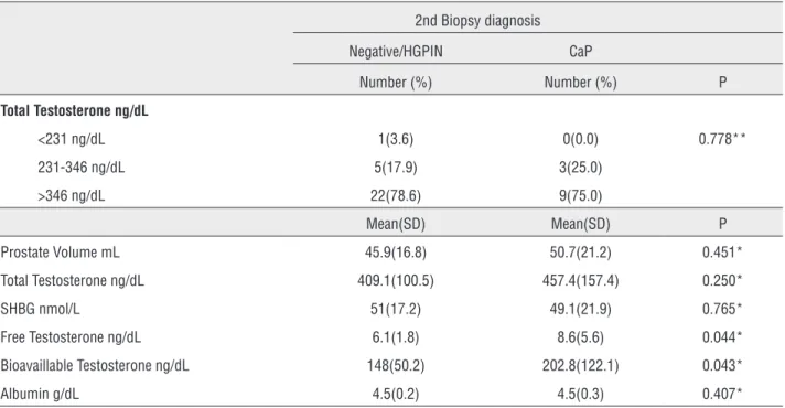
of the second biopsy was HGPIN in 14 patients (35%) and CaP in 12 patients (30%). From the 14 patients with HGPIN 8 had a negative first biopsy and 6 had a HGPIN at first biopsy, while from the 12 patients with CaP, 6 had a negative first biop-sy and 6 had a HGPIN result in their first biopbiop-sy. When prostatic volume (Vp), total testosterone, SHBG, free testosterone, bioavailable testosterone and albumin were compared between patients that underwent a second biopsy and those who did not, no statistically significant differences were found (Table-2). The comparison of the aforementioned indices between patients with a cancer diagnosis and those with negative or a HGPIN result in the second biopsy (Table-3) showed that patients with prostatic cancer (CaP) had significantly higher levels of free testosterone (p=0.043) and bioavail-able T (p=0.049).
DISCUSSION
In men, testosterone is predominantly pro-duced by Leydig cells of the testes; only a lesser amount (<10%) is produced in the adrenal glands. Testosterone plays a key role in the development of male reproductive tissues (10). It stimulates the prostate gland to grow both early in puberty, when the prostate doubles in size, and around age 25, when the gland begins to grow again. Tes-tosterone is converted into dihydrotesTes-tosterone (DHT), which is the androgen receptor’s major activator (11, 12). After DHT binds to androgen receptors, it translocates into the nucleus, where it mediates the transcriptional activation of target genes (13). Through androgen-stimulated changes in gene expression cellular growth occurs, even-tually leading to benign prostatic hyperplasia in elderly men.
Great controversy has risen in the last years regarding the exact relation between prostate can-cer and androgens. The first published work on this subject goes many years back in 1941, when for the first time Huggins and Hodges proved that testosterone deprivation leads to prostate cancer regression (7). Since then many studies tried to elucidate the relation between prostate cancer and testosterone. Many groups reported that the risk of prostate cancer was higher in men with lower Table 1 - Sample characteristics.
Mean(SD) Total sample (N=320)
Age 67(8.1)
PSA ng/mL 8.3(4.3)
Sample with 2nd biopsy (N=40)
Age 66.4(8.8)
PSA ng/mL 8.0(3.7)
2nd Biopsy diagnosis, N (%)
Negative 14(35.0)
HGPIN 14(35.0)
Prostate Cancer 12(30.0)
Table 3 - Comparative analysis of patients with and without CaP after rebiopsy, regarding measured variables.
2nd Biopsy diagnosis
Negative/HGPIN CaP
Number (%) Number (%) P
Total Testosterone ng/dL
<231 ng/dL 1(3.6) 0(0.0) 0.778**
231-346 ng/dL 5(17.9) 3(25.0)
>346 ng/dL 22(78.6) 9(75.0)
Mean(SD) Mean(SD) P
Prostate Volume mL 45.9(16.8) 50.7(21.2) 0.451*
Total Testosterone ng/dL 409.1(100.5) 457.4(157.4) 0.250*
SHBG nmol/L 51(17.2) 49.1(21.9) 0.765*
Free Testosterone ng/dL 6.1(1.8) 8.6(5.6) 0.044*
Bioavaillable Testosterone ng/dL 148(50.2) 202.8(122.1) 0.043*
Albumin g/dL 4.5(0.2) 4.5(0.3) 0.407*
*Student’s t-test **Fisher’s exact test
SD = Standard Deviation
Table 2 - Comparative analysis of patients with and without rebiopsy regarding measured variables.
2nd Biopsy
No Yes
Number (%) Number (%) P
Total Testosterone ng/dL
<231 ng/dL 28(10) 1(2.5) 0.217**
231-346 ng/dL 39(13.9) 8(20)
>346 ng/dL 213(76.1) 31(77.5)
Mean(SD) Mean(SD) P
Prostate Volume mL 49.7(22.5) 47.4(18.1) 0.534*
Total Testosterone ng/dL 410.9(145.2) 423.6(120.3) 0.597*
SHBG nmol/L 49.9(20.8) 50.4(18.4) 0.873*
Free Testosterone ng/dL 6.6(2.7) 6.9(3.5) 0.569*
Bioavaillable Testosterone ng/dL 161.6(65.2) 164.4(81.2) 0.807*
Albumin g/dL 4.5(0.3) 4.5(0.2) 0.640*
*Student’s t-test **Chi-square test
total testosterone levels (14, 15) and bioavailable testosterone level (16). An important role seems to be played by SHBG, which in turn determines the levels of free and bioavailable testosterone. A recent study showed that there is a relation between SHBG level and prostate cancer espe-cially in younger patients (17). Nonetheless, an article by Roddam et al. reported no association between blood levels of total testosterone and prostate cancer risk based on pooled analysis of 18 prospective studies (18). The pooled analy-sis included 3886 men with prostate cancer and 6438 controls. It is the largest serum based study with the most elegant and comprehensive analy-sis to date to test a central hypotheanaly-sis in pros-tate cancer etiology. It is important to note, that the pooled analysis did not find a positive link between circulating levels of total testosterone and prostate cancer risk, but few of the eighteen studies included reported a substantial positive association. On the other hand, fear that andro-gen supplementation could cause prostate cancer arousal in males, especially after prostate biopsy findings of HGPIN, has not been proven (19).
As shown in other studies, we also found no association between serum testosterone level and prostate cancer diagnosis at biopsy (17, 20, 21). Furthermore, more recent studies continuous-ly provide controversial results regarding serum androgens and prediction of prostate cancer. Regis et al. in their review of 124 publications came up to the conclusion that due to the heterogeneity of the studies they cannot recommend testosterone level measurement in order to predict prostate cancer and its aggressiveness (22). On the other hand, the correlation between testosterone and PSA remains (23). Additionally experimental in vitro and mice data show that testosterone might be used to help prediction of CaP in low PSA level measurements (24), as Usoro et al. showed that patients with CaP have higher estradiol levels while presenting no difference in testosterone levels (25). Focusing on this idea Black et al. suggest that the ratio estro-gen/androgen may be important in the develop-ment of CaP (26).
In our study we attempt to determine, whether testosterone level is associated with a pos-itive second prostate biopsy, both in patients that
There are several limitations to the present study. It is a single-centre, small series, and patient number undergoing re-biopsy was low. Moreover, we did not directly calculate the free Testosterone level. Lastly, the re-biopsy in our series occurred early, within the time span of our study (one year). The rates of cancer detection at re-biopsy rise with time, so some patients with negative biopsy might be diagnosed with cancer in the future. Larger prospective studies should be designed to eluci-date the exact relation between testosterone and prostate cancer, as well as the possible use of total, free and bioavailable testosterone as a marker for prostate cancer diagnosis at the second biopsy.
CONCLUSIONS
In our study, serum concentration of total testosterone was not associated with the risk of prostate cancer in men who underwent a second prostatic biopsy. However, higher free testosterone and bioavailable testosterone levels were associ-ated with a cancer diagnosis at re-biopsy. Further research is required to define the complex inter-play between sex steroids, genetic and lifestyle factors in prostate cancer aetiology. Our results indicate a possible role of free and bioavailable testosterone in predicting the presence of prostate cancer in patients considered for re-biopsy.
CONFLICT OF INTEREST
None declared.
REFERENCES
1. Ferlay J, Bray F, Pisani P, Parkin DM. GLOBOCAN 2002: Cancer Incidence, Mortality and Prevalence Worldwide. Version 2.0 Lyon, France: IARC Press; 2004
2. Bostwick DG, Burke HB, Djakiew D, Euling S, Ho SM, Landolph J, et al. Human prostate cancer risk factors. Cancer. 2004;101:2371-490.
3. Hsing AW. Hormones and prostate cancer: what’s next? Epidemiol Rev. 2001;23:42-58.
4. Platz EA, Giovannucci E. The epidemiology of sex steroid hormones and their signaling and metabolic pathways in the etiology of prostate cancer. J Steroid Biochem Mol Biol. 2004;92:237-53.
5. Noble RL. The development of prostatic adenocarcinoma in Nb rats following prolonged sex hormone administration. Cancer Res. 1977;37:1929-33.
6. Henderson BE, Ross RK, Pike MC, Casagrande JT. Endogenous hormones as a major factor in human cancer. Cancer Res. 1982;42:3232-9.
7. Huggins C, Hodges CV. Studies on prostatic cancer. I. The effect of castration, of estrogen and androgen injection on serum phosphatases in metastatic carcinoma of the prostate. CA Cancer J Clin. 1972;22:232-40.
8. Vermeulen A, Verdonck L, Kaufman JM. A critical evaluation of simple methods for the estimation of free testosterone in serum. J Clin Endocrinol Metab. 1999;84:3666-72.
9. Giton F, Fiet J, Guéchot J, Ibrahim F, Bronsard F, Chopin D, et al. Serum bioavailable testosterone: assayed or calculated? Clin Chem. 2006;52:474-81.
10. Carson C 3rd, Rittmaster R. The role of dihydrotestosterone in benign prostatic hyperplasia. Urology. 2003;61:(4 Suppl 1):2-7. 11. Heemers HV, Tindall DJ. Androgen receptor (AR)
coregulators: a diversity of functions converging on and regulating the AR transcriptional complex. Endocr Rev. 2007;28:778-808.
12. Deslypere JP, Young M, Wilson JD, McPhaul MJ. Testosterone and 5 alpha-dihydrotestosterone interact differently with the androgen receptor to enhance transcription of the MMTV-CAT reporter gene. Mol Cell Endocrinol. 1992;88:15-22. 13. Schalken JA. Molecular aspects of hormone-independent
prostate cancer. BJU Int. 2007;100:52-5.
14. Yano M, Imamoto T, Suzuki H, Fukasawa S, Kojima S, Komiya A, Naya Y, Ichikawa. T. The clinical potential of pretreatment serum testosterone level to improve the efficiency of prostate cancer screening. Eur Urol. 2007;51:375-80.
15. Sofikerim M, Eskicorapci S, Oruç O, Ozen H. Hormonal predictors of prostate cancer. Urol Int. 2007;79:13-8. 16. García-Cruz E, Huguet J, Piqueras M, Márquez MP, Peri L,
Izquierdo L, et al. Low testosterone bioavailability is related to prostate cancer diagnose in patients submitted to prostate biopsy. World J Urol. 2012;30:361-5.
17. Gann PH, Hennekens CH, Ma J, Longcope C, Stampfer MJ. Prospective study of sex hormone levels and risk of prostate cancer. J Natl Cancer Inst. 1996;88:1118-26.
18. Endogenous Hormones and Prostate Cancer Collaborative Group, Roddam AW, Allen NE, Appleby P, Key TJ. Endogenous sex hormones and prostate cancer: a collaborative analysis of 18 prospective studies. J Natl Cancer Inst. 2008;100:170-83. 19. Rhoden EL, Morgentaler A. Testosterone replacement
therapy in hypogonadal men at high risk for prostate cancer: results of 1 year of treatment in men with prostatic intraepithelial neoplasia. J Urol. 2003;170:2348-51. 20. Mearini L, Costantini E, Zucchi A, Mearini E, Bini V, Cottini E,
21. Morote J, Planas J, Ramirez C, Gómez E, Raventós CX, Placer J, et al. Evaluation of the serum testosterone to prostate-specific antigen ratio as a predictor of prostate cancer risk. BJU Int. 2010;105:481-4.
22. Regis L, Planas J, Celma A, de Torres IM, Ferrer R, Morote J. Behavior of total and free serum testosterone as a predictor for the risk of prostate cancer and its aggressiveness. Actas Urol Esp. 2015;39:573-81.
23. Peskoe SB, Joshu CE, Rohrmann S, McGlynn KA, Nyante SJ, Bradwin G, et al. Circulating total testosterone and PSA concentrations in a nationally representative sample of men without a diagnosis of prostate cancer. Prostate. 2015;75:1167-76.
24. Song W, Soni V, Khera M. Combined tests of prostate specific antigen and testosterone will improve diagnosis and monitoring the progression of prostate cancer. Asian J Androl. 2015;17:807-10.
25. Usoro AJ, Obot AS, Ekaidem IS, Akaiso OE, Udoh AE, Akinloye O. Serum Testosterone, 17β-Estradiol and PSA Levels in Subjects with Prostate Disorders. Indian J Clin Biochem. 2015;30:59-65.
26. Black A, Pinsky PF, Grubb RL 3rd, Falk RT, Hsing AW, Chu L, et al. Sex steroid hormone metabolism in relation to risk of aggressive prostate cancer. Cancer Epidemiol Biomarkers Prev. 2014;23:2374-82.
27. García-Cruz E, Piqueras M, Ribal MJ, Huguet J, Serapiao R, Peri L, et al. Low testosterone level predicts prostate cancer in re-biopsy in patients with high grade prostatic intraepithelial neoplasia. BJU Int. 2012;110:E199-202. 28. Schatzl G, Madersbacher S, Thurridl T, Waldmüller J, Kramer
G, Haitel A, et al. High-grade prostate cancer is associated with low serum testosterone levels. Prostate. 2001;47:52-8. 29. Ribeiro M, Ruff P, Falkson G. Low serum testosterone and
a younger age predict for a poor outcome in metastatic prostate cancer. Am J Clin Oncol. 1997;20:605-8.
30. Hoffman MA, DeWolf WC, Morgentaler A. Is low serum free testosterone a marker for high grade prostate cancer? J Urol. 2000;163:824-7.
31. Massengill JC, Sun L, Moul JW, Wu H, McLeod DG, Amling C, et al. Pretreatment total testosterone level predicts pathological stage in patients with localized prostate cancer treated with radical prostatectomy. J Urol. 2003;169:1670-5. 32. Teloken C, Da Ros CT, Caraver F, Weber FA, Cavalheiro AP,
Graziottin TM. Low serum testosterone levels are associated with positive surgical margins in radical retropubic prostatectomy: hypogonadism represents bad prognosis in prostate cancer. J Urol. 2005;174:2178-80.
33. Morgentaler A. Testosterone deficiency and prostate cancer: emerging recognition of an important and troubling relationship. Eur Urol. 2007;52:623-5.
34. Morgentaler A. Rapidly shifting concepts regarding androgens and prostate cancer. ScientificWorldJournal. 2009;9:685-90.
35. Pichon A, Neuzillet Y, Botto H, Raynaud JP, Radulescu C, Molinié V, et al. Preoperative low serum testosterone is associated with high-grade prostate cancer and an increased Gleason score upgrading. Prostate Cancer Prostatic Dis. 2015;18:382-7.
36. Khera M, Crawford D, Morales A, Salonia A, Morgentaler A. A new era of testosterone and prostate cancer: from physiology to clinical implications. Eur Urol. 2014;65:115-23.
_______________________ Correspondence address: