Mycoplasma pneumoniae
-related community-acquired
pneumonia and parapneumonic pleural effusion
in children and adolescents*
Pneumonia adquirida na comunidade e derrame pleural parapneumônico relacionados a Mycoplasma pneumoniae em crianças e adolescentes
Letícia Alves Vervloet, Vitor Earl Cardoso Vervloet, Mário Tironi Junior, José Dirceu Ribeiro
Abstract
Objective: To determine the prevalence and the characteristics of Mycoplasma pneumoniae-related community-acquired pneumonia (CAP) and parapneumonic pleural effusion (PPE) in children and adolescents. Methods: This was a retrospective observational study involving 121 patients with CAP/PPE hospitalized in a tertiary referral hospital between 2000 and 2008, divided into six groups according to the etiologic agent (G1 to G6, respectively): M. pneumoniae with or without co-infection, in 44 patients (group 1); etiologic agents other than M. pneumoniae, in 77 (group 2); M. pneumoniae without co-infection, in 34 (group 3); Streptococcus pneumoniae, in 36 (group 4); Staphylococcus aureus, in 31 (group 5); and M. pneumoniae/S. pneumoniae co-infection, in 9 (group 6). Results: In comparison with group 2, group 1 showed higher frequencies of females, dry cough, and previous use of beta-lactam antibiotics; longer duration of symptoms prior to admission; and lower frequencies of use of mechanical ventilation and chest tube drainage. In comparison with groups 4 and 5, group 3 showed higher frequencies of previous use of beta-lactam antibiotics and dry cough; longer duration of symptoms prior to admission; a lower frequency of use of chest tube drainage; a higher mean age and a lower frequency of nausea/vomiting (versus group 4 only); and a lower frequency of use of mechanical ventilation (versus group 5 only). M. pneumoniae/S. pneumoniae co-infection increased the duration of symptoms prior to admission. Conclusions: In this sample, the prevalence of M. pneumoniae-related CAP/PPE was 12.75%. Although the disease was milder than that caused by other microorganisms, its course was longer. Our data suggest that M. pneumoniae-related CAP/PPE in children and adolescents should be more thoroughly investigated in Brazil. Keywords: Pleural effusion; Empyema, pleural; Pneumonia; Mycoplasma pneumoniae.
Resumo
Objetivo: Determinar a prevalência e as características da pneumonia adquirida na comunidade (PAC) e derrames pleurais parapneumônicos (DPP) relacionados a Mycoplasma pneumoniae em um grupo de crianças e adolescentes. Métodos: Estudo observacional retrospectivo com 121 pacientes hospitalizados com PAC e DPP em um hospital de referência terciária, entre 2000 e 2008, divididos em seis grupos (G1 a G6) segundo o agente etiológico: M. pneumoniae com ou sem coinfecção, em 44 pacientes; outros agentes que não M. pneumoniae, em 77; M. pneumoniae sem coinfecção, em 34; Streptococcus pneumoniae, em 36; Staphylococcus aureus, em 31; e coinfecção M. pneumoniae/S. pneumoniae, em 9, respectivamente. Resultados: Na comparação entre os grupos, G1 apresentou frequências maiores em gênero feminino, tosse seca, uso prévio de beta-lactâmicos e na duração dos sintomas até a admissão, assim como menor uso de assistência ventilatória e de drenagem torácica que G2, enquanto G3 teve maiores frequências em uso prévio de beta-lactâmicos e tosse seca, maior duração dos sintomas antes da admissão e menor frequência de uso de drenos torácicos que G4 e G5, ao passo que G3 teve média de idade maior e menor frequência de náuseas/vômitos que G4, assim como menor uso de assistência ventilatória que G5. A coinfecção M. pneumoniae/S. pneumoniae aumentou a duração dos sintomas até a admissão. Conclusões: Nesta amostra, a prevalência de PAC e DPP por M. pneumoniae foi de 12,75%. Embora a doença apresentasse quadros mais leves que aquela por outros organismos, a evolução foi mais prolongada. Nossos dados sugerem a necessidade de uma maior diligência na investigação de M. pneumoniae em crianças e adolescentes com PAC e DPP em nosso meio.
Descritores: Derrame pleural; Empiema pleural; Pneumonia; Mycoplasma pneumoniae.
* Study carried out at the Federal University of Espírito Santo and the Nossa Senhora da Glória Children’s Hospital, Vitória, Brazil, and at the State University at Campinas, Campinas, Brazil.
Correspondence to: Letícia Alves Vervloet. Rua Eugenio Netto 488, sala 712, Edifício Praia Office, Praia do Canto, CEP 29065-270, Vitória, ES, Brasil.
Tel. 55 27 3325-0489. E-mail: [email protected] Financial support: None.
cystic fibrosis, or tuberculosis, as well as those whose data were incomplete.
During the study period, we included all of the HINSG inpatients aged 3 months to 16 years with clinical and radiological diagnosis of CAP/ PPE (routine posteroanterior, lateral, and lateral decubitus chest X-rays). We included only those patients in whom the etiologic agents were found to be typical bacteria or M. pneumonia and in whom no other atypical bacteria were isolated and identified as being the sole etiologic agents.
The diagnosis of M. pneumoniae-related pneumonia was based on the finding of M. pneumoniae-positive IgM in peripheral blood by ELISA. The kit used (ImmunoWELL; GenBio, San Diego, CA, USA) has a specificity of 90% and a sensitivity of 92% (IgM in the acute phase),
when compared with PCR.(10) The diagnosis of
pneumonia due to typical bacteria was based on positive blood culture results or isolation of the pathogens from pleural fluid.
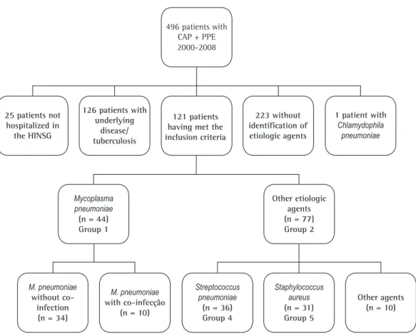
Between 2000 and 2008, 496 patients with CAP/PPE were admitted to the HINSG. After having applied the exclusion criteria, we initially evaluated 345 children and adolescents hospitalized for CAP/PPE; of those, 121 were included in the study on the basis of the etiologic agent, which was identified as typical bacteria or M. pneumonia, no other atypical bacteria having been isolated and identified as the sole etiologic agents (Figure 1). The patients were divided into six groups according to the diagnosis:
• group 1—M. pneumoniae-related CAP/PPE with or without co-infection with other etiologic agents
• group 2—CAP/PPE due to etiologic agents
other than M. pneumoniae
• group 3—M. pneumoniae-related CAP/PPE without co-infection
• group 4—Streptococcus pneumoniae-related CAP/PPE
• group 5—Staphylococcus aureus-related CAP/PPE
• group 6— CAP/PPE due to
M. pneumoniae/S. pneumoniae co-infection Patient data were encoded and entered into 2007 Microsoft Office Excel spreadsheets, having been analyzed with the Epi Info software, version 6.0. We used the chi-square test and Fisher’s exact test in order to evaluate categorical and ordinal variables, as well as using ORs in order to evaluate the magnitude of the associations.
Introduction
In Brazil, the prevalence of Mycoplasma pneumoniae as the etiologic agent of parapneumonic pleural effusion (PPE) remains unknown. This is due to the difficulty in correlating M. pneumoniae with clinical characteristics; to the fact that M. pneumoniae grows little in culture media; and to the lack of rapid, specific tests in the early phase of community-acquired
pneumonia (CAP) with PPE.(1)
The clinical presentation of M. pneumoniae infection varies widely. The clinical course is
generally moderate and self-limiting,(2-4) and
pneumonia occurs in 3-10% of patients. However, severe cases of pneumonia have been described, PPE having been reported in 4-20%; although PPE is generally small and unilateral (on the side of the pulmonary infiltrate), it can be massive and bilateral.(5-7)
The importance of investigating M. pneumoniae-related CAP/PPE (with or without co-infection) lies in the fact that the microorganism does not respond to the antibiotics that are typically used in the treatment of CAP/ PPE, among which are beta-lactam antibiotics.
(8,9) Appropriate treatment prevents prolonged
clinical manifestations and pulmonary sequelae.(6)
The objective of the present study was to compare the characteristics of M. pneumoniae-related PPE with those of PPE pneumoniae-related to other etiologic agents in a group of children and adolescents treated at a regional referral center in Brazil.
Methods
This was a retrospective observational study involving 121 children and adolescents with CAP/ PPE hospitalized in the Hospital Infantil Nossa Senhora da Glória (HINSG, Nossa Senhora da Glória Children’s Hospital), located in the city of Vitória, Brazil, between 2000 and 2008. The HINSG is a regional tertiary referral teaching hospital affiliated with the Espírito Santo State Department of Health. The present study was approved by the HINSG Research Ethics Committee (Protocol no. 34/04).
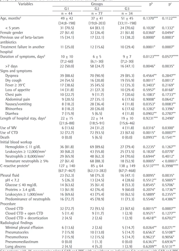
and group 2 patients (with other etiologic agents) in terms of age. The number of patients younger than 5 years of age was found to be 31 (70.5%) in group 1 and 64 (83.1%) in group 2. The mean age was found to be 4.1 years in group 1 and 3.1 years in group 2. Regarding gender, 27 (61.4%) of the patients in group 1 were female, as were 32 (41.6%) of those in group 2, the difference being statistically significant (p = 0.03). Previous use of beta-lactam antibiotics and the need for being transferred to our facility because of treatment failure were more common in group 1 (p < 0.01 for both). In comparison with group 2, group 1 showed longer duration of symptoms prior to admission, higher frequency of dry cough, and lower frequency of nausea or vomiting (p < 0.01; p < 0.01; and p = 0.03, respectively). In contrast, the use of mechanical ventilation and chest tube drainage during hospitalization was more common in group 2 (p = 0.03 and p < 0.01, respectively). Deaths In addition, we used the Student’s t-test in order
to compare continuous variables and ANOVA in order to compare more than two continuous variables. Descriptive statistical analysis results are presented as means, medians, and standard deviations. For all tests, the level of significance required in order to reject the null hypothesis was set at 5% (p < 0.05).
Results
Of the 345 HINSG inpatients diagnosed with CAP/PPE, M. pneumoniae was found in 44 (12.75%). The final analysis of the clinical, radiological, and hematological profiles of the children and adolescents with PPE due to M. pneumoniae (44 patients) or other agents (77 patients) was performed by comparing the six groups.
We found no statistically significant differences between group 1 patients (with M. pneumoniae)
Figure 1 - Flowchart of inclusions, losses, and exclusions among the study population. Study comparing patients with community-acquired pneumonia (CAP) and parapneumonic pleural effusion (PPE) due to
statistically insignificant (p = 0.86). Previous use of beta-lactam antibiotics prior to hospitalization and the need for being transferred to our facility because of treatment failure were significantly more common in group 3 (p < 0.01 for both). In addition, group 3 showed significantly longer duration of symptoms prior to admission and higher frequency of dry cough (p = 0.04 and p = 0.01, respectively). The use of mechanical ventilation during hospitalization was more common in group 5, and blood workup showed a larger quantity (over 5%) of immature neutrophils in that group (p = 0.04 and p < 0.01, respectively). We found no significant differences between the two groups in terms of C-reactive protein levels. Closed chest tube drainage was performed more frequently in group 5 (p < 0.01). Although open chest tube drainage was also more common in that group, the difference was not statistically significant. Although there were no significant differences between the two groups in terms of the frequency of chest X-ray changes, liver abscess was more common in group 5 (p = 0.02; Table 3).
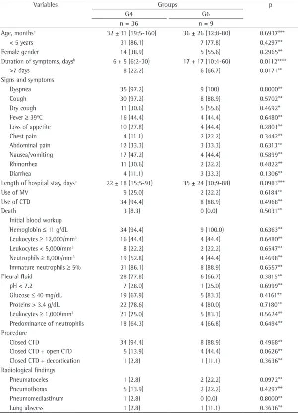
When we compared the patients in group 6 (M. pneumoniae/S. pneumoniae co-infection) with those in group 4, we found that the mean age was 3.0 years in the former and 2.5 years in the latter, the difference being statistically insignificant. We also found that M. pneumoniae/S. pneumoniae co-infection prolonged the duration of symptoms prior to admission, the mean duration being 17 days in group 6 and 6 days in group 4 (p = 0.01; Table 4).
Discussion
In our study sample, M. pneumoniae was found in 12.75% of the cases, similar proportions having been reported in the literature. In a study published in 2006 and involving 81 children with pleural
effusion, Shen et al.(11) found S. pneumoniae,
M. pneumoniae, Pseudomonas aeruginosa, S. aureus, Haemophilus influenzae, and other etiologic agents in 20%, 18%, 10%, 3%, 3%, and 6% of the cases, respectively. In an 11-year review published in 2006 in Spain, Deiros Bronte et al.
(12) found 130 patients younger than 15 years
of age with PPE, the etiologic agent having been documented in 42 (32.3%). Among those agents, the most common was S. pneumoniae (found in 42.8% of the patients), followed by M. pneumoniae (in 19%), S. aureus (in 9.5%), occurred only in group 2, 5 patients having
died. Regarding ancillary tests, blood workup showed a predominance of immature neutrophils in group 2 (p < 0.01). In contrast, there were no statistically significant differences between the two groups in terms of C-reactive protein levels or biochemical parameters of pleural fluid. The chest X-ray finding of minimal pleural effusion (PPE < 10 mm) was more common in group 1 (p = 0.02; Table 1). In 12 of the patients in group 1, we found co-infection with other bacteria, the most common being S. pneumoniae, in 9 patients (20.5%). In group 2, S. pneumoniae was the most common etiologic agent, having been found in 36 patients (46.8%), followed by S. aureus, in 31 (40.3%; Table 2).
When we compared the patients in group 3 (M. pneumoniae without co-infection) with those in group 2, we found that the removal of the 10 cases of co-infection with typical bacteria from group 1 had virtually no impact on the variables that showed statistically significant differences (Table 1).
When we compared the patients in group 3 with those in group 4 (S. pneumoniae), we found that the mean age was 4.25 years in the former and 2.58 years in the latter (p = 0.01). Previous use of beta-lactam antibiotics prior to hospitalization and the need for being transferred to our facility because of treatment failure were more common in group 3 (p < 0.01 for both). In comparison with group 4, group 3 showed longer duration of symptoms prior to admission, higher frequency of dry cough, and lower frequency of nausea or vomiting (p = 0.02; p = 0.01; and p < 0.01, respectively). Leukocytosis was more common in group 3 (p = 0.01), whereas immature neutrophils were more common in group 4 (p < 0.01). There were no significant differences between the two groups in terms of C-reactive protein levels. Closed chest tube drainage was performed more frequently in group 4 (p < 0.01). Although open chest tube drainage was also more common in that group, the difference was not statistically significant. We found no statistically significant differences between the two groups in terms of radiological and extrapulmonary changes (Table 3).
Table 1 - Comparison between the group of patients with Mycoplasma pneumoniae-related community-acquired pneumonia (CAP) and parapneumonic pleural effusion (PPE) with or without co-infection and the group of patients with CAP and PPE due to other etiologic agents.a
Variables Groups pb pc
G1 G2 G3
n = 44 n = 77 n = 34
Age, monthsd 49 ± 42
(34;8-198)
37 ± 41 (19;0-203)
51 ± 45 (33;11-198)
0.1370*** 0.1122***
< 5 years 31 (70.5) 64 (83.1) 24 (70.6) 0.1028* 0.1332* Female gender 27 (61.4) 32 (26.4) 21 (61.8) 0.0360* 0.0494* Previous use of beta-lactam
antibiotics
15 (34.1) 17 (22.1) 13 (38.2) 0.0000* 0.0003*
Treatment failure in another hospital
11 (25.0) 12 (15.6) 10 (29.4) 0.0001* 0.0007*
Duration of symptoms, daysd 10 ± 10
(7;2-60)
6 ± 5 (6;1-30)
9 ± 7 (7;2-30)
0.0123**** 0.0577****
>7 days 22 (50.0) 58 (24.7) 16 (47.1) 0.0046* 0.0035* Signs and symptoms
Dyspnea 39 (88.6) 70 (90.9) 29 (85.3) 0.4564** 0.2847** Dry cough 24 (54.5) 16 (20.8) 19 (55.9) 0.0011* 0.0013* Fever ≥ 39°C 17 (38.6) 35 (45.5) 12 (35.3) 0.3403* 0.2169* Loss of appetite 14 (31.8) 21 (27.3) 10 (29.4) 0.5957* 0.8168* Chest pain 10 (22.7) 9 (11.7) 7 (20.6) 0.1083* 0.1733** Abdominal pain 9 (20.5) 27 (35.1) 6 (17.6) 0.9008* 0.0642* Nausea/vomiting 8 (18.2) 28 (36.4) 4 (11.8) 0.0353* 0.0083** Rhinorrhea 8 (18.2) 20 (26.0) 6 (17.6) 0.3282* 0.3396*
Diarrhea 7 (15.9) 5 (6.5) 4 (11.8) 0.0902** 0.2787**
Length of hospital stay, daysd 22 ± 15
(21;6-88)
22 ± 14 (18;5-91)
19 ± 10 (15;6-46)
0.9231*** 0.2490*
Use of MV 6 (13.6) 24 (31.2) 4 (11.8) 0.0316* 0.0300*
Use of CTD 32 (72.7) 72 (93.5) 23 (67.6) 0.0015* 0.0007**
Death 0 (0.0) 5 (6.5) 0 (0.0) 0.0993** 0.1541**
Initial blood workup
Hemoglobin ≤ 11 g/dL 36 (81.8) 69 (89.6) 27 (79.4) 0.2235* 0.1267** Leukocytes ≥ 12,000/mm3 30 (68.2) 43 (55.8) 25 (73.5) 0.1820* 0.0778*
Neutrophils ≥ 8,000/mm3 29 (65.9) 48 (62.3) 24 (70.6) 0.6944* 0.4012*
Immature neutrophils ≥ 5% 27 (61.4) 68 (88.3) 18 (52.9) 0.0005* < 0.0001* C-reactive proteind 127 ± 140
(67;7-467)
85 ± 64 (62;13-282)
138 ± 149 (67;7-468)
0.5137**** 0.3998****
Pleural fluid 23 (52.3) 58 (75.3) 16 (47.1) 0.0095* 0.0035* pH < 7.2 5 (26.3) 13 (24.5) 4 (28.6) 0.5512** 0.5005** Glucose ≤ 40 mg/dL 14 (63.6) 35 (61.4) 8 (53.3) 0.8545* 0.5706* Proteins > 3.4 g/dL 13 (61.9) 42 (76.4) 9 (60.0) 0.2074* 0.1736** Leukocytes ≥ 1,000/mm3 16 (69.6) 43 (75.4) 10 (62.5) 0.5889* 0.2355**
Predominance of neutrophils 16 (72.7) 45 (78.9) 11 (73.3) 0.5546* 0.4386** Procedure
Closed CTD 32 (72.7) 72 (93.5) 23 (67.6) 0.0015** 0.0007** Closed CTD + open CTD 5 (11.4) 9 (11.7) 1 (2.9) 0.9571* 0.1277** Closed CTD + decortication 2 (4.5) 2 (2.6) 1 (2.9) 0.4618** 0.6702** Radiological findings
Minimal pleural effusion 6 (13.6) 2 (2.6) 5 (14.7) 0.0264** 0.0271** Pneumatoceles 7 (15.9) 10 (13.0) 5 (14.7) 0.6563* 0.5108** Pneumothorax 7 (15.9) 12 (15.5) 5 (14.7) 0.9623* 0.9056* Pneumomediastinum 0 (0.0) 1 (1.3) 0 (0.0) 0.6363** 0.6936** Lung abscess 2 (4.5) 4 (5.2) 1 (2.9) 0.6209** 0.5131** G1: patients with M. pneumoniae-related CAP and PPE with or without co-infection; G2: patients with CAP and PPE due to other etiologic agents; G3: patients with M. pneumoniae-related CAP and PPE without co-infection; MV: mechanical ventilation; and CTD: chest tube drainage. aValues expressed as n (%), except where otherwise indicated. bComparison between G1 and G2. cComparison between G2 and G3. dValues expressed as mean ± SD (median;range).
were identified. This would have increased, first and foremost, the positivity for S. pneumoniae. In childhood pneumonia, S. pneumoniae is the most common bacterial agent and rarely causes bacteremia, positive blood culture for
S. pneumoniae being found in < 5% of cases.(16)
When we evaluated the clinical characteristics of the patients under study, we found that the mean age was higher in group 1 (M. pneumoniae) than in group 2 (other etiologic agents), i.e., 4.1 years vs. 3.1 years. Age is an important predictor of
the etiologic agent in CAP.(17) According to the
British Thoracic Society,(13) bacterial pneumonia
is more common in children over 3 years of age; viral pneumonia is more common in younger children; and M. pneumoniae-related pneumonia is more common in school-age children. In Finland, among children hospitalized for pneumonia, the mean age of those infected with typical bacteria, M. pneumonia, and virus was found to be 39.5 months, 60.2 months, and 18.5 months, respectively.(18)
Although M. pneumoniae infection is more common in patients in the 5-25 year age bracket,
it can affect patients of all ages. Waris et al.(4)
reported that 21% of patients with M. pneumoniae-related pneumonia were younger than 5 years of age. Although M. pneumoniae-related pneumonia is less common in patients in that age bracket, the clinical profile tends to be more severe, the number of hospitalizations being highest among Streptococcus pyogenes (in 7.1%), H. influenzae
(in 7.1%), Mycobacterium tuberculosis (in 4.8%), Klebsiella pneumoniae (in 2.3%), mixed anaerobic flora (in 2.3%), Coxiella burnetii (in 2.3%), and Chlamydophila pneumoniae (in 2.3%).
Pleural effusions constitute a complication of pneumonia in children and adolescents, leading to
increased morbidity and mortality.(13) Despite the
introduction of vaccination against S. pneumoniae, the incidence of PPE has increased fivefold in
the pediatric population.(14) The causes of this
increase are unknown, meaning that PPE was not caused by S. pneumoniae or S. aureus, and PPE cultures were negative for other microorganisms.
(15) Therefore, it is important to understand
the role of atypical microorganisms, such as M. pneumoniae, in CAP/PPE.
One limitation of the present study was that typical bacteria were identified by blood and pleural fluid culture, the sensitivity of which is far lower than is that of ELISA, which was used for the diagnosis of M. pneumonia. The sensitivity of cultures is commonly low, principally in patients who are under treatment with antimicrobial agents. In CAP, the sensitivity of blood culture has been reported to be lower than 10%, whereas in CAP/ PPE the sensitivity of pleural fluid culture has
been reported to be approximately 9%.(16) Had
we used other diagnostic methods, we would probably have detected microorganisms in several of the 223 patients in whom no etiologic agents
Table 2 - Comparison between the group of patients with community-acquired pneumonia (CAP) and parapneumonic pleural effusion (PPE) due to Mycoplasma pneumoniae and another microorganism and the group of patients with CAP and PPE due to etiologic agents other than M. pneumoniae in terms of the bacteria identified.
Variables Groups
G1 G2
n = 44 n = 77
n % n %
With typical bacteria 10 22.8 77 100
Streptococcus pneumoniae 9 20.5 36 46.8
Streptococcus pyogenes 0 0.0 3 3.9
Other streptococci 0 0.0 2 2.6
Staphylococcus aureus 1 2.3 31 40.3
Staphylococcus epidermidis 0 0.0 1 1.3
Klebsiella pneumoniae 0 0.0 3 3.9
Haemophilus influenzae 0 0.0 1 1.3
With atypical bacteria 2 4.6 0 0.0
Chlamydia sp. 2 4.6 0 0.0
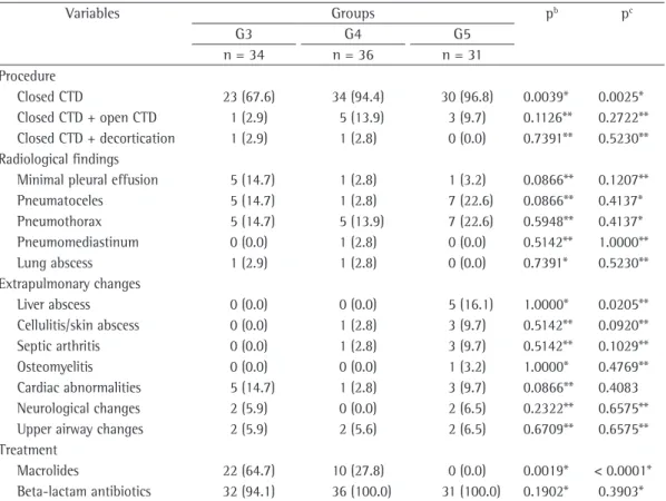
Table 3 - Comparison among the group of patients with community-acquired pneumonia (CAP) and parapneumonic pleural effusion (PPE) due to Mycoplasma pneumoniae without co-infection, the group of patients with CAP and PPE due to Streptococcus pneumoniae, and the group of patients with CAP and PPE due to Staphylococcus aureus.
Variables Groups pb pc
G3 G4 G5
n = 34 n = 36 n = 31
Age, monthsd 51 ± 45
(33;11-198)
32 ± 31 (19;5-160)
49 ± 54 (24;0-203)
0.0111**** 0.8684***
< 5 years 24 (70.6) 31 (86.1) 23 (74.2) 0.1136* 0.7456* Female gender 21 (61.8) 14 (38.9) 15 (48.4) 0.0557* 0.2785* Previous use of beta-lactam
antibiotics
13 (38.2) 6 (16.7) 9 (29.0) 0.0021* 0.0014*
Treatment failure in another hospital
10 (29.4) 4 (11.1) 5 (16.1) 0.0080* 0.0014*
Duration of symptoms, daysd 9 ± 7 (7;2-30) 6 ± 5 (6;2-30) 6 ± 5 (6;1-30) 0.1823**** 0.0450***
>7 days 16 (47.1) 8 (22.2) 7 (22.6) 0.0286* 0.0392* Signs and symptoms
Dyspnea 29 (85.3) 35 (97.2) 26 (83.9) 0.0866** 0.5715** Cough 24 (70.58) 30 (83.3) 14 (45.2) 0.2043* 0.0377* Dry cough 19 (55.9) 11 (30.6) 4 (12.9) 0.0104* 0.0043* Fever ≥ 39°C 12 (35.3) 16 (44.4) 14 (45.2) 0.4630* 0.4868* Loss of appetite 10 (29.4) 10 (27.8) 8 (25.8) 0.8797* 0.7456* Chest pain 7 (20.6) 4 (11.1) 5 (16.1) 0.2761* 0.6434* Abdominal pain 6 (17.6) 12 (33.3) 12 (38.7) 0.1334* 0.0580* Nausea/vomiting 4 (11.8) 17 (47.2) 8 (25.8) 0.0012* 0.1450* Rhinorrhea 6 (17.6) 11 (30.6) 6 (19.4) 0.2081* 0.8593*
Diarrhea 4 (11.8) 4 (11.1) 0 (0.0) 0.6120** 0.0684**
Length of hospital stay, daysd 19 ± 10
(15;6-46)
22 ± 18 (15;5-91)
21 ± 10 (21;8-50)
0.6506**** 0.2987***
Use of MV 4 (11.8) 9 (25.0) 10 (32.3) 0.1546* 0.0447*
Use of CTD 23 (67.6) 34 (94.4) 30 (96.8) 0.0039* 0.0025*
Death 0 (0.0) 3 (8.3) 2 (6.5) 0.1304** 0.2235**
Initial blood workup
Hemoglobin ≤ 11 g/dL 27 (79.4) 34 (94.4) 25 (80.6) 0.0629* 0.9011* Leukocytes ≥ 12,000/mm3 25 (73.5) 16 (44.4) 21 (67.7) 0.0135* 0.6083*
Leukocytes < 5,000/mm3 0 (0.0) 8 (22.2) 3 (9.7) 0.0032** 0.1029**
Neutrophils ≥ 8,000/mm3 24 (70.6) 19 (52.8) 23 (74.2) 0.1260* 0.7456*
Immature neutrophils ≥ 5% 18 (52.9) 31 (86.1) 28 (90.3) 0.0024* 0.0009* C-reactive protein, mg/dLd 138 ± 149
(67;7-468)
72 ± 47 (60;47-248)
111 ± 89 (67;13-282)
0.2493**** 0.6034***
Pleural fluid 16 (47.1) 28 (77.8) 22 (71.0) 0.0078* 0.0507* pH < 7.2 4 (28.6) 7 (28.0) 5 (22.7) 0.6242** 0.4938** Glucose ≤ 40 mg/dL 8 (53.3) 19 (67.9) 13 (59.1) 0.3476** 0.7285* Proteins > 3.4 g/dL 9 (60.0) 22 (78.6) 16 (76.2) 0.1738** 0.2394** Leukocytes ≥ 1,000/mm3 10 (62.5) 21 (75.0) 17 (77.2) 0.2950** 0.2497**
Predominance of neutrophils 11 (73.3) 18 (64.3) 21 (95.4) 0.4016** 0.0757** G3: patients with M. related CAP and PPE without co-infection; G4: patients with Streptococcus pneumoniae-related CAP and PPE; G5: patients with Staphylococcus aureus-pneumoniae-related CAP and PPE; MV: mechanical ventilation; and CTD: chest tube drainage. aValues expressed as n (%), except where otherwise indicated. bComparison between G3 and
G4. cComparison between G3 and G5. dValues expressed as mean ± SD (median;range). *Chi-square test. **Fisher’s exact
and S. aureus. M. pneumoniae/S. pneumoniae co-infection increased the duration of symptoms prior to admission, the mean of which was found to be 17 days for those with M. pneumoniae/S. pneumoniae co-infection and 6 days for those with S. pneumoniae alone. Therefore, prolonged PPE should raise the hypothesis of M. pneumoniae with or without co-infection.
In the present study, the bacterium that was most commonly found in association with M. pneumoniae was S. pneumoniae, having been found in 9 patients (20.5%). It has been reported that up to 30% of patients with CAP can present with M. pneumoniae/S. pneumoniae co-infection.(18)
Among the signs and symptoms, dry cough was found to be more common in the group of patients with M. pneumoniae, whereas nausea/ vomiting were found to be more common in the group of patients with other etiologic agents. such patients. In 2004 in Finland, Korppi et al.
(18) found hospitalization rates of 67%, 5%, and
9% in patients aged < 4 years, 5-9 years, and 10-14 years, respectively. It is of note that more than 70% of the patients with M. pneumonia in our study were younger than 5 years of age. The abovementioned data underscore the need for including M. pneumoniae in the differential diagnosis of PPE not only in individuals in the 5-25 year age bracket but also in those who are younger than 5 years of age.
The duration of symptoms prior to admission plays an important role in differentiating PPEs. In patients with M. pneumoniae, the onset of symptoms is generally gradual (i.e., over a period of several days), and the symptoms can persist
for weeks or months.(19) In the present study, the
duration of symptoms prior to hospitalization was found to be longer in the patients with M. pneumoniae than in those with other etiologic agents, specifically S. pneumoniae
Variables Groups pb pc
G3 G4 G5
n = 34 n = 36 n = 31
Procedure
Closed CTD 23 (67.6) 34 (94.4) 30 (96.8) 0.0039* 0.0025* Closed CTD + open CTD 1 (2.9) 5 (13.9) 3 (9.7) 0.1126** 0.2722** Closed CTD + decortication 1 (2.9) 1 (2.8) 0 (0.0) 0.7391** 0.5230** Radiological findings
Minimal pleural effusion 5 (14.7) 1 (2.8) 1 (3.2) 0.0866** 0.1207** Pneumatoceles 5 (14.7) 1 (2.8) 7 (22.6) 0.0866** 0.4137* Pneumothorax 5 (14.7) 5 (13.9) 7 (22.6) 0.5948** 0.4137* Pneumomediastinum 0 (0.0) 1 (2.8) 0 (0.0) 0.5142** 1.0000** Lung abscess 1 (2.9) 1 (2.8) 0 (0.0) 0.7391* 0.5230** Extrapulmonary changes
Liver abscess 0 (0.0) 0 (0.0) 5 (16.1) 1.0000* 0.0205** Cellulitis/skin abscess 0 (0.0) 1 (2.8) 3 (9.7) 0.5142** 0.0920** Septic arthritis 0 (0.0) 1 (2.8) 3 (9.7) 0.5142** 0.1029** Osteomyelitis 0 (0.0) 0 (0.0) 1 (3.2) 1.0000* 0.4769** Cardiac abnormalities 5 (14.7) 1 (2.8) 3 (9.7) 0.0866** 0.4083 Neurological changes 2 (5.9) 0 (0.0) 2 (6.5) 0.2322** 0.6575** Upper airway changes 2 (5.9) 2 (5.6) 2 (6.5) 0.6709** 0.6575** Treatment
Macrolides 22 (64.7) 10 (27.8) 0 (0.0) 0.0019* < 0.0001* Beta-lactam antibiotics 32 (94.1) 36 (100.0) 31 (100.0) 0.1902* 0.3903* G3: patients with M. related CAP and PPE without co-infection; G4: patients with Streptococcus pneumoniae-related CAP and PPE; G5: patients with Staphylococcus aureus-pneumoniae-related CAP and PPE; MV: mechanical ventilation; and CTD: chest tube drainage. aValues expressed as n (%), except where otherwise indicated. bComparison between G3 and
G4. cComparison between G3 and G5. dValues expressed as mean ± SD (median;range). *Chi-square test. **Fisher’s exact
Table 4 - Comparison between the group of patients with community-acquired pneumonia (CAP) and parapneumonic pleural effusion (PPE) due to Streptococcus pneumoniae and the group of patients with CAP and PPE due to co-infection with Mycoplasma pneumoniae and Streptococcus pneumoniae.
Variables Groups p
G4 G6
n = 36 n = 9
Age, monthsb 32 ± 31 (19;5-160) 36 ± 26 (32;8-80) 0.6937***
< 5 years 31 (86.1) 7 (77.8) 0.4297**
Female gender 14 (38.9) 5 (55.6) 0.2965**
Duration of symptoms, daysb 6 ± 5 (6;2-30) 17 ± 17 (10;4-60) 0.0112****
>7 days 8 (22.2) 6 (66.7) 0.0171**
Signs and symptoms
Dyspnea 35 (97.2) 9 (100) 0.8000**
Cough 30 (97.2) 8 (88.9) 0.5702**
Dry cough 11 (30.6) 5 (55.6) 0.4692*
Fever ≥ 39°C 16 (44.4) 4 (44.4) 0.6480**
Loss of appetite 10 (27.8) 4 (44.4) 0.2801**
Chest pain 4 (11.1) 2 (22.2) 0.3442**
Abdominal pain 12 (33.3) 3 (33.3) 0.6313**
Nausea/vomiting 17 (47.2) 4 (44.4) 0.5899**
Rhinorrhea 11 (30.6) 2 (22.2) 0.4822**
Diarrhea 4 (11.1) 3 (33.3) 0.1306**
Length of hospital stay, daysb 22 ± 18 (15;5-91) 35 ± 24 (30;9-88) 0.0983***
Use of MV 9 (25.0) 2 (22.2) 0.6184**
Use of CTD 34 (94.4) 8 (88.9) 0.4968**
Death 3 (8.3) 0 (0.0) 0.5031**
Initial blood workup
Hemoglobin ≤ 11 g/dL 34 (94.4) 9 (100.0) 0.6363** Leukocytes ≥ 12,000/mm3 16 (44.4) 4 (44.4) 0.6480**
Leukocytes < 5,000/mm3 8 (22.2) 2 (22.2) 0.6547**
Neutrophils ≥ 8,000/mm3 19 (52.8) 4 (44.4) 0.4698**
Immature neutrophils ≥ 5% 31 (86.1) 8 (88.9) 0.6557**
Pleural fluid 28 (77.8) 6 (66.7) 0.3815**
pH < 7.2 7 (28.0) 1 (25.0) 0.6999**
Glucose ≤ 40 mg/dL 19 (67.9) 5 (83.3) 0.4161**
Proteins > 3.4 g/dL 22 (78.6) 4 (80.0) 0.7180**
Leukocytes ≥ 1,000/mm3 21 (75.0) 5 (83.3) 0.5624**
Predominance of neutrophils 18 (64.3) 4 (66.8) 0.6494** Procedure
Closed CTD 34 (94.4) 8 (88.9) 0.4968**
Closed CTD + open CTD 5 (13.9) 4 (44.4) 0.0626**
Closed CTD + decortication 1 (2.8) 1 (11.1) 0.3636** Radiological findings
Pneumatoceles 1 (2.8) 2 (22.2) 0.0972**
Pneumothorax 5 (13.9) 2 (22.2) 0.4297**
Pneumomediastinum 1 (2.8) 0 (0.0) 0.8000**
Lung abscess 1 (2.8) 1 (11.1) 0.3636**
G4: patients with CAP and PPE due to Streptococcus pneumoniae; G6: patients with CAP and PPE due to Mycoplasma pneumoniae and S. pneumoniae; MV: mechanical ventilation; and CTD: chest tube drainage. aValues expressed as n (%), except where
otherwise indicated. bValues expressed as mean ± SD (median;range). *Chi-square test. **Fisher’s exact test. ***Student’s
in the group of patients with S. aureus, confirming hematogenous dissemination in those cases.
The usual treatment for PPE does not include drugs against M. pneumoniae, which is naturally resistant to beta-lactam antibiotics (such as penicillins, penicillin derivatives, and cephalosporins) because it has no cell wall.(23)
Beta-lactam antibiotics were used prior to hospitalization in 38.0% of the patients with M. pneumoniae, in 16.7% of those with S. pneumoniae, and in 29% of those with S. aureus. Therefore, the use of beta-lactam antibiotics in children with CAP/PPE prior to hospitalization, with no improvement in the symptoms and with persistent fever, constitutes evidence for a differential diagnosis of atypical bacteria.
Our data suggest that M. pneumoniae-related CAP/PPE in children and adolescents should be more thoroughly investigated in Brazil. Prospective longitudinal studies should be able to explain the role of M. pneumoniae in PPE, principally in children younger than 5 years of age, as demonstrated in the present study.
Acknowledgements
We would like to thank Décio Sesquim, thoracic surgeon in the HINSG Department of Pulmonology, for his aid in monitoring the cases. We would also like to thank Adyléia Aparecida Dalbo Contrera Toro, pediatric pulmonologist in the Pediatrics Department of the State University at Campinas, and Antonia Terezinha Tresoldi, tenured professor at the State University at Campinas, for their suggestions, which enriched the present study. Finally, we would like to thank the HINSG medical record department staff for their dedication and excellence in providing service.
References
1. Vervloet LA, Camargos PA, Soares DR, Oliveira GA, Oliveira JN. Clinical, radiographic and hematological characteristics of Mycoplasma pneumoniae pneumonia. J Pediatr (Rio J). 2010;86(6):480-7. PMid:21069252. http://dx.doi.org/10.1590/S0021-75572010000600006 2. Matas Andreu L, Molinos Abós S, Fernández Rivas G,
González Soler V, Ausina Ruiz V. Serologic diagnosis of Mycoplasma pneumoniae infections [Article in Spanish]. Enferm Infecc Microbiol Clin. 2006;24 Suppl 1:19-23. http://dx.doi.org/10.1157/13094274
3. Ferwerda A, Moll HA, de Groot R. Respiratory tract infections by Mycoplasma pneumoniae in children: a review of diagnostic and therapeutic measures. Eur J Pediatr. 2001;160(8):483-91. PMid:11548186. http:// dx.doi.org/10.1007/s004310100775
According to the literature, if M. pneumoniae infects the trachea, bronchi, and bronchioles, patients can experience dry cough, which is constant and uncontrollable and can cause
nocturnal awakenings.(19) However, the absence of
that symptom does not rule out M. pneumoniae infection.
We found that 12% of the patients with M. pneumoniae required mechanical ventilation during hospitalization, as did 25% of those with S. pneumoniae and 32.3% of those with S. aureus. In the patients with M. pneumoniae, PPE was found to be smaller, and the use of chest tube drainage was less common. These findings confirm that cases of infection with typical bacteria are more severe.
By definition, PPE is pneumonia-related pleural effusion, which can be complicated or uncomplicated. Complicated PPE is an exudate that can be purulent, culture or Gram staining revealing bacteria and biochemical analysis revealing pH < 7, glucose < 40 mg/dL, and lactate
dehydrogenase > 1,000 IU/L.(20) Mocelin et al.
(21) found that pleural fluid glucose < 40 mg/dL
and pleural fluid pH < 7.0 had a sensitivity of 76% and 55%, respectively, for the diagnosis of complicated PPE, the sensitivity of pleural fluid pH having increased to 79% when the cut-off point was increased to 7.2. In 2010,
Maranhão et al.(22) quantified total proteins
in pleural fluid in order to diagnose pleural exudates. Using a cut-off point of 3.4 g/dL for total proteins in pleural fluid, the sensitivity, specificity, and accuracy for the diagnosis of exudates were, respectively, 99.4%, 72.6%, and 99.2%. In our hospital, determination of lactate dehydrogenase in pleural fluid is a test that is not routinely performed.
In the present study, the characteristics of pleural fluid were useless in differentiating among the groups. Neither pleural fluid pH nor pleural fluid glucose showed statistically significant differences among the groups. The same was true for the finding of pleural fluid total proteins > 3.4 g/dL, which was less common in the patients with M. pneumoniae (60.0%) than in those with S. pneumoniae (78.6%) or S. aureus (76.2%).
15. Hendrickson DJ, Blumberg DA, Joad JP, Jhawar S, McDonald RJ. Five-fold increase in pediatric parapneumonic empyema since introduction of pneumococcal conjugate vaccine. Pediatr Infect Dis J. 2008;27(11):1030-2. PMid:18845981. http://dx.doi. org/10.1097/INF.0b013e31817e5188
16. Harris M, Clark J, Coote N, Fletcher P, Harnden A, McKean M, et al. British Thoracic Society guidelines for the management of community acquired pneumonia in children: update 2011. Thorax. 2011;66 Suppl 2:ii1-23. PMid:21903691. http://dx.doi.org/10.1136/ thoraxjnl-2011-200598
17. O’Handley JG, Gray LD. The incidence of Mycoplasma pneumoniae pneumonia. J Am Board Fam Pract. 1997;10(6):425-9. PMid:9407483.
18. Korppi M, Heiskanen-Kosma T, Kleemola M. Incidence of community-acquired pneumonia in children caused by Mycoplasma pneumoniae: serological results of a prospective, population-based study in primary health care. Respirology. 2004;9(1):109-14. PMid:14982611. http://dx.doi.org/10.1111/j.1440-1843.2003.00522.x 19. Waites KB. New concepts of Mycoplasma pneumoniae
infections in children. Pediatr Pulmonol. 2003;36(4):267-78. PMid:12950038. http://dx.doi.org/10.1002/ppul.10346 20. Moreira GO, Ribeiro JD, Tresoldi AT. Utility of a scoring
system and indicative variables for assessing the need for pleural drainage in pediatric patients with parapneumonic pleural effusion. J Bras Pneumol. 2005;31(3):205-11. http://dx.doi.org/10.1590/S1806-37132005000300005 21. Mocelin HT, Fischer GB. Fatores preditivos para drenagem
de derrames pleurais parapneumônicos em crianças. J Pneumol. 2001;27(4):177-84. http://dx.doi.org/10.1590/ S0102-35862001000400003
22. Maranhão BH, Silva Junior CT, Chibante AM, Cardoso GP. Determination of total proteins and lactate dehydrogenase for the diagnosis of pleural transudates and exudates: redefining the classical criterion with a new statistical approach. J Bras Pneumol. 2010;36(4):468-74. PMid:20835594.
23. van de Garde EM, Endeman H, van Hemert RN, Voorn GP, Deneer VH, Leufkens HG, et al. Prior outpatient antibiotic use as predictor for microbial aetiology of community-acquired pneumonia: hospital-based study. Eur J Clin Pharmacol. 2008;64(4):405-10. PMid:18060396. PMCid:2254473. http://dx.doi. org/10.1007/s00228-007-0407-0
4. Waris ME, Toikka P, Saarinen T, Nikkari S, Meurman O, Vainionpää R, et al. Diagnosis of Mycoplasma pneumoniae pneumonia in children. J Clin Microbiol. 1998;36(11):3155-9. PMid:9774556. PMCid:105292. 5. John SD, Ramanathan J, Swischuk LE. Spectrum of
clinical and radiographic findings in pediatric mycoplasma pneumonia. Radiographics. 2001;21(1):121-31. PMid:11158648.
6. Cohen M, Sahn SA. Resolution of pleural effusions. Chest. 2001;119(5):1547-62. PMid:11348966. http:// dx.doi.org/10.1378/chest.119.5.1547
7. Neumayr L, Lennette E, Kelly D, Earles A, Embury S, Groncy P, et al. Mycoplasma disease and acute chest syndrome in sickle cell disease. Pediatrics. 2003;112(1 Pt 1):87-95. PMid:12837872. http://dx.doi.org/10.1542/ peds.112.1.87
8. Hammerschlag MR. Mycoplasma pneumoniae infections. Curr Opin Infect Dis. 2001;14(2):181-6. PMid:11979130. http://dx.doi.org/10.1097/00001432-200104000-00012 9. Korppi M. Community-acquired pneumonia in children:
issues in optimizing antibacterial treatment. Paediatr Drugs. 2003;5(12):821-32. PMid:14658923. http:// dx.doi.org/10.2165/00148581-200305120-00005 10. Petitjean J, Vabret A, Gouarin S, Freymuth F. Evaluation
of four commercial immunoglobulin G (IgG)- and IgM-specific enzyme immunoassays for diagnosis of Mycoplasma pneumoniae infections. J Clin Microbiol. 2002;40(1):165-71. PMid:11773112. PMCid:120121. http://dx.doi.org/10.1128/JCM.40.1.165-171.2002 11. Shen YH, Hwang KP, Niu CK. Complicated parapneumonic
effusion and empyema in children. J Microbiol Immunol Infect. 2006;39(6):483-8. PMid:17164951.
12. Deiros Bronte L, Baquero-Artigao F, García-Miguel MJ, Hernández González N, Peña García P, del Castillo Martín F. Parapneumonic pleural effusion: an 11-year review [Article in Spanish]. An Pediatr (Barc). 2006;64(1):40-5. http://dx.doi.org/10.1016/S1695-4033(06)70007-8 13. British Thoracic Society Standards of Care Committee.
British Thoracic Society Guidelines for the Management of Community Acquired Pneumonia in Childhood. Thorax. 2002;57 Suppl 1:i1-24. PMid:11994552. PMCid:1765993. 14. Hernández-Bou S, García-García JJ, Esteva C, Gené A,
Luaces C, Muñoz Almagro C. Pediatric parapneumonic pleural effusion: epidemiology, clinical characteristics, and microbiological diagnosis. Pediatr Pulmonol. 2009;44(12):1192-200. PMid:19911359. http://dx.doi. org/10.1002/ppul.21114
About the authors
Letícia Alves Vervloet
Assistant Professor. Department of Pediatrics, Federal University of Espírito Santo, Vitória, Brazil.
Vitor Earl Cardoso Vervloet
Attending Physician. Pulmonology Ward, Nossa Senhora da Glória Children’s Hospital, Vitória, Brazil.
Mário Tironi Junior
Coordinator. Pulmonology Ward, Nossa Senhora da Glória Children’s Hospital, Vitória, Brazil.
José Dirceu Ribeiro