Dental Materials
Farid Mohammed Sabry El-Askary(a)
Mohammed Salah Abdelaziz Nassif(b)
Andrea Mello Andrade(c) Alessandra Reis(d)
Alessandro Dourado Loguercio(d)
(a) Operative Dentistry Department, Faculty of Dentistry, Ain Shams University, Cairo, Egypt.
(b) Dental Biomaterials Department, Faculty of Dentistry,Ain Shams University, Cairo, Egypt.
(c) Department of Dental Materials, School of Dentistry, University of São Paulo - USP, São Paulo, SP, Brazil.
(d) Department of Restorative Dentistry, School of Dentistry, Universidade Estadual de Ponta Grossa, Ponta Grossa, PR, Brazil.
Corresponding Author:
Farid Mohammed Sabry El-Askary E-mail: [email protected]
Submitted: Mar 09, 2012
Accepted for publication: May 15, 2012 Last revision: May 24, 2012
Effect of surface area and air-drying
distance on shear bond strength of
etch-and-rinse adhesive
Abstract: We evaluated the effects of air-drying distance and bond sur-face area on the shear bond strength of a 2-step etch-and-rinse adhe-sive. A total of 120 bovine anterior teeth were equally divided into 6 main groups based on bonding surface area. The main groups were di-vided into sub-groups (n = 5) according to air-drying distance. The shear strength was determined using a universal testing machine at a crosshead speed of 0.5 mm/min. The averaged results were subjected to two-way ANOVA and Tukey’s test (α = 0.05). Two-way ANOVA testing identiied no signiicant cross-product interactions (p > 0.05), but the main factors of area (p < 0.0001) and air-drying distance (p < 0.00001) signiicantly affected the mean bond strength. Shorter air-drying distances improved bond strength, and increased surface area decreased the bond strength.
Descriptors: Dentin-Bonding Agents; Shear Strength; Material Testing.
Introduction
Shear and tensile tests are the most common methods of evaluating the bond strength of adhesives. The shear test is considered the most useful test,1 and, in a survey of recent literature, it was used in 26% of
the papers.2 Despite this, there are disadvantages to macro-shear bond
strength testing.3-5 In order to combine ease of manipulation with the
ability to test multiple specimens on a single tooth, the micro-shear test was developed.6
The average bonding surface area in macro-shear tests ranges from 3.14 to 12.56 mm2 (corresponding to a 2–4 mm specimen diameter). The
relationship between bonding area and strength has received more atten-tion with the development of micro-tensile and micro-shear tests,7-10 and
the inluence of bonding area on macro-shear bond strength has been evaluated in one study.11
Excessive adhesive removal, air entrapment within the adhesive, and improper solvent evaporation12,13 are the most common clinical problems
during air-drying of adhesives. These problems may be related to the air-drying distance from the dentin surface, and this distance must be considered an important clinical variable during adhesive performance testing. To our knowledge, only two studies have examined the effect of air-drying distance on bond strength. One of these studied the effect on the bond strength of a simpliied etch-and-rinse adhesive,14 while the
other evaluated a one-step self-etching adhesive.12 Both of these studies Declaration of Interests: The authors
indicated that air-drying distance had a signiicant effect on the bond strength. In addition, due to the lack of a recommended distance in the adhesive in-structions, various air-drying distances have been used in previous studies to evaporate solvent from etch-and-rinse adhesives.15,16
Therefore, the aim of this study was to evaluate the effect of very short (1 cm), intermediate (5 cm), long (10 cm), and very long (20 cm) air-drying dis-tances on the shear bond strength of a 2-step etch-and-rinse adhesive applied to bovine teeth. The ef-fect of bond surface area was also investigated.
Methodology
A total of 120 bovine anterior teeth were cleaned and stored in 0.1% thymol solution for not more than 1 month. Before preparation, the teeth were washed under abundant running water and the roots were removed using a diamond saw at slow speed (Isomet, Buehler, Lake Bluff, USA) with copi-ous water cooling. The pulp was removed and the crowns were sawn in half in the mesio-distal direc-tion. Each half was ground on wet 180-grit SiC pa-per to remove the labial enamel and expose a lat dentin surface.
Following enamel removal, the pulp chambers were illed with cotton and both tooth halves were ixed using double-faced adhesive tape in a cylin-drical polyvinyl chloride (PVC) mold, which was then illed with a self-curing acrylic resin (Jet, Ar-tigos Odontológicos Clássico, São Paulo, Brazil). The molds were immersed in tap water to minimize the temperature increase during curing. The mating halves of each tooth were similarly marked. After curing of the acrylic resin, the dentin surfaces were wet-ground for 60 s using 600-grit SiC paper to cre-ate a standardized smear layer. The specimens were ultrasonically cleaned in distilled water for 5 min prior to the bonding procedure to remove any re-maining silicon carbide particles.17
Specimen grouping
The teeth were equally divided into 6 groups (n = 20) according to the amount of surface area bonded: 0.44, 0.82, 1.98, 3.14, 7.06, or 12.56 mm2
(corresponding to mold diameters of 0.75, 1, 1.59,
2, 3, or 4 mm). Each main group was divided into 4 subgroups (n = 5) based on air-drying distances of 1, 5, 10, or 20 cm.
Bonding procedure
An etch-and-rinse 2-step adhesive (Adper Single Bond 2, 3MESPE, St. Paul, USA) and a hybrid com-posite material (Filtek Z250, 3MESPE, St. Paul, USA) were used in this study. The dentin surfaces were acid-etched for 15 s, rinsed for 15 s, and blot-dried using sterile cotton, leaving the dentin surface moist. The adhesive was applied in 2 consecutive layers, gently agitated for 15 s, and gently air-dried for 10 s using compressed air.15 The air-drying
pressure was adjusted to 1 bar12,18 using a pressure
regulator, and the air nozzle was held at 45° to the dentin surface. The adhesive was light-cured for 10 s using an LED curing unit (LEDition, Ivoclar Vivadent, Schaan, Liechtenstein) with an output of 600 mW/cm2.
For the 0.44, 0.82, and 1.98 mm2 surface areas,
each tooth received 6 specimens. For the 3.14 mm2
surface area, each tooth received 5 specimens. For 7.06 mm2, each tooth received 3 specimens. For
12.56 mm2, each tooth received 2 specimens. The
specimens were distributed over the two halves us-ing polyethylene tubes. The polyethylene tubes (TY-GON Medical Tubing Formulations 54-HL, Saint Gobain Performance Plastics, Akron, USA) were applied before light-curing of the adhesive. After adhesive light curing, resin composite was careful-ly packed inside each tube, and a clear cellophane sheet was placed over the illed tubes and pressed gently into place. The resin samples were light-cured for 20 s. The plastic tubes were carefully removed and the specimens were examined using a stereo-microscope at 10× magniication to identify those with evidence of air bubbles or gaps at the interface. Residual adhesive extending beyond the base of the resin cylinder was carefully removed from each sample using a blade. The specimens were stored in distilled water for 24 h at 37°C.
Shear bond strength testing
was then placed in a universal testing machine (Model 5565, Instron, Canton, USA). The samples were shear tested using a chisel-bladed metallic at-tachment with a 0.2-mm tip for micro-shear test-ing and a 0.4-mm tip for macro-shear testtest-ing. The attachment was positioned as close as possible to the composite/dentin interface. The tests were per-formed at a crosshead speed of 0.5 mm/min. The force required for failure (Newton) was divided by the surface area (mm2) to determine the shear bond
strength in MPa.
SEM evaluation
The fractured specimens were mounted on alu-minum stubs, sputter-coated, and examined using SEM (JEOL 5600 LVj, JEOL Ltd., Tokyo, Japan). Failure modes were evaluated and classiied as mixed, cohesive, or adhesive.
Statistical analysis
Each experimental condition was represented by ive teeth (10 tooth halves). Each half-tooth
sup-ported 1–3 resin cylinders depending on the adhe-sive area. The overall bond strength values were av-eraged and subjected to 2-way analysis of variance (ANOVA) and Tukey’s test at a signiicance level of 0.05.
Results
The number of specimens undergoing each type of fracture is listed in Table 1. Figures 1–3 are mi-crographs of the fracture zones depicting each fail-ure type.
Two-Way ANOVA did not identify statistically signiicant cross-product interactions, but both area and distance signiicantly affected the bond strength (p < 0.00001). Regardless of air drying distance, signiicantly higher strengths were observed in the 0.44 mm2-area specimens (Table 2). Larger bond
areas (7.06 and 12.56 mm2) exhibited signiicantly
lower bond strength. Regardless of the surface area, signiicantly higher bond strengths were developed in specimens dried at a distance of 1 cm (Table 2). Signiicantly lower bond strength was obtained in
Area (mm2)
0.44 0.82 1.98 3.14 7.06 12.56
Fracture Pattern Distance
(cm) A M C A M C A M C A M C A M C A M C
1 24 6 0 20 10 0 15 15 0 7 18 0 6 9 0 1 9 0
5 21 9 0 14 16 0 15 15 0 7 18 0 4 10 1 2 5 3
10 24 6 0 16 14 0 11 19 0 9 16 0 7 7 1 3 6 1
20 19 11 0 13 17 0 11 19 0 10 15 0 10 5 0 6 3 1
Fracture pattern: A = adhesive; M = mixed; C = cohesive.
Table 1 - Number of specimens for each fracture pattern in each experimental condition.
Table 2 - Means ± standard deviation in MPa for each experimental condition.
Distance (cm) Area (mm2) Total distance
0.44 0.82 1.98 3.14 7.06 12.56
1 34.2 ± 11.7 25.0 ± 9.2 23.6 ± 8.9 24.3 ± 9.9 18.0 ± 4.6 14.6 ± 2.0 23.3 ± 10.3 a 5 28.4 ± 12.5 20.7 ± 8.0 20.7 ± 10.9 18.9 ± 7.5 13.3 ± 4.3 11.2 ± 4.7 18.9 ± 9.7 b 10 20.1 ± 6.4 18.9 ± 12.9 19.4 ± 4.9 22.3 ± 8.0 16.5 ± 6.7 12.2 ± 5.7 18.2 ± 8.4 b 20 16.3 ± 8.9 13.5 ± 5.6 12.9 ± 6.7 18.2 ± 9.1 8.2 ± 5.8 8.7 ± 3.3 13.0 ± 8.1 c Total area 24.5 ± 12.1 A 19.5 ± 10.1 B 19.1 ± 8.9 B 20.8 ± 8.9 B 13.7 ± 6.5 C 11.7 ± 4.6 C
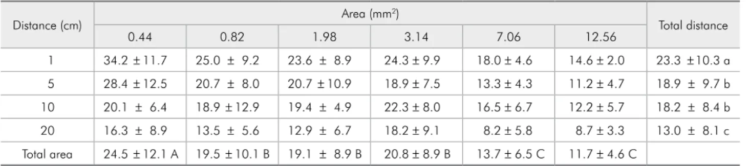
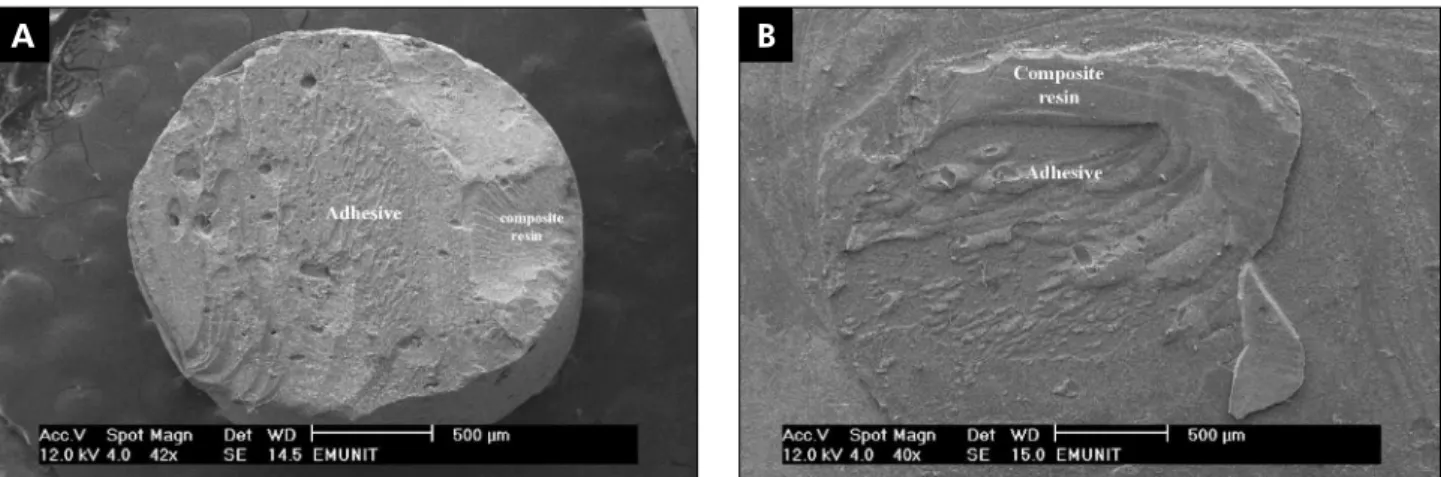
Figure 1 - SEM photomicrographs indicating cohesive failure within both the adhesive and the composite resin. A: composite resin side; B: dentin side.
Figure 2 - SEM photomicrographs depicting adhesive failure between the adhesive and dentin. A: composite resin side with a thick cracked adhesive layer. The thick adhesive layer might be due to evaporation of solvent at a longer distance (20 cm) and
the crack within the adhesive layer could be due to dehydration during SEM sample preparation. B: dentin side.
Figure 3 - SEM photomicrographs depicting a mixed failure type. A: composite resin side revealing a large area of dentin attached to the composite resin cylinder. This could be due to shear bond strength setup and the large bonding surface area (12.56 mm2). B: dentin side.
B
B
B A
A
specimens dried at 20 cm.
Discussion
The rapid growth in restorative dentistry has generated an increased need for in vitro mechani-cal testing. The many clinimechani-cal variables affecting the success of adhesive applications must also be evalu-ated. One important factor affecting adhesive per-formance is the air-drying step. Several parameters control the eficiency of air-drying, including drying time, pressure, and nozzle-tooth distance.
In this study, air-drying distance exerted a sig-niicant effect on bond strength. The adhesive con-tained a water/ethanol solvent, and the low vapor pressure of the water component hindered removal during the air-drying step.19 The formulation also
contained HEMA, which could interfere with the removal of residual water20 and (to a lesser extent)
ethanol from the adhesive.21 Increasing the
air-dry-ing distance decreased the effectiveness, particularly at low pressures.12 The reduced air-drying
effective-ness resulted in decreased shear bond strength. SEM photomicrographs (Figure 2) of samples dried at long distances revealed a very thick adhesive layer. A thick adhesive layer might improve resistance to debonding stresses during composite polymeriza-tion,19 but the unnecessary increase in the thickness
of the adhesive layer might be considered a weak junction at the composite/dentin interface. The ex-cessive increase in the adhesive layer thickness could be related to the air-drying distance.
Our results were in disagreement with those of Kanca,14 who used an acetone-based 2-step
etch-and-rinse adhesive. Acetone has a high vapor pres-sure (approximately 4 times that of ethanol)19 and
is therefore easier to remove. Nevertheless, future studies should be performed to evaluate the effect of air-drying distance on the bonding performance of contemporary simpliied etch-and-rinse adhesives containing various solvents.
Specimens with very small surface areas (0.44 mm2) had signiicantly greater shear bond
strength, and the strength decreased in larger speci-mens (7.06 and 12.65 mm2). This was similar to the
indings of Phrukkanon et al.22 and Watanabe et
al.,11 who reported that increasing the bond surface
area led to a signiicant decrease in bond strength. Based on results obtained using a different test methodology, El Zohairy et al.8 described an
in-verse relationship between the micro-tensile bond strength and the specimen dimensions. Grifith23
observed that large specimens contained a greater number of defects. Upon loading during testing, stresses were concentrated at the defects, resulting in crack formation.
The fracture analysis in the current study re-vealed a tendency (Table 1) toward an increase in mixed-type failures with increasing bond surface area, ranging from 26.7% for 0.44 mm2 to 57.5%
for 12.56 mm2. Previous studies have reported
pull-out of dentin when shear tests were performed on high-strength adhesives, preventing calculation of shear bond strength from load and area measure-ments.24 There was no correlation between the
pres-ence of cohesive failure in dentin and bond strength.5
It is obvious that increasing the bonded surface area decreases the bond strength, and that the di-ameter of the bonded surface must be taken into consideration when comparing shear bond strength results between studies. Furthermore, additional ef-forts should be made by both manufacturers and scientists to evaluate the technique sensitivity of cur-rently used adhesives. As the air-drying distance ap-pears to be a very important variable, information about the optimum air-drying distance should be included in the material proiles.
Conclusions
The bond strength was adversely affected by in-creased air-drying distances, and the bond strength decreased with increased surface area. The failure type was relatively consistent, with mixed failure occurring more often with increasing surface area.
Acknowledgment
References
1. Braga R, Meira J, Boaro L, Xavier T. Adhesion to tooth struc-ture: a critical review of “macro” test methods. Dent Mater. 2010 Feb;26(2):e38-49.
2. Van Meerbeek B, Peumans M, Poitevin A, Mine A, Van Ende A, Neves A, et al. Relationship between bond-strength tests and clinical outcomes. Dent Mater. 2010 Feb;26(2):e100-21. 3. Della Bona A, van Noort R. Shear vs. tensile bond strength
of resin composite bonded to ceramic. J Dent Res. 1995 Sep;74(9):1591-6.
4. van Noort R, Noroozi S, Howard IC, Cardew G. A critique of bond strength measurements. J Dent. 1989 Apr;17(2):61-7. 5. Versluis A, Tantabirojn D, Douglas W. Why do shear bond
strength test pulls out dentin? J Dent Res. 1997 Jun;76(6):1298-307.
6. Armstrong S, Geraldeli S, Maia R, Raposo L, Soares C, Yamagawa J. Adhesion to tooth structure: A critical review of “micro” bond strength test methods. Dent Mater. 2010 Feb;26(2)e50-62.
7. Pashley D, Sano H, Ciucchi B, Yoshiyama M, Carvalho R. Adhesion testing of dentin bonding agents: a review. Dent Mater. 1995 Mar;11(2):117-25.
8. El-Zohairy A, de Gee A, de Jager N, van Ruijven N, Feilzer A. The Influence of Specimen Attachment and Dimension on Microtensile Strength. J Dent Res. 2004 May;83(5):420-4. 9. Sano H, Shono T, Sonoda H, Tatatsu T, Ciucchi B, Carvalho
R, et al. Relationship between surface area for adhesion and tensile bond strength-Evaluation of a microtensile bond test. Dent Mater.1994 Jul;10(4):236-40.
10. Shono Y, Terashita M, Pashely E, Brewer P, Pashley D. Effect of cross-sectional area on resin-enamel tensile bond strength. Dent Mater. 1997 Sep;13(5):290-95.
11. Watanabe L, Marshall G, Marshall S. Variables influence on shear bond strength testing to dentin. In: Advanced Adhesive Dentistry-3rd International Kuraray Symposium. 1999:75-90. 12. El-Askary F, Van Noort R. Effect of air-drying pressure and
distance on µTBS of a self-etching adhesive. J Adhes Dent. 2011 Apr;13(2):147-53.
13. Spreafico D, Semeraro S, Mezzanzanica D, Re D, Gagliani M, Tanaka T, et al. The effect of the air-blowing step on the
technique sensitivity of four different adhesive systems. J Dent. 2006 Mar;34(3):237-44.
14. Kanca J 3rd. Wet bonding: effect of drying time and distance.
Am J Dent. 1996 Dec;9(6):273-6.
15. Reis A, Klein-Jùnior C, Accorinte M, Grande R, dos Santos C, Loguercio A. Effects of adhesive temperature on the early and 6-month dentin bonding. J Dent. 2009 Oct; 37(10):791-8 16. Reis A, Klein-Júnior C, Coelho de Souza F, Stanislawczuk R,
Loguercio A. The use of warm air stream for solvent evapo-ration: effects on the durability of resin-dentin bonds. Oper Dent. 2010 Jan-Feb, 35(1):29-36
17. El-Askary F, Nassif M. Bonding nano-filled resin modified glass ionomer to dentin using different self etch adhesives. Oper Dent. 2011 Jul-Aug;36(4):413-21.
18. De Munck J, Arita A, Shirai K, Van Landuyt K, Coutinho E, Poitevin A, et al. Microrotary fatigue resistance of a HEMA-free All-in-one adhesive bonded to dentin. J Adhes Dent. 2007 Aug;9(4):373-9.
19. Van Landuyt K, Snauwaert J, De Munck J, Peumans M, Yo-shida Y, Poitevin A, et al. Systematic review of the chemical composition of contemporary dental adhesives. Biomaterials. 2007 Sep;28(26):3757-85.
20. Sadr A, Shimada Y, Tagami J. Effects of solvent drying time on micro-shear bond strength and mechanical proper-ties of two self-etching adhesive systems. Dent Mater. 2007 Sep;23(9):1114-19.
21. Pashley E, Zhang Y, Lockwood P, Rueggeberg F, Pashley D. Effects of HEMA on water evaporation from water–HEMA mixtures. Dent Mater. 1998 Jan;14(1):6-10.
22. Phrukkanon S, Burrow M, Tyas M. Effect of cross-sectional surface area on bond strengths between resin and dentin. Dent Mater. 1998 Mar;14(2):120-28.
23. Griffith A. The phenomena of rupture and flow in solids. Philosophical Transactions of the Royal Society of London. 1921; SeriesA221:163-98.