Key words: PESA; Epididymis;
Vasovasostomy; Spermatozoa; Vas Deferens
Int Braz J Urol. 2013; 39: 720-6
__________________ Submitted for publication: May 24, 2013
__________________ Accepted after revision: August 13, 2013 Objectives: Evaluation of the presence of spermatozoa in vas deferens fluid after a long
interval of unilateral and homolateral percutaneous epididymal sperm aspiration (PESA) in vasectomized men. When found, the spermatozoa were evaluated including concentra-tion and motility, in order to verify the patency of the epididymal tubules.
Materials and Methods: Four patients, numbered in a progressive order, from one to four, with 38, 40, 48 and 51 years old and vasectomy interval of 10, 10, 25 and 11 years, respectively, whose wives did not get pregnant using intracytoplasmic sperm injection of sperm obtained by unilateral PESA and decided to try only natural conception, were submitted to intrasurgical sperm analysis of the vas deferens fluid (ISAVDF) during mi-crosurgery for reconstruction of the seminal tract.
Results: Time interval between PESA and ISAVDF was 13.75 ± 11.12 months (x ± s) varying from 3 to 29 months. Homolateral ISAVDF and PESA showed the presence of spermatozoa. Patients 1, 2 and 4 had a high concentration of 10 x 106, 64 x 106 and 45
x 106 spermatozoa/ mL; the first two had motile sperms and patient 3 had no sperms.
Conclusions: Three of four patients showed spermatozoa in the vas deferens fluid after a long interval of unilateral and homolateral PESA with high concentration, including motile forms. These findings support the concept that PESA may not result in late epipi-dymal tubule obstruction in vasectomized patients.
INTRODUCTION
Although most men at the time of vasec-tomy are sure of the choice of surgical contra-ception, around 4 to 6% will desire posteriorly to father children using their own spermatozoa (1). In those cases the treatment options include mi-crosurgical reconstruction of seminal tract and in-tracytoplasmic sperm injection (ICSI).
In order to obtain spermatozoa to ICSI some surgical techniques are employed, as
percu-taneous epididymal sperm aspiration (PESA), tes-ticular sperm aspiration (TESA), testes-ticular sperm extraction (TESE) and microsurgical dissection of seminiferous tubules (microTESE) (2-4). It is still possible to use microsurgical epididymal sperm aspiration (MESA) (5-7), during which it is per-formed a microsurgical repair of the epididymal tubule in order to avoid posterior obstructive scar-ring of the tubules keeping them patent.
Classically, PESA is considered a recovery technique that causes obstruction of epididymal
Sperm analysis of the vas deferens fluid after a long
interval of unilateral percutaneous epididymal sperm
aspiration in vasectomized patients
_______________________________________________
Fernando Lorenzini, Eduardo Zanchet, Mariana Lorenzini
Department of Urology, Federal University of Parana and Department of Urology, Hospital Cruz Vermelha, Curitiba, Brazil
ABSTRACT
ARTICLE
INFO
tubules (3,8). The mechanism of obstruction is rela-ted to scarring after the use of the aspiration needle and leakage of spermatozoa.
In the vasectomized patient, the spermato-zoa flow freely inside the epididymal tubules until the place of the vasectomy with natural processes of production, death and absorption of sperm. On the other hand, it was believed that in the vasectomized patients submitted to PESA, there would be an air--tight space between the place of PESA and the site of vasectomy where spermatozoa would only suffer death and absorption, and after a period of time, this site would not present sperms.
The presence of high concentration of sper-ms including motile forsper-ms during intrasurgical analysis of vas deferens fluid (ISAVDF) after a long interval of time of PESA, homolateral to ISAVDF, would definitely demonstrate that PESA do not cau-se epididymal tubule obstruction. From a practical point of view, these ISAVDF findings would allow homolateral vasovasoanatomosis (VV) to PESA ins-tead of vasoepididymal anastomosis (VE), that is more troublesome and more complex, with the need of great microsurgical ability and results of preg-nancy and patency inferior to VV.
The objetive of the present study was to determine the presence of spermatozoa in the vas deferens fluid after a long period of the uni-lateral and homouni-lateral PESA in vasectomized patientes, and, if affirmative, to determine their concentration and motility in order to infer epi-didymal tubule patency.
MATERIALS AND METHODS
The study was performed in four patients with azoospermia due to bilateral vasectomy who were submitted to unilateral PESA and ICSI, whose wives did not get pregnant. These patients further decided to father children spontaneously and de-cided to be submitted to ISAVDF during bilateral microsurgical reconstruction of seminal tract. It was proposed vasoepididymal anastomosis in the homolateral side submitted to PESA and VV or VE on the contralateral side of unilateral PESA, accor-ding to the results of ISAVDF.
VE was proposed using termino-lateral mi-crosurgical anastomosis with single stitches of
mo-nonylon 10-0 and VV termino-terminal anastomosis with single stitches of mononylon 9-0, single plan under surgical microscope. The fluid from the pro-ximal vas deferens stumps in relation to the epidi-dymus was aspirated in natura with a 26G needle attached to a 1 mL syringe that was send to the cli-nical laboratory. After surgery, patients were evalu-ated every three months. Table-1 shows the patients age as well as their wives age, time since vasectomy, side, place and number of aspirative punctures with 26G needles during unilateral PESA and the time be-tween unilateral PESA and the microsurgical recons-tructive procedure.
RESULTS
The median age of the patients was 44.25 ± 6.24 years (medium ± standard deviation) and of the wives 33.00 ± 1.63 years. The median time of in-terval between vasectomy and microsurgical recons-truction was 14.00 ± 7.35 years and between uni-lateral PESA and microsurgical reconstruction was 13.75 ± 11.12 months, varying from 3 to 29 months.
In all patients, the fluid from the vas defe-rens stump during ISAVDF homolateral and con-tralateral to unilateral PESA was whitish or trans-parent serous.
ISAVDF homolateral to unilateral PESA showed the presence of a high number of spermato-zoa: 10 x 106, 64 x 106 and 45 x 106 sperms/mL, with motility of 30%, 1% and 0% in patients 1, 2 and 4, respectively. Patient 3 showed no sperms.
Contralateral ISAVDF in relation to unila-teral PESA showed the presence of high number of spermatozoa, of 8 x 10⁶ and 54 x 106 sperms/mL and motility of 0% and 32% in patients 1 and 2, respec-tively; patients 3 and 4 had no sperms.
Tables 2 and 3 show the results of homola-teral and contralahomola-teral ISAVDF in relation to PESA, respectively. All patients were submitted to VV. The follow-ups every three months of patients are des-cribed in Table-4.
DISCUSSION
techni-que, but with the inconvenience of contraindicating a future VV (3,8).
Marmar et al. (9) described the first VV af-ter bilaaf-teral PESA in eight vasectomized patients, and during the intrasurgical microsurgical proce-dure they evaluated the presence of spermatozoa in the vas deferens fluid proximal stump in rela-tion to epididymis. They reported that these pa-tients had been submitted to one to four bilateral PESA; seven patients showed spermatozoa at least in one of the stumps intrasurgically and among
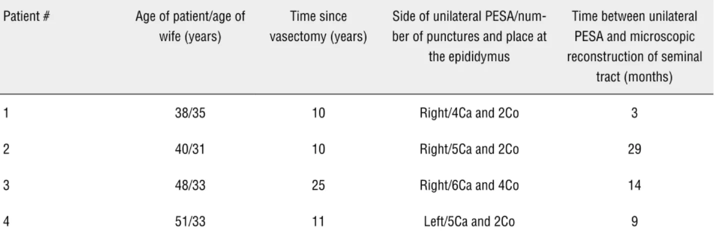
Table 1 - Distribution of patients, time since vasectomy and unilateral PESA data.
Patient # Age of patient/age of wife (years)
Time since vasectomy (years)
Side of unilateral PESA/num-ber of punctures and place at
the epididymus
Time between unilateral PESA and microscopic reconstruction of seminal
tract (months)
1 38/35 10 Right/4Ca and 2Co 3
2 40/31 10 Right/5Ca and 2Co 29
3 48/33 25 Right/6Ca and 4Co 14
4 51/33 11 Left/5Ca and 2Co 9
PESA: percutaneous epididymal sperm aspiration; nCa: number of aspirative punctures on the head of the epididymus with a 26G diameter needle, nCo: number of aspirative punctures of the body of the epididymus with a 26G diameter needle.
Table 2 - Results of intrasurgical analysis of vas deferens fluid homolateral to unilateral PESA.
Patient # Macroscopic aspect of the vas deferens fluid: consistency/color
Microscopic analysis of the vas deferens fluid
1 Serous/whitish 10 x 106 sperms/mL
30% motile
2 Serous/transparent 64 x 106 sperms/mL
1% motile
3 Serous/whitish Abasence of spermatozoa
4 Serous/transparent 45 x 106 sperms/mL
100% non-motile
three patients submitted to bilateral VV two of their wives got pregnant naturally.
Table 3 - Results of intrasurgical analysis of the vas deferens fluid contralateral to unilateral PESA.
Patient # Macroscopic aspect of the vas deferens fluid: con-sistency/color
Microscopic analysis of the vas deferens fluid
1 Serous/whitish 8 x 10⁶ sperms/mL
100% non-motile
2 Serous/transparent 54 x 106 sperms/mL
32% motile
3 Serous/whitish Absence of spermatozoa
4 Serous/transparent Absence of spermatozoa
Table 4 - Post-surgical results of microscopic reconstruction of seminal tract (bilateral microsurgical vasovasoanastomosis).
Patient # Post-operatory sperm analysis* Natural pregnancy of wife
1 25,6 x 10⁶ sperms/mL
Progressive motility = 20% Strict forms = 14%
Present: after 5 months of microsurgery Born of a healthy baby
2 99,0 x 10⁶ sperms/mL
Progressive motility = 48% Strict forms = 19%
Present: after 6 months of microsurgery Born of a healthy baby
3 Rare spermatozoa evolving to azoospermia Absence
4 6,8 x 10⁶ spems/mL
Progressive motility = 13% Strict forms = 11%
Absence
* Arithmetic media of three post-operatory sperm analysis performed every three months.
This study presents four vasectomized patients submitted to unilateral PESA that poste-riorly were submitted to microsurgical reconstruc-tion of the seminal tract. In three patients it was observed the presence of spermatozoa with high concentration at the vas deferens stumps homola-teral to unilahomola-teral PESA, with motility in two pa-tients. Since the interval time between PESA and ISAVDF of all patients was equal or superior to three months, enough time for death and tubu-lar absorption of remaining sperms between the
PESA and vasectomy sites, based on the results of ISAVDF, PESA did not cause late obstruction of the epididymal tubules. Macroscopic evaluations of the fluids collected during ISAVDF at the time of the microscopic reconstruction was coincident with the sperm analysis, since the fluids were serous, whitish or transparent, as shown in Tables 2 and 3.
were submitted to microscopic reconstruction af-ter PESA and with ISAVDF results showing matozoa (9). Second, patient 3 did not have sper-matozoa in both sides during ISAVDF. However, due to the bilateral macroscopic characteristics of the fluids in the vas deferens stumps (Tables 2 and 3), and although with a long interval time betwe-en vasectomy and reconstruction, it was perfor-med bilateral VV. Table-4 shows that this patient presented rare spermatozoa in the post-operatory period, that evolved to azoospermia, meaning that at least in one side there was epididymal patency. This patient was maintained in the study since he matched the inclusion methodological criteria. Third, patient 4 had spermatozoa present during ISAVDF at the homolateral side of PESA and no sperms on the contralateral side. This aspect could be explained by the characteristics of the vas defe-rens fluid at the contralateral side of PESA. Maybe it did not represent all tubular fluid of the vas deferens stump and the epididymis, being only the most proximal fluid at the site of vasectomy whe-re possibly thewhe-re wewhe-re no sperms. At the homo-lateral side in relation to PESA, all fluid or most of it was present. Fourth, although the results of sperm analysis and pregnancies at Table-4 could not be related exclusively to microsurgical recons-truction homo or contralateral to PESA, these data were presented since they were part of the post--operatory follow-up of patients.
In the present study it was also quoted the diameter of the needle and the number of aspi-rative punctures of epididymus during unilateral PESAs (Table-1). Accordingly, Saade et al. (11) after histologic analysis of epididymal tissue, after one to five percutaneous aspirative punctures of sperm of epididymal rats, with a 25G needle, they conclu-ded that there is a cumulative effect of the amount of lymphoplasmocitary infiltrate, local fibrosis and volumetric augmentation of the connective tissue, directly proportional to the number of punctures.
The hypothesis why PESA do not cause epididymal tubule obstruction after PESA had already been quoted by Marmar et at (9). They are based on the possibility of self-repair of
epi-didymal tissue and that there are anatomically 10 to 15 efferent ducts that converge to a single epididymal tubule.
Finally, we suggest that multicentric and prospective studies with the same characteristics of the present must be realized, as well as studies in animals, in order to obtain a greater popula-tion analysis.
CONCLUSIONS
Three out of four patients showed sperm at the vas deferens fluid after a long period of the uni-lateral and homouni-lateral PESA procedure, with high concentration and motile forms in two patients. It is possible to infer that PESA may not cause epididy-mal tubule obstruction in vasectomized patients.
CONFLICT OF INTEREST
None declared.
REFERENCES
1. Belker AM, Thomas AJ Jr, Fuchs EF, Konnak JW, Sharlip ID: Results of 1,469 microsurgical vasectomy reversals by the Vasovasostomy Study Group. J Urol. 1991; 145: 505-11. 2. Glina S, Fragoso JB, Martins FG, Soares JB, Galuppo AG,
Wonchockier R: Percutaneous epididymal sperm aspiration (PESA) in men with obstructive azoospermia. Int Braz J Urol. 2003; 29: 141-5; discussion 145-6.
3. Esteves SC, Miyaoka R, Agarwal A: Sperm retrieval techniques for assisted reproduction. Int Braz J Urol. 2011; 37: 570-83. 4. Schlegel PN: Testicular sperm extraction: microdissection
improves sperm yield with minimal tissue excision. Hum Re-prod. 1999; 14: 131-5.
5. Temple-Smith PD, Southwick GJ, Yates CA, Trounson AO, de Kretser DM: Human pregnancy by in vitro fertilization (IVF) using sperm aspirated from the epididymis. J In Vitro Fert Em-bryo Transf. 1985; 2: 119-22.
7. Palermo G, Joris H, Devroey P, Van Steirteghem AC: Pregnan-cies after intracytoplasmic injection of single spermatozoon into an oocyte. Lancet. 1992; 340: 17-8.
8. Chan PT, Libman J: Feasibility of microsurgical reconstruction of the male reproductive tract after percutaneous epididymal sperm aspiration(PESA). Can J Urol. 2003; 10: 2070-3. 9. Marmar JL, Sharlip I, Goldstein M: Results of vasovasostomy
or vasoepididymostomy after failed percutaneous epididymal sperm aspirations. J Urol. 2008; 179: 1506-9.
10. van Roijen JH: Two cases of delayed patency following “failed” epididymovasostomy and subsequent percutaneous epididy-mal spermaspiration. Can J Urol. 2010; 17: 5022-5; discus-sion 5025.
11. Saade RD, Neves PA, Glina S, D’Ancona CA, Dambros M, Lú-cio MA: Quantitative (stereological) and qualitative study of rat epididymis after vasectomy and percutaneous epididymal spermaspiration. J Urol. 2008; 179: 381-4.
_____________________
Correspondence address:
Dr. Fernando Lorenzini, MD, PhD Universidade Federal do Paraná Serviço de Urologia do Hospital de Clínicas
EDITORIAL COMMENT
The authors present an interesting study on the presence of viable sperm in the proximal vas deferens end of vasectomized patients who underwent PESA previously. This is not a frequen-tly seen situation, but may be a challenging case in the office of those who treat Male Infertility.
PESA has long been admitted to induce epidydimal obstruction at the puncture site which would demand a vasoepidydimal reconstruction in cases where vas reconstruction was elected.
This work reveals this may not be true and vasovasostomy may be a viable choice in most
cases. Some points deserve to be highlighted: sperm appearance in the ejaculate may not oc-cur immediately after vas reconstruction, specially when sperm is not detected intraoperatively; this doesn’t mean, however, it will not appear at all. Besides, sperm may appear only temporarily befo-re vanishing again. So cryopbefo-reservation should be considered if ART is still a future possibility which would avoid a novel scrotal intervention.
PESA can be performed using an insulin needle instead of a 25G needle. Our personal expe-rience shows that viable sperm in adequate amount may be retrieved in most cases for IVF/ ICSI tre-atment. It shoud be true the thinner the needle the lower the trauma caused in the epidydimis.
Dr. Ricardo Miyaoka
Androfert - Referral Center for Male Infertility Av. Dr. Heitor Penteado, 1464 - Taquaral 13075-460, Campinas, Sao Paulo, Brasil