Autores
Natália Novaretti1 Gyl Eanes Barros Silva2 Roberto Silva Costa2 Miguel Moysés Neto3 Osvaldo Merege Vieira Neto3
Elen Almeida Romão3 Eduardo Barbosa Coelho4 Márcio Dantas4
1 Hospital das Clínicas da FMRP- USP.
2 Departamento de Patologia,
FMRP-USP.
3 Nefrologia, Departamento de Clínica Médica, HCFMRP-USP. 4 Departamento Clínica Médica, FMRP-USP.
Data de submissão: 26/09/2012. Data de aprovação: 14/12/2012.
Correspondência para: Márcio Dantas.
Hospital das Clínicas da FMRP-USP Departamento Internacional de Medicina (Nefrologia). Av. Bandeirantes, nº 3900, Ribeirão Preto, SP, Brasil. CEP: 14048-900.
E-mail: [email protected] Tel: 55 (16) 3602-2543. Fax: 55 (16) 3633-6695.
Introduction: Some beneficial effects from long-term use of corticosteroids have been reported in patients with IgA ne-phropathy. Objective: This retrospective study aimed to evaluate the outcome of proteinuria and renal function according to a protocol based on a 6-month cour-se of steroid treatment. Method: Twelve patients were treated with 1 g/day intra-venous methylprednisolone for 3 conse-cutive days at the beginning of months 1, 3, and 5 plus 0.5 mg/kg oral prednisone on alternate days for 6 months (treated group). The control group included 9 untreated patients. Results: Proteinuria (median and 25th and 75th percentiles) at
baseline in the treated group was 1861 mg/24h (1518; 2417 mg/24h) and was 703 mg/24h (245; 983) and 684 mg/24h (266; 1023) at the 6th (p < 0.05 vs.
baseli-ne) and 12th months (p < 0.05 vs. baseline),
respectively. In the control group the pro-teinuria was 1900 mg/24h (1620; 3197) at baseline and was 2290 mg/24h (1500; 2975) and 1600 mg/24h (1180; 2395) at the 6th and 12th months, respectively (not
significant vs. baseline). When compared with the control group, the treated group showed lower proteinuria (p < 0.05) du-ring the follow-up and a higher number of patients in remission (p < 0.05) at the 6th and 12th months. Renal function did
not change during the follow-up and the adverse effects were mild in most of the patients. Conclusion: The 6-month course of steroid treatment was effective in redu-cing proteinuria during the 12 months of the follow-up, and was well-tolerated by most of the patients.
Introdução: Tem sido sugerido que tratamento mais prolongado com corti-costeroides pode ser benéfico em paci-entes com nefropatia da IgA primária. Objetivo: Neste estudo retrospectivo avaliamos os efeitos na proteinúria e na função renal após 12 meses do protocolo baseado no uso por 6 meses de cortico-steroides. Método: Doze pacientes rece-beram pulsos de 1 g/dia de metilprednis-olona intravenosa por 3 dias consecutivos no início dos meses 1, 3 e 5, seguidos por prednisona (0,5 mg/kg) por via oral em dias alternados após cada pulso du-rante 6 meses (grupo tratado). O grupo controle foi composto por nove pacientes não tratados. Resultados: A proteinúria (mg/24h; mediana; 25º; 75º percentis) no período basal no grupo tratado foi de 1861 (1518; 2417) e de 703 (245; 983) e de 684 (266; 1023) nos 6º (p < 0,05 vs.
basal) e 12º (p < 0,05 vs. basal) meses, respectivamente. No grupo controle, a proteinúria foi de 1900 (1620; 3197) no período basal e de 2290 (1500; 2975) e de 1600 (1180; 2395) nos 6º e 12º me-ses, respectivamente (não significantes vs.
basal). Comparado com o grupo controle, o grupo tratado teve menor proteinúria (p < 0,05) e número maior de pacientes em remissão (p < 0,05) nos 6º e 12º meses. A função renal não teve alteração signifi-cante e eventos adversos foram de peque-na intensidade peque-na maioria dos pacientes. Conclusão: O protocolo terapêutico base-ado no uso por 6 meses de corticosteroi-des foi efetivo em reduzir a proteinúria durante os 12 meses de seguimento e foi bem tolerado pela maioria dos pacientes.
Resposta à corticoterapia na nefropatia da IgA primária
Corticotherapy response in primary IgA nephropathy
A
BSTRACTR
ESUMODOI: 10.5935/01012800.20130005
Keywords: glomerular filtration rate, glo-merulonephritis, IgA, proteinuria, steroids.
I
NTRODUCTIONPrimary IgA nephropathy (IgAN) is considered to be the most common primary glomerulopathy worldwide and is a significant cause of chronic kidney disease (CKD).1 Patients with IgA
nephro-pathy presenting with normal renal function, per-sistent microscopic hematuria, and minimal or no proteinuria have excellent long-term prognosis.2
However, sustained high proteinuria levels during follow-up in IgA nephropathy is accepted to be the most important predictor of the rate of renal function decline.3 For these patients, rate of
pro-gression to CKD stage V is estimated nearly 15% at 10 years and 20% to 30% at 20 years.4-7 On
the other hand, patients with proteinuria higher than 3 g/24h at presentation who achieved protei-nuria lower than 1 g/24h with treatment showed a similar clinical course to those patients who had proteinuria lower than 1 g/24h.3 The real incidence
of primary IgAN is unknown, but it was recently demonstrated to be at least 2.5/100,000/year in adults8. It is note-worthy that this disease can exist
sub clinically, and it is therefore only detected by chance in some patients. In Brazil, the São Paulo Registry of Glomerulopathies reported that 17.8% of all biopsies of native kidneys were diagnosed as IgAN,9 and one nephropathology center reported
from a total of 4,619 renal biopsies with primary glomerulopathies, IgAN corresponded to 20.1%.10
Proteinuria is an important and independent predictor of adverse outcomes in patients with glomerulopathies, including IgAN.3,11-13 While
ne-phrotic range proteinuria levels are necessary to confirm a poor prognosis in most glomerular di-seases, this seems to happen at much lower levels in primary IgAN.3,11,12 The goal of any treatment
of IgAN aims to decrease the proteinuria levels in order to provide a cure or at least achieve better long-term renal survival.
There is no established therapy for this disea-se, although some treatments according to spe-cific classes of severity have been discussed.14-16
Treatment based on low-dose corticosteroids has no effect on kidney survival.17 However, the use of
relatively higher doses of corticosteroids over se-veral months has shown better results.18,19 A 1999
controled and randomized study, by Pozzi et al.20
investigated patients with proteinuria < 3.5 g/24h and a relatively well-preserved renal function.
Patients were submitted to a 6-month course of ste-roid treatment, and the authors showed renal sur-vival was significantly better in the steroid-treated group than in the untreated group after 5 years.
Although randomized controlled trials (RCTs) are the gold standard in evaluating the effects of IgAN treatment, results must be relevant to specific patient populations in different clinical settings. In this context, studies measuring the degree of bene-ficial effect of treatment under “real world” clini-cal settings could generate new hypothesis or con-firm previously result obtained in RCT. The aim of the present study was to evaluate the outcome of proteinuria levels and renal function during treat-ment with Pozzi’s protocol in patients with primary IgAN from a single nephrology center.
M
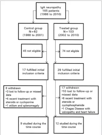
ETHODSIn this retrospective study, we reviewed the medical records of 165 patients (all at least 15 years old) with biopsy-proven IgAN from 1988 to 2010 at our Institution in order to select those who fulfil-led the inclusion criteria. We assigned the patients to groups according to the period of diagnosis: the non-treated group (control group) consisted of pa-tients diagnosed between 1988 and 2001, a period during which supportive therapy alone was used for patients with non-nephrotic proteinuria; and the treated group consisted of patients diagnosed between 2002 and 2010 and treated with steroids according to the Pozzi’s protocol.20 The supportive
therapy was applied to both groups and was based mainly on ACE-inhibitors or angiotensin receptor blockers (ARB) in order to decrease proteinuria, ar-terial blood pressure control with other anti-hyper-tensive drugs and low salt diet, and statin therapy if necessary. The study was approved by the local Research Ethics Committee and it was performed in accordance with the ethical standards laid down in the 2000 Declaration of Helsinki as well as the Declaration of Istanbul 2008.
neoplasia, active peptic-ulcer disease, viral hepati-tis, or other infectious or inflammatory diseases.
The onset of IgA nephropathy was arbitrarily defined as the first macroscopic hematuria episo-de or the episo-detection of urinalysis abnormalities by chance or edema noticed by the patient. The glo-merular filtration rate was estimated by the abbre-viated MDRD formula21 (eGFR). All of the
diag-noses of IgA nephropathy were confirmed by the
presence of typical predominant or codominant
mesangial deposits of IgA by immunofluorescence
microscopy. Histologic grading of the renal biop-sy samples was performed by a central pathologist in accordance with the classification published by Haas.22 One non-treated patient’s renal biopsy
specimen for histologic grading was unavailable. However, this 25-year-old patient had the typical mesangial IgA deposition upon immunofluorescent microscopy, recurrent macroscopic hematuria as clinical presentation of IgA nephropathy, and a se-rum creatinine concentration of 0.8 mg/dL.
Of the 17 eligible patients in the control group, nine were studied and eight were withdrawn; of the 29 eligible patients in the treated group, 12 were studied and 17 were withdrawn (Figure 1). Baseline levels of the control group were those at the time of biopsy. For the treated group, baseline levels were those at the beginning of the steroid tre-atment that was no longer than 3 months from the biopsy in 10 patients and 6 months and 36 months in the remaining two patients, respectively.
The treatment protocol was based on 1 g intra-venous methylprednisolone per day for three con-secutive days at the beginning of months 1, 3, and 5 plus 0.5 mg/kg oral prednisone on alternate days for 6 months according to Pozzi’s protocol.20
The primary objective was to assess whether the 6-month course of corticosteroids improved protei-nuria at the 6th and 12th months after the beginning
of the steroid treatment. Remission was defined by the presence of both proteinuria less than 1000 mg/24h and reduction of proteinuria to at least 50% of the baseline level. The secondary objective was to assess whether the steroid treatment preser-ved renal function (measured by serum creatinine levels and eGFR). The systolic and diastolic blood pressure and the total and relative (percentage) bo-dy weight increase from baseline to the 6th month
was evaluated.
Once the statistical tests do not have enough po-wer to assure Gaussian distribution of the samples, we used only non-parametric statistical tests. The Mann-Whitney U test was employed to evaluate the variables at the baseline. The Kruskal-Wallis test was employed to evaluate the proteinuria, serum creatinine, eGFR, and systolic and diasto-lic blood pressure during follow-up. Changes in proteinuria and eGFR over time between the two groups were also compared using Friedman test for repeated measures. Categorical variables (gender and number of remissions at different time points between the two groups) were compared using a Fisher’s exact test. Data are expressed as median and 25th and 75th percentiles. All p values were
two-tailed and p < 0.05 was considered statistically significant.
R
ESULTSAs shown in Table 1, the control and treated groups at the baseline had no statistically significant di-fferences related to age and gender distributions, duration of IgA nephropathy from clinical presen-tation to renal biopsy, systolic and diastolic blood
pressure, body weight, serum albumin concentra-tion, proteinuria, and grade of renal histology.22
However, the treated group showed a statistical tendency of increased serum creatinine levels and a statistically significant lower levels of the eGFR (p = 0.009).
The most common clinical presentation of the primary IgAN was recurrent visible hematuria in six patients of the control group (associated with upper respiratory tract infection in two patients and diarrhea in one patient) and in six patients of the treated group (also associated with upper respi-ratory tract infection in two patients and diarrhea in one patient). Other clinical presentation patterns included: microscopic hematuria associated with asymptomatic proteinuria, which was identified by chance in a urine screening of one patient in the control group and four in the treated group, follo-wed by edema in two patients in the control group, and isolated foamy urine in two patients in the tre-ated group.
Seven patients in the control group were tated with a low dose ACE-inhibitor, and two re-ceived no drugs. In the treated group, all patients were treated with either an ACE-inhibitor or ARB, five of them at low dose and seven at high dose. Only one patient was treated with an ACE-inhibitor and ARB in combination. No patient in the control group and only one patient in the trea-ted group had previously received a course of oral prednisone up to two years before beginning the current 6-month course of steroid treatment.
As shown in Table 2 and Figure 2A, the protei-nuria level in the treated group was significantly lower at the 6th and 12th months compared to
base-line, while the control group showed no statistical differences at these same time points. Proteinuria was significantly lower in the treated group when compared to control group at the 6th and the 12th
months (Table 2 and Figure 2A). Remission occur-red in nine patients of the treated group vs. none in the control group at the 6th month (p < 0.01), and
in nive patients vs. one (p < 0.01) at the 12th month
in the treated and control groups, respectively.
TABLE 1 BASELINEDEMOGRAPHICANDCLINICALFEATURESOFTHECONTROL (UNTREATED) ANDTREATED (CORTICOSTEROIDS)
GROUPSOFPATIENTSWITHPRIMARY IGA NEPHROPATHY
Clinical features Control group (n = 9) Treated group (n = 12) p
Gender (male/female) 7/2 4/8 0.085
Age (years) 32.0 (21.0; 34.0) 35.0 (30.0; 38.0) 0.188 Time from clinical presentation to biopsy (months) 36.0 (27.0; 56.0) 24.0 (7.5; 57.0) 0.372 Systolic blood pressure (mmHg) 130.0 (120.0; 140.0) 128.0 (112.5; 137.5) 0.490 Diastolic blood pressure (mmHg) 80.0 (75.0; 90.0) 80.0 (70.0; 90.0) 0.825 Body weight (kg) 75.0 (72.4; 81.25) 74.5 (63.7; 87.8) 0.859 Proteinuria (mg/24h) 1900 (1620; 3197) 1861 (1518; 2417) 0.374 Serum creatinine (mg/dL) 0.90 (0.80; 1.05) 1.20 (1.00; 1.30) 0.057 Estimated GFR* (mL/min/1.73 m2) 114.3 (72.5; 126.8) 66.2 (55.6; 76.3) 0.009
Serum albumin (g/dL) 4.1 (3.9; 4.7) 3.8 (3.5; 4.1) 0.123 Grade of histologic classification [1] 2.5 (2.0; 3.0) 2.0 (2.0; 2.7) 0.298
* Estimated GFR: glomerular filtration rate evaluated by abbreviated MDRD formula[2]; Mann-Whitney U test; data are presented as median (25th percentile; 75th percentile).
Proteinuria (mg/24h; median and 25th and
75th percentiles)
Groups Baseline 6th month 12th month
Control (n = 9)
1900 (1620; 3197)
2290 (1500; 2975)
1600 (1180; 2395) Treated
(n = 12)
1861 (1518; 2417)
703*,**
(245; 983)
684*,**
(266; 1023)
TABLE 2 PROTEINURIAOFTHECONTROL (UNTREATED) AND
TREATED (CORTICOSTEROID) GROUPSOFPATIENTS
WITHPRIMARY IGA NEPHROPATHYATBASELINE
ANDDURINGTHE 12 MONTHSOFFOLLOW-UP
* p< 0.05 versus baseline; ** p < 0.05 control versus treated groups at the same occasion; (Kruskal-Wallis test); data are presented as median (25th percentile; 75th percentile).
In the control group, serum creatinine levels were 0.90 mg/dL (0.80; 1.05 mg/dL) at baseline (Table 1) and 1.00 mg/dL (0.90; 1.10 mg/dL), and 1.20 mg/dL (0.90; 1.20 mg/dL) at the 6th and 12th
In the treated group, serum creatinine levels were 1.20 mg/dL (1.00; 1.30 mg/dL) at baseline (Table 1) and 1.15 mg/dL (0.82; 1.35 mg/dL) and 1.05 mg/ dL (0.80; 1.40 mg/dL) at the 6th and 12th months,
respectively, and no statistical difference was ob-served during the follow-up compared to baseline. Serum creatinine levels of the control vs. treated groups at the 6th and 12th months also showed no
statistical difference. Estimated GFR was lower at baseline in the treated group [66.2 mL/min/1.73 m2
(55.6; 76.3)] compared to the control group [114.3 mL/min/1.73 m2 (72.5; 126.8)] (Table 1 and Figure
2B). However, eGFR (mL/min/1.73 m2) in the
tre-ated group was 75.2 (61.5; 82.3) and 74.7 (56.7; 89.3) at 6th and 12th months, respectively, which did
not reach statistical significance (Figure 2B). The eGFR in the control group was 75.8 (58.9; 125.5) and 77.1 (55.7; 112.0) at the 6th and 12th months,
respectively, and also showed no statistically signi-ficant difference (Figure 2B). During the follow-up, eGRF did not show statistically significant differen-ces between control and treated groups at the 6th
and 12th months.
Most patients of the treated group showed good tolerance to steroid treatment after the 6-month course of steroid therapy. Two patients showed mild hypertension in the treated group, but there was no statistical difference in systolic and diastolic blood pressures between groups. Only one patient presented cushingoid facies, acne and striae rubrae, and another one presented moderate cutaneous mycosis. Most patients in the treated group pre-sented weight gain with a median of 4.55 kg (0.62;
7.12 kg), but it was not statistically different com-pared with the control group, which had a weight gain of 3.55 kg (2.75; 3.55 kg). The relative weight gain was 6.0% (3.1%; 8.7%) in the treated group and 4.6% (2.6; 4.8%) in the control group (not statistically significant).
D
ISCUSSIONIn the present study, a 6-month course of steroid treatment caused a significant reduction in the pro-teinuria levels and higher frequency of remission during the first year compared to control group. These results are similar to the original study by Pozzi et al.,20 who showed proteinuria excretion
decreases in the steroid-treated group and remains unchanged in the control group. Later, the same re-searchers demonstrated that after a median follow--up of 7 years, proteinuria remains lower in the steroid group.23 Moreover, this 6-month course of
steroid treatment protects against deterioration in renal function after 5 and 10 years of follow-up.20,23
Patients in the present study were followed-up for only 12 months, which is not long enough to detect changes in glomerular filtration rate.
Although uncommon, spontaneous remission can also happen in the non-treated patients. However, the 6-month steroid course used in the present study was much more effective to indu-ce remission. Sinindu-ce relapses are expected, other strategies must be defined. In an attempt to re-duce the loss of renal function in high-risk-adult patients with IgAN, Pozzi et al.24 added a
6-month course of azathioprine to a 6-month corticosteroid treatment. No difference between the two treatment groups was found concerning maintaining renal function or decrease in protei-nuria, and adverse effects were significantly mo-re common in the combined corticosteroid and azathioprine group.24
In this study, the treated group showed a lower estimated GFR at baseline compared to control group. However, the treated group that was expec-ted to have the worst prognosis,25-27 had a
signifi-cant reduction of proteinuria levels.
One of the limitations of this study is the sam-ple size of each group. In fact, we selected the patients available at our institution according to the inclusion and exclusion criteria. Even so, these sample sizes show very consistent results,
Figure 2. Graphics depict the (A) proteinuria levels and (B) estimated glomerular filtration rate during the 12 months of follow-up of patients with primary IgA nephropathy who were untreated (control) or treated with corticosteroids. The lines in the middle and those limiting the boxes indicate the median and 25th and 75th percentile values, respectively. Whiskers show
and we believed there were enough patients to show the beneficial effects of this treatment. Another limitation is that this is a retrospective and non-randomized study. However, prospecti-ve, randomized controlled trials to evaluate tre-atment of primary IgA nephropathy are few, and most studies have used approaches similar to the present study.27 Finally, the present study focused
on the effects during the first 12 months of treat-ment, and this time is not long enough to study the efficiency of this treatment to preserve renal function. For this effect would be necessary 5 to 10 years of follow-up.
There is a consensus that the three major in-dependent consensual risk factors predictive of progression toward CKD stage V are arterial hypertension, proteinuria with a usual cut-off over 1 g/24h, and the presence of severe lesions on initial renal biopsy such as crescents, abundant obsolescent glomeruli, segmental hyalinosis, and tubulointerstitial fibrosis.28,29 In our study, the use
of an ACE-inhibitor or ARB seemed to be more intensive in the treated group compared to the control group. This difference could have some effect on the proteinuria levels observed in this group. Indeed, a retrospective study found pa-tients followed-up from 1996 through 2006 had higher renal survival rates than those followed-up before 1995.27 This difference was attributed to
the more intensive use of the renin-angiotensin inhibitors and corticosteroid therapy in the more recently followed-up group. Using a multivariate model, it has been demonstrated that proteinuria together with high serum creatinine and histologi-cal severity at clinihistologi-cal presentation are independent risk factors.27 During analysis of another study of
Pozzi et al.,24 which reported that the addition of
azathioprine does not improve long-term renal sur-vival in patients treated with the 6-month course of corticosteroid therapy, an editorial16 considered
it a limitation of the study that fewer than half of the patients received an ACE-inhibitor or ARB at baseline. According to the 2011 Kidney Disease: Improving Global Outcomes (KDIGO) Clinical Practice Guideline for Glomerulonephritis, optimi-zation of supportive care before considering steroi-ds or other immunosupressive agents is recommen-ded unless the patient shows a rapidly progressive disease course.30 We also agree that a reduction in
proteinuria with ACE-inhibitors or ARB in renal diseases, including IgAN, improves the renal sur-vival.3,13,31 However, it has also been demonstrated
that combining corticosteroid and ACE-inhibitors is superior to ACE-inhibitors alone in preventing the progression of renal disease in proteinuric IgAN patients.32,33
Most patients in the treated group showed good tolerance to steroid treatment after a 6-month course of therapy. Even the weight gain and change in the blood pressure were not statistically higher compared to the control group. This good toleran-ce to steroids is similar to the study of Pozzi et al., which used the same corticosteroid protocol.20
The pathogenesis of IgA nephropathy has been recently reviewed,34,35 and the several cells such as
macrophages,36 myofibroblasts, and mast cells37 and
several mediators including monocyte chemoattrac-tant protein-138 and nuclear factor κ-B39 have been
shown to participate in the inflammatory response. The effects of corticosteroids on this disease remain to be clarified, but some may be related to its ac-tion on the inflammatory response and cellular signaling. One question that arises is whether it is necessary to use intravenous pulse of methylpred-nisolone or whether oral corticosteroids are suffi-cient. One study suggests that pulse steroid therapy decreases the risk of CKD stage V compared to oral steroid therapy;40 however, this study used
differ-ent schedules than Pozzi et al.20,23 and that used
in the present study. In spite of our choice to use Pozzi’s protocol, other treatment regiments with oral corticosteroids have been successful.18,19,32,33 A
recent meta-analysis concluded that steroid therapy was associated with a decrease in proteinuria and with a statistically significant reduction of the risk in CKD stage V.41 Furthermore, the 2011 Kidney
Disease: Improving Global Outcomes (KDIGO) Clinical Practice Guideline for Glomerulonephritis30
suggests that corticosteroid regimens based on ei-ther Pozzi’s protocol or a 6-month regime of oral prednisone should be used.
was well-tolerated and resulted only in mild adver-se effects in most of the patients.
C
ONFLICTS OF INTERESTThe authors declare that have no potential conflicts of interest related with the content of this study.
A
CKNOWLEDGMENTSResearch partially supported by Fundação de Apoio ao Ensino, Pesquisa e Assistência (FAEPA) do HCFMRP-USP, and by Fundação de Apoio à Pesquisa do Estado de São Paulo (FAPESP). Roberto S. Costa, Eduardo B. Coelho and Márcio Dantas were the recipients of CNPq, DF, Brazil, fellowships.
R
EFERENCES1. Maisonneuve P, Agodoa L, Gellert R, Stewart JH, Buccianti G, Lowenfels AB, et al. Distribution of primary renal diseases lea-ding to end-stage renal failure in the United States, Europe, and Australia/New Zealand: results from an international compara-tive study. Am J Kidney Dis 2000;35:157-65.
2. Gutierrez E, Zamora I, Ballarín JA, Arce Y, Jimenez S, Quere-da C, et al.; Grupo de Estudio de EnfermeQuere-dades Glomerulares de la Sociedad Española de Nefrología (GLOSEN). Long-term outcomes of IgA nephropathy presenting with minimal or no proteinuria. J Am Soc Nephrol 2012;23:1753-60.
3. Reich HN, Troyanov S, Scholey JW, Cattran DC; Toron-to Glomerulonephritis Registry. Remission of proteinuria improves prognosis in IgA nephropathy. J Am Soc Nephrol 2007;18:3177-83.
4. Bartosik LP, Lajoie G, Sugar L, Cattran DC. Predicting progres-sion in IgA nephropathy. Am J Kidney Dis 2001;38:728-35. 5. Johnston PA, Brown JS, Braumholtz DA, Davison AM.
Clinico--pathological correlations and long-term follow-up of 253 Uni-ted Kingdom patients with IgA nephropathy. A report from the MRC Glomerulonephritis Registry. Q J Med 1992;84:619-27. 6. Manno C, Strippoli GF, D’Altri C, Torres D, Rossini M, Schena
FP. A novel simpler histological classification for renal survival in IgA nephropathy: a retrospective study. Am J Kidney Dis 2007;49:763-75.
7. Rekola S, Bergstrand A, Bucht H. Deterioration of GFR in IgA nephropathy as measured by 51Cr-EDTA clearance. Kidney Int 1991;40:1050-4.
8. McGrogan A, Franssen CF, de Vries CS. The incidence of pri-mary glomerulonephritis worldwide: a systematic review of the literature. Nephrol Dial Transplant 2011;26:414-30.
9. Malafronte P, Mastroianni-Kirsztajn G, Betônico GN, Romão JE Jr., Alves MA, Carvalho MF, et al. Paulista Registry of glo-merulonephritis: 5-year data report. Nephrol Dial Transplant 2006;21:3098-105.
10. Polito MG, de Moura LA, Kirsztajn GM. An overview on fre-quency of renal biopsy diagnosis in Brazil: clinical and patholo-gical patterns based on 9,617 native kidney biopsies. Nephrol Dial Transplant 2010;25:490-6.
11. Donadio JV, Bergstralh EJ, Grande JP, Rademcher DM. Protei-nuria patterns and their association with subsequent end-stage renal disease in IgA nephropathy. Nephrol Dial Transplant 2002;17:1197-203.
12. Eiro M, Katoh T, Kuriki M, Asano K, Watanabe K, Wata-nabe T. The product of duration and amount of proteinu-ria (proteinuproteinu-ria index) is a possible marker for glomerular and tubulointerstitial damage in IgA nephropathy. Nephron 2002;90:432-41.
13. Ruggenenti P, Perna A, Gherardi G, Garini G, Zoccali C, Sal-vadori M, et al. Renoprotective properties of ACE-inhibition in non-diabetic nephropathies with non-nephrotic proteinuria. Lancet 1999;354:359-64.
14. Ballardie FW. Quantitative appraisal of treatment options for IgA nephropathy. J Am Soc Nephrol 2007;18:2806-9.
15. Eitner F, Floege J. Glomerular disease: ACEIs with or wi-thout corticosteroids in IgA nephropathy? Nat Rev Nephrol 2010;6:252-4.
16. Floege J, Eitner F. Combined immunosuppression in high-risk pa-tients with IgA nephropathy? J Am Soc Nephrol 2010;21:1604-6. 17. Katafuchi R, Ikeda K, Mizumasa T, Tanaka H, Ando T, Yanase
T, et al. Controlled, prospective trial of steroid treatment in IgA nephropathy: a limitation of low-dose prednisolone therapy. Am J Kidney Dis 2003;41:972-83.
18. Kobayashi Y, Fujii K, Hiki Y, Tateno S. Steroid therapy in IgA nephropathy: a prospective pilot study in moderate proteinuric cases. Q J Med 1986;61:935-43.
19. Kobayashi Y, Hiki Y, Kokubo T, Horii A, Tateno S. Steroid therapy during the early stage of progressive IgA nephropathy. A 10-year follow-up study. Nephron 1996;72(2):237-42. 20. Pozzi C, Bolasco PG, Fogazzi GB, Andrulli S, Altieri P,
Pontice-lli C, et al. Corticosteroids in IgA nephropathy: a randomised controlled trial. Lancet 1999;353:883-7.
21. Levey AS, Bosch JP, Lewis JB, Greene T, Rogers N, Roth D. A more accurate method to estimate glomerular filtration rate from serum creatinine: a new prediction equation. Modifica-tion of Diet in Renal Disease Study Group. Ann Intern Med 1999;130:461-70.
22. Haas M. Histologic subclassification of IgA nephropathy: a clinicopathologic study of 244 cases. Am J Kidney Dis 1997;29:829-42.
23. Pozzi C, Andrulli S, Del Vecchio L, Melis P, Fogazzi GB, Al-tieri P, et al. Corticosteroid effectiveness in IgA nephropathy: long-term results of a randomized, controlled trial. J Am Soc Nephrol 2004;15:157-63.
24. Pozzi C, Andrulli S, Pani A, Scaini P, Del Vecchio L, Fogazzi G, et al. Addition of azathioprine to corticosteroids does not benefit patients with IgA nephropathy. J Am Soc Nephrol 2010;21:1783-90.
25. Barratt J, Feehally J, Smith AC. Pathogenesis of IgA nephropa-thy. Semin Nephrol 2004;24:197-217.
26. D’Amico G. Natural history of idiopathic IgA nephropathy and factors predictive of disease outcome. Semin Nephrol 2004;24:179-96.
27. Komatsu H, Fujimoto S, Hara S, Fukuda A, Fukudome K, Yamada K, et al. Recent therapeutic strategies improve renal outcome in patients with IgA nephropathy. Am J Nephrol 2009;30:19-25.
28. Berthoux F, Mohey H, Laurent B, Mariat C, Afiani A, Thibau-din L. Predicting the risk for dialysis or death in IgA nephropa-thy. J Am Soc Nephrol 2011;22:752-61.
29. Berthoux FC, Mohey H, Afiani A. Natural history of primary IgA nephropathy. Semin Nephrol 2008;28:4-9.
30. KDIGO Clinical Practice Guideline for Glomerulonephritis. Im-munoglobulin A nephropathy. Kidney Int Suppl 2012;2:209-17. 31. Praga M, Gutiérrez E, Gonzalez E, Morales E, Hernández E.
Treatment of IgA nephropathy with ACE inhibitors: a randomi-zed and controlled trial. J Am Soc Nephrol 2003;14:1578-83. 32. Lv J, Zhang H, Chen Y, Li G, Jiang L, Singh AK, et al.
Combi-nation therapy of prednisone and ACE inhibitor versus ACE--inhibitor therapy alone in patients with IgA nephropathy: a randomized controlled trial. Am J Kidney Dis 2009;53:26-32. 33. Manno C, Torres DD, Rossini M, Pesce F, Schena FP.
Rando-mized controlled clinical trial of corticosteroids plus ACE-inhi-bitors with long-term follow-up in proteinuric IgA nephropa-thy. Nephrol Dial Transplant 2009;24:3694-701.
35. Lai KN. Pathogenesis of IgA nephropathy. Nat Rev Nephrol 2012;8:275-83.
36. Rifai A. IgA nephropathy: immune mechanisms beyond IgA mesangial deposition. Kidney Int 2007;72:239-41.
37. Silva GE, Costa RS, Ravinal RC, dos Reis MA, Dantas M, Coimbra TM. Mast cells, TGF-beta1 and alpha-SMA expres-sion in IgA nephropathy. Dis Markers 2008;24:181-90. 38. Dantas M, Romão EA, Costa RS, dos Reis MA, Vieira Neto
OM, Ribeiro RA, et al. Urinary excretion of monocyte che-moattractant protein-1: a biomarker of active tubulointerstitial damage in patients with glomerulopathies. Kidney Blood Press Res 2007;30:306-13.
39. Silva GE, Costa RS, Ravinal RC, Ramalho LZ, Dos Reis MA, Coimbra TM, et al. NF-kB expression in IgA nephropathy out-come. Dis Markers 2011;31:9-15.
40. Katafuchi R, Ninomiya T, Mizumasa T, Ikeda K, Kumagai H, Nagata M, et al. The improvement of renal survival with ste-roid pulse therapy in IgA nephropathy. Nephrol Dial Trans-plant 2008;23:3915-20.