CASE REPORT
J Vasc Bras. 2012;11(4):320-323.
Introduction
he glomus tumor is a benign familial disorder. It originates from the glomus body, is oten smaller than 3 mm and is formed by nerve, muscle and arterial components. his rare tumor accounts for less than 5% of the hand tumors and is more frequent in young women in their 20 to 40 years1. It is oten found in subungual regions, and its clinical presentation includes local hemorrhage. Its main characteristic is intense local pain. he diagnosis is made according to clinical symptoms, and only one third of the cases show radiological abnormalities, such as bone erosion2-7. he diferential diagnosis should include other
hand tumors, such as neurilemmoma, hemangioma, osteoma and schwannoma. Surgical removal results in cure and immediate pain relief.
Case report
A 58-year-old man presented with a tumor with a 4-month history, measuring about 2 cm, extremely painful, located in the dorsal region of the right hand, between the irst and second ingers over the short lexor muscle of the thumb. He denied any local trauma, exhaustive physical activity or any other probable cause of tumor formation. His pain worsened gradually, and he had an enlarged Abstract
We report on a case of glomus tumor in the branch of the radial artery of the right thumb. he tumor had a 4-year clinical history. he patient reported the development of a 2.0-cm tumor in the interdigital region between the irst and second ingers of the right hand on the short lexor muscle of thumb. he patient also complained of severe and progressive pain. Tests using bidirectional Doppler and echo-color-Doppler revealed a presumptive diagnosis of arteriovenous malformation based on the turbulence of the low and absence of stenosis. he tumor was removed by open surgery and sent for histopathological examination, which showed a diagnosis of glomangioma. he present report describes a rare arterial disease causing extreme discomfort to the patient, which may be treated with surgical resection without sequelae.
Keywords:glomus tumor; glomangioma; radial artery.
Resumo
Os autores relatam um caso de tumor glômico em ramo da artéria radial que irriga o polegar direito, com história clínica de 4 meses. O doente referia o aparecimento de tumoração de aproximadamente 2,0 centímetros na região interdigital, entre o primeiro e o segundo quirodactilos da mão direita, sobre o músculo lexor curto do polegar, extremamente dolorosa e com dor em progressão. Os exames com aparelho de Doppler bidirecional e o eco-color-Doppler apresentaram, como diagnóstico presuntivo, malformação arteriovenosa, pelo turbilhonamento do luxo e ausência de estenoses. A tumoração foi retirada por cirurgia aberta e encaminhada para exame histopatológico, com diagnóstico de glomangioma. Este relato descreve uma doença arterial pouco frequente, que causa extremo desconforto ao seu portador, mas que é solucionada pela exerese cirúrgica, sem sequelas.
Palavras-chave: tumor glômico; glomangioma; artéria radial.
Glomus tumor of digital artery of thumb – Case Report
Glomangioma da artéria digital do polegar – Relato de um caso
Ana Terezinha Guillaumon1, Carla Aparecida Faccio Bosnardo2, Luciana Rodrigues de Meirelles3
Study carried out at the first author’s vascular surgery and angiology clinic.
1 Profa. Associada Livre Docente, Chefe da Disciplina de Moléstias Vasculares Departamento de Cirurgia e Chefe do Laboratório de Microprocedimentos e Pesquisas Vasculares - Núcleo de Medicina e Cirurgia Experimental – Faculdade de Ciências Médicas (FCM) da Universidade Estadual de Campinas (UNICAMP). Coordenadora do Centro de Referência de Alta Complexidade em Cirurgia Endovascular – Hospital de Clínicas (HC) da UNICAMP.
2 Doutora em Cirurgia pela Universidade Estadual de Campinas – UNICAMP. Médica da Disciplina de Moléstias Vasculares, Departamento de Cirurgia – Hospital das Clínicas da UNICAMP. 3 Professora Assistente Doutora do Departamento de Anatomia Patológica da Faculdade de Ciências Médicas (FCM) da Universidade Estadual de Campinas (UNICAMP).
Financial support: none.
Glomus tumor - Guillaumon AT et al. J Vasc Bras 2012, Vol. 11, Nº 4 321
Operative technique and results
he patient underwent regional anesthesia of the right upper extremity using 1% lidocaine, with no vasoconstrictive agents. A skin incision was made, tissue layers were separated, and the vessels were carefully dissected. A large number of collateral veins were seen, and some had thrombosis7,8. he tumor was encapsulated in the artery, which was fully removed, and the pseudocapsule was preserved and sent to the laboratory for histopathology examination.
Immediately ater operation (24 hours), pain disappeared. At follow–up, the patient was asymptomatic and had returned to work5,9.
Histopathology
Tumor made up of polygonal cells with small and regular nuclei, some in solid groups, but others in regularly organized cell threads. Few mitotic igures were found. Dilated and congested blood capillaries proliferated in the middle of the tumor cells, containing recent thrombi and covered by a single layer of endothelial cells (Figure 2).
Discussion
A neuromyoarterial glomus and its tumor were irst described by Masson10 in 1924, and in 1972 Martorrel classiied it as a glomangioma, or glomus tumor11. It is formed by nervous ibers, muscle cells and vascular components and, for that reason, it was called glomangioma11,12.
It is a rare tumor and accounts for less than 2% of all tumors that afect sot tissues17. Its appearance and clinical history almost always involve some type of trauma. Pain tumor mass and hyperesthesia in the irst inger. He had
been unsuccessfully treated with several non-steroid anti-inlammatory drugs and powerful analgesics.
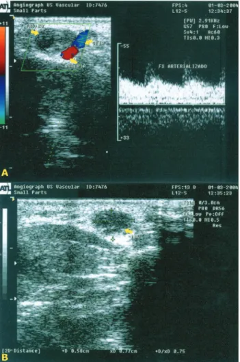
Physical examination revealed an extremely painful tumor of about 2.0 cm over the short lexor muscle of the thumb in the dorsal region of the right hand. Pulses were normal, no abnormalities were found in the right upper extremity, and there were no thrills or murmurs in the arteries. he artery of the irst inger was dilated and markedly pulsatile, which suggested an aneurism. Bidirectional Doppler scanning (Figure 1) of the radial artery of the thumb detected turbulent low. Eco-color Doppler scanning revealed the presence of an arteriovenous malformation, a large number of collateral vessels and thrombophlebitis in the dorsal vein of the thumb.
Preoperative blood counts and clotting tests were normal: erythrocytes =5.05 × 10⁶; hemoglobin = 11.1 g/dL; hematocrit = 47.30%; platelets = 198000; leukocytes = no abnormalities; TTPA = 28 seconds; and INR = 1.0.
Ultrasound HE, 400 times magniication.
Glomus tumor - Guillaumon AT et al. J Vasc Bras 2012, Vol. 11, Nº 4
322
to avoid the rupture of the capsule that contained it, the implantation of glomus cells and tumor recurrence24,in agreement with treatment data in the literature25.
References
1. Kamarashev LE, French R, Dummer K. Symplastic glomus tumor - a rare but distinct begnin histological variant with analogy to other ancient begnin skin neoplasms. J Cutan Pathol. 2009;36:1099-1102. http://dx.doi.org/10.1111/j.1600-0560.2008.01232.x
2. Ponnelle T, Gounny P, Boudghène F, et al. [Glomus tumor of the extremities]. J Mal Vasc. 1999; 24(5):364-7.
3. Wong CH, Chow L, Yen CH, Ho PC, Yip R, Hung LK. Uncommon hand tumours. Hand Surg. 2001;6:67-80. http://dx.doi.org/10.1142/ S0218810401000564
4. Robbins SL, Cotran RS, Kumar V. Patologia estrutural e functional. Vasos sanguíneos. Rio de Janeiro: Guanabara; 1986. p. 522-3.
5. Pulitzer DR, Martin PC, Reed RJ. Epithelloid glomus tumor. Hum Pathol. 1995;26:1022-7. http://dx.doi.org/10.1016/0046-8177(95)90093-4
6. Debol SM, Stanley MW, Mallery S, Sawinski E, Bardales RH. Glomus tumor of the stomach: cytologic diagnosis by endoscopic ultrasound-guided ineneedle aspiration. Diagn Cytopathol. 2003;28:316-21. http://dx.doi.org/10.1002/dc.10294
7. Mentzel T, Hugel H, Kutzner H. CD34- positive glomus tumor: clinicopathologic and immunohistochemical analysis of six cases with myxoid stromal changes. J Cutan Pathol. 2002;29:421-5. http:// dx.doi.org/10.1034/j.1600-0560.2002.290706.x
8. Keefer CJ, Brantley B, DeLozier JB. Familial iniltrative glomangiomas: dignosis and treatment. J Craniofac Sur. 1996;7:145-7. http://dx.doi. org/10.1097/00001665-199603000-00014
9. Kim SW, Jung SN. Glomus tumour within digital nerve: A case report. J Plast Reconstr Aesthet Surg. 2011;64:958-60. http://dx.doi. org/10.1016/j.bjps.2010.11.012
10. Masson P. Le glomus neuro-mio-artériel dés régions tactiles et sés tumeurs. Lyon Chirurgical. 1924;21:257.
11. Martorrel F. Angiologia – Enfermidades Vasculares. Glomangiomas e tumores glômicos. Santiago de Chile: Salvat Editores S/A; 1972. p. 470-477.
12. Bayley OT. he cutaneus glomus and its tumors-glomangiomas. Am J Pathol. 1935;11:915. Pmid:19970241. PMCid:1910995.
13. Chong Y, Eom M, Min HJ, Kim S, Chung YK, Lee KG. Symplastic glomus tumor: a case report. Am J Dermatopathol. 2009;31:71-3. http://dx.doi.org/10.1097/DAD.0b013e31818d3ac7
14. Fujioka H, Kokubu T, Akusue T, et al. Treatment of subungual glomus tumor. Kobe J Med Sci. 2009;55:E1-E4.
15. Monacelli G, Cascioli I, Mongardini M, Corelli S, Cigna E, Spagnoli AM. [Glomus tumor and neovascular syndrome of the arm: a clinical case]. G Chir. 2003;24:235-8. [Article in Italian]. PMid:14569920.
16. Kamano M, Kazuki K. Glomangioma arising from the supericial palmar arch in the hand: case report. Clin Orthop Relat Res. 2004;(419):162-4. http://dx.doi.org/10.1097/00003086-200402000-00026
is reported by all patients. In the beginning, pain is mild, but later becomes intense and unbearable. here is some regularity in time between pain episodes, but, as the disease progresses, pain frequency increases and the simple touch of clothing is oten enough to trigger a crisis. Pain usually occurs at night, which makes it impossible for the patient to sleep. One of the hypotheses to explain pain intensity is based on tumor expansion, that is, as it is contained in a pseudocapsule, its growth is restricted, which leads to the necrosis of the central cells and their replacement by connective tissue. Another hypothesis to justify pain is tumor site, as it arises in the myoneural junction13,15,16.
he region most frequently afected by an extremity glomangioma is the dermis, the subcutaneous cell tissue, particularly in the nail bed17, because there is little resistance to its development there, and not in small-caliber arteries, as Kamarashev et al1 used to believe. hese tumors may also be found in other sites, such as the stomach, knees, shoulder and mediastinum, as well as in the middle ear, where it may lead to serious balance disorders and hearing loss. hey are benign tumors with a well deined oval shape, and they are never larger than 5 mm, regardless of the region where they arise or its progression time11,18,19. When it is subungual and does not have space to grow, it loses its oval shape and may cause bone erosion, which may give the false impression of a malignant iniltrating tumor18.
Imaging studies, such as arteriography and echo-color Doppler ultrasound, are useful to evaluate glomangiomas of the extremities, particularly to make the diferential diagnosis with other tumors18-21, but images are not conclusive.
Its diagnosis, in general, is made early on due to pain, but studies in the literature describe cases that had a history of 40 years. Some authors suggest that cell atypia is a result of the accumulation of heterochromatin associated with DNA inactivation. Moreover, the increased capillarity, the local architecture in the arrangement of tumor cells with uniform sizes and forms, and the uniformity of base cells suggest the diagnosis of glomus tumor. From the moment the diagnosis of a glomangioma is made, no other case of such tumor goes unnoticed by the examiner because of its particular characteristics.
Glomus tumor - Guillaumon AT et al. J Vasc Bras 2012, Vol. 11, Nº 4 323
24. Dahlin LB, Besjakov J, Veress B. Scand A glomus tumour: classic signs without magnetic resonance imaging indings. J Plast Reconstr Surg Hand Surg. 2005;39:123-5. PMid:16019743. http:// dx.doi.org/10.1080/02844310410002993
25. Falleti J, Vita G, De Cecio R, et al. Symplatic glomus tumor: Report of a chalenging lesion with literature review. Pathol Res Pract. 2012;208:372-375. PMid:22572036. http://dx.doi. org/10.1016/j.prp.2012.04.001
Correspondence
Ana Terezinha Guillaumon Rua Hermantino Coelho, 901/11 CEP 13087-500 – Campinas (SP), Brazil Fone: 55 (19) 3296-1986 Fax: 55 (19) 3521-7442 E-mail: [email protected]
Authors’ contributions
Conception and design: ATG Analysis and interpretation: ATG, CAFB, LRM Data collection: ATG Writing the article: ATG, CAFB, LRM Critical revision of the article: ATG Final approval of the article*: ATG, LRM, CAFB Statistical analysis: none. Overall responsibility: ATG *All authors have read and approved the inal version submitted to J Vasc Bras.
17. Fletcher CDM, Unni KK, Mertens F. WHO classiication of tumors in: Pahology and Genetics of Tumors of Soft tissue and bone. Lyon: IARC Press; 2002.
18. Tsuneyosshi M, Enjoji M. Glomus Tumor: a clinicopathologic and electron microscopic study. Cancer. 1982;50:1601-7. http:// dx.doi.org/10.1002/1097-0142(19821015)50:8%3C1601::AID-CNCR2820500823%3E3.0.CO;2-5
19. De Candia A, Como G, Passon P, Pedace E, Bazzocchi M. Sonographic indings in glomus tynpanicum tumor. J Clin Ultrasound. 2002;30:236-40. http://dx.doi.org/10.1002/jcu.10058
20. Vogl TJ, Juergens M, Balzer JO, et al. Glomus tumors of the skull base: combined use of MR angiography and spin-echo imaging. Radiology. 1994;192:103-10. PMid:8208919.
21. Arnold SM, Strecker R, Schefer K, et al. Dynamic contrast enhancement of paragangliomas of head and neck: evaluation with time-resolved 2D MR projection angiography. Eur Radiol. 2003;13:1608-11. http://dx.doi.org/10.1007/s00330-002-1717-3
22. Sharma JK, Miller R. Treatment of multiple glomangioma with tuneable dye laser. J Cutan Med Surg1999;3:167-8. PMid:10082598.