239 1. Cardiovascular Surgery Specialist. Cardiovascular Surgeon of the
Hospital dos Fornecedores de Cana de Piracicaba (HFC). 2. PhD. Full Professor of FAMERP e UNICAMP; Director of the
Postgraduation Department of FAMERP. Editor of the Brazilian Journal of Cardiovascular Surgery.
3. Cardiovascular Surgery Specialist (BSCVC). Cardiovascular Surgeon of the HFC.
4. Cardiovascular Surgery Specialist. Cardiovascular Surgeon of the HFC; Professor of the Cardiovascular Surgery and Cardiology Department of FAMERP; Cardiovascular Physician Surgeon (FAMERP).
Marcos Rogério JOAQUIM1,Domingo Marcolino BRAILE2,Marcus Vinícius Ferraz de ARRUDA3, Marcelo José Ferreira SOARES4
Rev Bras Cir Cardiovasc 2009; 24(2): 239-241 CASE REPORT
RBCCV 44205-1082
Ressecção de um lipoma de átrio direito e reconstrução parcial com pericárdio bovino
Right atrial lipoma resection and partial
reconstruction using bovine pericardium
Abstract
The primary heart tumors have an incidence varying from 0.001% to 0.28% of all tumors, reported in necropsies series. Lipoma consists of a benign tumour corresponding to about 8% of all primary heart tumors. When present, the symptoms are related to the size and location of such tumor. We report a case of a 27-year-old man with a lipoma in the right atrium who underwent surgical treatment with tumor resection and partial reconstruction of the right atrium using bovine pericardium.
Descriptors: Lipoma. Heart neoplasms. Heart atria/
surgery.
Resumo
Os tumores primários do coração têm uma incidência variando de 0,001% a 0,28% dentre todos os tumores, relatados em séries de necropsias. Lipoma consiste de um tumor benigno que corresponde a cerca de 8% de todos os tumores cardíacos primários. Os sintomas, quando presentes, estão relacionados ao tamanho e à localização do tumor. Apresentamos o caso de um homem com 27 anos, com um lipoma no átrio direito que foi submetido a tratamento cirúrgico, com ressecção tumoral e reconstrução parcial do átrio direito com pericárdio bovino.
Descritores: Lipoma. Neoplasias cardíacas. Átrios do
coração/cirurgia.
This study was carried out at the Hospital dos Fornecedores de Cana de Piracicaba (HFCP), Piracicaba, SP, Brazil.
Correspondence address: Marcos Rogério Joaquim.
Avenida Barão de Valença, 716 - 3º andar – Piracicaba, SP, Brazil. CEP 13405-126.
E-mail: [email protected]
Article received on October 8th, 2008 Article accepted on February 9th, 2009 INTRODUCTION
Cardiac lipomas are rare with incidence of 8.4% among all primary tumors [1]. In turn, the secondary tumors are 30 to 40 times more frequent than the primary ones [2].
Lipoma is composed of fat cells, originated from the epimyocardium and can attach both to the fibrous tissue (pericardium), as the muscle tissue (subepicardial) [1].
The clinical changes are generally mechanical and result of the location of the tumor, and this may cause valve
dysfunction or heart compression, thus compromising the cardiac dynamic.
The therapy indicated for the treatment of primary tumors of the heart is the total resection of the tumor in order to avoid primary and secondary complications, which are the obstruction and embolization [3].
The first report of success in resection of epicardial lipoma occurred in 1954 by Maurer et al. [4].
240
JOAQUIM, MR ET AL - Right atrial lipoma resection and partial reconstruction using bovine pericardium
Rev Bras Cir Cardiovasc 2009; 24(2): 239-241
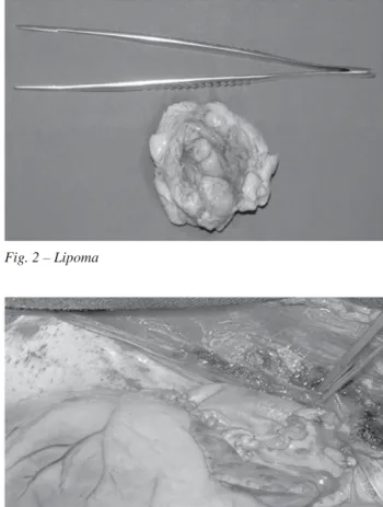
The pathological examination confirmed it was fibrolipoma of 5.5 x 5.3 cm, encapsulated with striated muscle tissues and soft tissues, with proliferation of mature adipose tissue and the presence of fibrous tissue bundles. The postoperative period progressed without complications (Figure 4), and the patient was discharged from hospital on the 7th day, under use of oral anticoagulant due to the right atrial repair. performed and revealed a mass in the right atrium – or for
clearing. The patient underwent resection of this mass, with histopathological diagnosis of fibrolipoma.
CASE REPORT
27-year-old male patient, born in Americana, SP. Asymptomatic patient until 7 years ago, when he present progressive fatigue associated with episodes of tachycardia. The patient sought a medical service of his city and, on that occasion, echocardiogram was requested, which showed mass in right atrium, measuring around 4.5 x 3.8 cm in diameter and also a Holter monitoring (24-hour), which showed ventricular and supraventricular extrasystoles and non-sustainable paroxysmal atrial tachycardia.
The patient was referred to reference service and remained without resolution of the case until early 2007, when he presented two syncopal episodes: the first at rest and the second driving his vehicle.
The patient sought our service, where he underwent a new echocardiogram, confirming the presence of a mass with increased echotexture and fixed on the right atrial wall, in its mid-basal portion with a diameter of about 3.5 cm (Figure 1).
Surgical treatment was indicated. The surgery was performed through a median sternotomy with aorto-bicaval cardiopulmonary bypass, antegrade and retrograde normothermic blood cardioplegia, incision in the right atrium, visualization of mass adhered to the atrial side wall. Tumor resection (Figure 2) was performed, in addition to the partial withdrawal of the atrial wall on which the tumor was adhered, respecting the safety margin of 0.5 cm from the pedicle. Partial reconstruction of the right atrium was also performed using bovine pericardium (Figure 3).
Fig. 1 – Preoperative echocardiogram with visualization of mass in the right atrium
Fig. 2 – Lipoma
Fig. 3 – Partial reconstruction of the right atrium using pericardium
241
REFERENCES
1. Silveira WL, Nery WM, Soares ECG, Leite AF, Nazzetta H, Batista MAL, et al. Lipoma de átrio direito. Arq Bras Cardiol. 2001;77(4):361-4.
2. Heath D. Pathology of cardiac tumors. Am J Cardiol. 1968;21(3):315-27.
3. Fernandes F, Soufen HN, Lanni BM, Arteaga E, Ramires FJA, Mady C. Neoplasias primárias do coração. Apresentação clínica e histológica de 50 casos. Arq Bras Cardiol. 2001;76(3):231-4.
4. Silverman NA. Primary cardiac tumors. Ann Surg. 1980;191(2):127-38.
5. Lima I PRL, Crotti II PLR. Tumores cardíacos malignos. Rev Bras Cir Cardiovasc. 2004;19(1):64-73.
6. Lion MF, Moreira AELC, Silva MVB. Inversão de aurícula esquerda por lipoma epicárdico. Comportamento atípico de um tumor levando a obstrução de veias pulmonares. Arq Bras Cardiol. 1994;62(3):207-10.
7. Arslan S, Gundogdu F, Acikel M, Kantarci AM. Asymptomatic cardiac lipoma originating from the interventricular septum diagnosed by multi-slice computed tomography. Int J Cardiovasc Imaging. 2007;23(2):277-9.
8. Mousseaux E, Idy-Peretti I, Bittoun J, Diebold B, Paulylaubry C, Carpentier A, et al. MR tissue characterization of a right atrial mass: diagnosis of a lipoma. J Comput Assist Tomogr. 1992;16(1):148-51.
9. Rubino M, Hamad AM, Rea F, Gerosa G. Reconstruction of the right atrium with pulmonary artery homograft after resection of right atrial lipomatosis. Interact Cardiovasc Thorac Surg. 2007;6(6):826-7.
DISCUSSION
Among the benign tumors, myxomas are the most frequent. There is a primary tumor of the heart for every 5,000 non-selected autopsies. The incidence of tumors of the heart is less than 0.2% among the tumors that can be found in the body. Despite this fact, myxomas represent approximately half of benign tumors of the heart [5].
Lipomas were first described by Orth in 1886 [6], and the first resection of an epicardial lipoma was successfully performed in 1954 by Maurer et al. [1].
Lipoma is composed of fat cells originated from the epimyocardium, with macroscopic appearance similar to that observed in other areas, promoting, in its turn, adherence to the fibrous connective tissue (pericardium) and muscular tissue (subepicardium). It presents forms of lipomatous degeneration and also fat necrosis and calcification. About 50% of these tumors are of subendocardial origin, and 25%, intramyocardial, most commonly affecting the left ventricle, right atrium and interatrial septum – in this sequence of events - and the remaining 25% in the pericardium [1].
The intracardiac tumors may provoke disorders of atrioventricular or intraventricular conduction, which are manifested by arrhythmias, interfering in the cardiac dynamic, leading to sudden death [1,7]. Many cases are data from autopsy findings, since the disease progressed asymptomatic in some cases, resulting from size and location of the tumors [6].
Recent diagnostic methods positively lead to early detection of these cardiac disorders. Tomography, magnetic resonance and transesophageal echocardiography provide a reliable noninvasive diagnosis in relation to the location of the tumor [8].
Careful maneuver of the heart and tumor structures during removal of the tumor reduces the possibility of fragmentation and the occurrence of embolic phenomena [3.9]. The resection of the implantation basis of the pedicle should be performed with small safety margin to prevent recurrence [8].
The diagnosis of tumors of the heart should be quick and accurate because they can progress silently and cause valvular dysfunction, arrhythmias and cardiac compression.
Once established the diagnosis of presence of cardiac mass, the patient should be referred immediately to a specialized center, and surgical resection should be indicated, aiming at histopathological diagnosis and elimination of the risk of cardiac compression and embolic accidents.
JOAQUIM, MR ET AL - Right atrial lipoma resection and partial reconstruction using bovine pericardium