Independent DNA Interstrand Crosslink Repair in Yeast
Thomas A. Ward1., Zuzana Duda´sˇova´2., Sovan Sarkar1
, Mangesh R. Bhide3, Danusˇa Vlasa´kova´2, Miroslav Chovanec2, Peter J. McHugh1*
1Department of Oncology, Weatherall Institute of Molecular Medicine, University of Oxford, John Radcliffe Hospital, Oxford, United Kingdom,2Laboratory of Molecular Genetics, Cancer Research Institute, Bratislava, Slovak Republic,3Department of Microbiology and Immunology, University of Veterinary Medicine, Kosˇice, Slovak Republic
Abstract
Fanconi anemia (FA) is a devastating genetic disease, associated with genomic instability and defects in DNA interstrand cross-link (ICL) repair. The FA repair pathway is not thought to be conserved in budding yeast, and although the yeast Mph1 helicase is a putative homolog of human FANCM, yeast cells disrupted forMPH1are not sensitive to ICLs. Here, we reveal a key role for Mph1 in ICL repair when the Pso2 exonuclease is inactivated. We find that the yeast FANCM ortholog Mph1 physically and functionally interacts with Mgm101, a protein previously implicated in mitochondrial DNA repair, and the MutSamismatch repair factor (Msh2-Msh6). Co-disruption ofMPH1,MGM101,MSH6,orMSH2withPSO2produces a lesion-specific increase in ICL sensitivity, the elevation of induced chromosomal rearrangements, and persistence of ICL-associated DNA double-strand breaks. We find that Mph1-Mgm101-MutSadirects the ICL-induced recruitment of Exo1 to chromatin, and we propose that Exo1 is an alternative 59-39 exonuclease utilised for ICL repair in the absence of Pso2. Moreover, ICL-induced Rad51 chromatin loading is delayed when both Pso2 and components of the Mph1-Mgm101-MutSa
and Exo1 pathway are inactivated, demonstrating that the homologous recombination stages of ICL repair are inhibited. Finally, the FANCJ- and FANCP-related factors Chl1 and Slx4, respectively, are also components of the genetic pathway controlled by Mph1-Mgm101-MutSa. Together this suggests that a prototypical FA–related ICL repair pathway operates in budding yeast, which acts redundantly with the pathway controlled by Pso2, and is required for the targeting of Exo1 to chromatin to execute ICL repair.
Citation:Ward TA, Duda´sˇova´ Z, Sarkar S, Bhide MR, Vlasa´kova´ D, et al. (2012) Components of a Fanconi-Like Pathway Control Pso2-Independent DNA Interstrand Crosslink Repair in Yeast. PLoS Genet 8(8): e1002884. doi:10.1371/journal.pgen.1002884
Editor:Gregory P. Copenhaver, The University of North Carolina at Chapel Hill, United States of America ReceivedDecember 20, 2011;AcceptedJune 22, 2012;PublishedAugust 9, 2012
Copyright:ß2012 Ward et al. This is an open-access article distributed under the terms of the Creative Commons Attribution License, which permits unrestricted use, distribution, and reproduction in any medium, provided the original author and source are credited.
Funding:Work in the laboratory of MC is supported by the VEGA Grant Agency of the Slovak Republic (grant no. 2/0165/09) (http://alipro.eurescom.de/page/et/ sk_prg_vega) and by the project TRANSMED, part of the Research and Development Operational Programme funded by the European Regional Development Fund. ZD was supported by European Social Fund (http://ec.europa.eu/esf/home.jsp?langId = en) – project code 13120200038. Work performed in the laboratory of PJM was supported by Cancer Research UK Programme Grant CA9047/A10111 (http://science.cancerresearchuk.org/). MRB was funded by the project INFEKTZOON (ITMS code – 26220120002) (http://infektzoon.uvm.sk/). The funders had no role in study design, data collection and analysis, decision to publish, or preparation of the manuscript.
Competing Interests:The authors have declared that no competing interests exist. * E-mail: [email protected]
.These authors contributed equally to this work.
Introduction
DNA interstrand cross-links (ICLs) represent an extremely toxic form of genomic damage; a consequence of their ability to inhibit basic cellular processes such as transcription and replication [1–3]. Eukaryotic ICL repair remains relatively poorly understood, although an initial ICL incision step followed by homologous recombination repair (HRR) and translesion DNA synthesis (TLS) steps have been implicated by genetic studies [4–7]. Molecular evidence for some of these events has been provided, although many mechanistic details remain obscure [8–11]. Patients suffering from the inherited condition Fanconi anemia (FA) are defective in ICL repair, where ICL incision and the subsequent TLS and HRR stages are all affected [12–14]. This repair defect is associated with bone marrow failure and a predisposition to haematological and solid malignancies [15]. Despite some differences in ICL processing between yeast and human cells, most notably the absence of readily identifiable homologs of many FA factors, yeast does possess a putative ortholog (Mph1) of the
protein mutated in FA complementation group M (FANCM) [16,17], indicating that yeast could be a powerful model for understanding some aspects of the repair defect in FA. Strikingly, however, and in contrast to higher eukaryoticfancmmutants, yeast mph1cells are not significantly ICL sensitive.
and is able to unwind a number of substrates including model replication forks, D-loops and Holliday junctions [21,25]. Recent studies demonstrated that the entire helicase domain of human FANCM and ATP binding by the Walker A motif is required for normal resistance to ICLs [26], although mutation of the Walker B motif suggests that ICL repair is not dependent upon functional helicase activity [27]. Finally, FANCM has a role in the ATR-mediated replication checkpoint in vertebrate cells [28–30].
Like human FA cells, budding yeastpso2mutants (originally also known assnm1) are highly sensitive to ICLs, but not to other forms of DNA damage [31–33]. Several groups have concluded that the initial unhooking incisions at ICLs are normal in pso2 mutants [34–36]. However, ICL-associated DNA double-strand breaks (DSBs) accumulate in pso2 cells [35–38]. It was subsequently demonstrated that such DSBs are the result of replication fork cleavage when an ICL stalls replication [6,11,39–43]. Recent studies confirm that a human homolog of Pso2, hSNM1A, also plays a key role in ICL repair during S-phase [43]. Finally, several lines of evidence also point to a role for mismatch repair (MMR) factors in ICL repair [44–46]. In budding yeast, Msh2 and Exo1 appear to play a role that is redundant with Pso2 in ICL repair during S-phase [38], although the basis for this remains unknown. The identification of novel Pso2- and Mph1-interacting factors will be critical to an improved understanding of ICL repair. Here, we demonstrate that Mgm101, a factor hitherto thought to only repair mitochondrial DNA, interacts with both Mph1 and MutSa (Msh2-Msh6). Our subsequent analysis reveals a critical role for Mph1-MutSa-Mgm101 and Exo1, as well as the FANCJ- and FANCP-related factors Chl1 and Slx4, respectively, in the Pso2-independent repair of ICLs, suggestive of an FA-like repair pathway in budding yeast.
Results
Mgm101 interacts with Mph1 and Msh2-Msh6
Large-scale protein-interactome studies suggested interactions between Pso2 and the Mgm101 protein [47]. Mgm101 was
originally identified as a factor required for the maintenance of the mitochondrial genome [48], and is a component of the mitochondrial nucleoid, where it co-localises and interacts with the mitochondrial genome maintenance factor, Mmm1 [49,50]. In addition to genome transmission, Mgm101 might be required for the repair of oxidative DNA lesions in mitochondria [49], as well as HR in mitochondria [51]. Outside of fungi and marine invertebrates Mgm101 is not clearly conserved in higher eukaryotes, and it has recently been noticed that Mgm101 has some similarity to Rad52 [51,52]. Our extensive yeast two-hybrid and co-immunoprecipitation studies failed to produce evidence of an interaction between Pso2 and Mgm101 (data not shown). However, when we employed FLAG-tagged Mgm101, separated Mgm101-FLAG associated proteins by liquid chromatography (LC) and determined their identity by MALDI-TOF mass-spectrometry (Figure 1A), three nuclear DNA repair proteins, Mph1, Msh2, Msh6 as well as Fkh1, the nucleo-cytoplasmic factor Lsm7 and a mitochondrial factor Mrp51 were identified.
We confirmed this pattern of interaction with the three DNA repair factors by performing reciprocal immunoprecipitations using C-terminally FLAG-tagged Mgm101 introduced into strains with C-terminal chromosomally 6xHis-tagged Msh2, Msh6 or Mph1. By incubating cell extracts with anti-FLAG coated agarose or nickel resin to immunoprecipitate the FLAG- and 6xHIS-tagged proteins, respectively, reciprocal interactions between Mgm101 and Msh2, Msh6 and Mph1 were all confirmed (Figure 1B). Note that in all immunoprecipitation experiments the cell extracts were treated with a nuclease mix, excluding the possibility that the interactions observed are mediatedviaDNA. To further explore the possibility of a complex containing the identified Mgm101-interacting factors, we tagged each factor with a unique epitope in a single strain and performed Superdex 200 gel filtration analysis, followed by immunoblotting to establish the elution profile of each factor. Consistent with recently published data suggesting that Mgm101 multimerises efficiently [51], much of the Mgm101 was found in fractions peaking at around 160 kDa (fractions 12–16). The major elution peaks for Msh2 and Msh6 were around 250 kDa (fractions 10–11), consistent with the majority of the cellular pool of these factors existing together in the MutSacomplex [53]. The presence of all these factors in a co-eluting high molecular weight fraction (.670 kDa, fractions 4–5) was also observed, consistent with a sub-population of each of these proteins residing in a high molecular weight complex with the same elution characteristics.
Since Msh2-Msh6 and Mph1 are primarily nuclear factors, our interaction results suggested that Mgm101 could, therefore, reside in the nucleus as well as the mitochondrial nucleoid [49]. Cellular fractionation of the nuclear and mitochondrial compartments confirmed that Mgm101-FLAG was present in both fractions, with enrichment in both the nucleus and mitochondria compared to levels in whole cell extracts (Figure S1A).
Mph1, MutSa, and Mgm101 are required for nuclear ICL repair in the absence of Pso2
When strains disrupted forPSO2,MSH2,MPH1andMGM101 were tested for sensitivity to a range of DNA damaging agents, including hydrogen peroxide (H2O2), ionising radiation (IR),
methyl methanesulfonate (MMS) and ultraviolet light (UV), no significant sensitivity was apparent (Figure S2, panels A–F and J– O). Moreover, none of the mph1, msh2, msh6 or mgm101 single mutants demonstrated any sensitivity to the cross-linking drug nitrogen mustard (HN2) (Figure 2A, B and D). Since Mgm101 interacts with both Mph1 and Msh2-Msh6, and our previous studies demonstrated a functional overlap between Pso2 and Msh2 Author Summary
Individuals with Fanconi anemia (FA) suffer from bone marrow failure and from elevated rates of haematological and solid malignancy. Moreover, FA patients exhibit extreme sensitivity to DNA interstrand cross-links (ICLs), but not other forms of DNA damage. Despite recent progress in identifying and characterising FA factors, little is known about the mechanistic basis of the ICL repair defect in FA. The identification and characterisation of FA– like pathways in simple model eukaryotes, amenable to genetic dissection, would clearly accelerate progress. Here, we have identified an ICL repair pathway in budding yeast that has significant similarities to the FA pathway and that acts in parallel to an established pathway controlled by the Pso2 exonuclease. We have discovered that a key component of this pathway, the FANCM-like helicase, Mph1, interacts and collaborates with a mismatch repair factor (MutSa) and a novel nuclear DNA repair factor Mgm101 to control ICL repair. We also found that a central role of these factors is to recruit Exonuclease 1 (Exo1) to ICL-damaged chromatin, and propose that this factor acts redundantly with Pso2 to execute the exonucleolytic processing of ICLs. Our findings reveal new mechanistic insights into the control of ICL repair by FA–like proteins in an important model organism.
in ICL repair [38], we deletedMGM101,MSH6andMPH1in a pso2mutant background. This revealed an approximately ten-fold increase in HN2 sensitivity for thepso2 mph1,pso2 mgm101andpso2 msh6 strains over the pso2 single mutant at doses over 10 mM (Figure 2A, B and D). This sensitisation to ICLs is highly specific, since it was also observed for another cross-linking drug cisplatin (CDDP, Figure S2G–I), but not other forms of DNA damage including those induced by H2O2, IR, UV or MMS treatment
(Figure S2A–F and J–O). Since Mgm101 and Mph1 phy-sically interact, and show an indistinguishable phenotype upon
co-deletion withPSO2, we asked whether they were epistatic for ICL repair. Creation of a pso2 mgm101 mph1 triple mutant confirmed this to be the case (Figure 2C).
The genetic relationship between pso2 andmph1/mgm101is strongly reminiscent of our previous observations, in a different strain background (W303), of the relationship between pso2 and msh2 double disruptant (recapitulated in the genetic background used in these studies, RDKY3615, Figure 2D and Figure S2G) [38]. It is clear (Figure 2E) that thepso2 msh2 mgm101andpso2 msh2 mph1triple disruptants show equivalent Figure 1. Mgm101 interacts with Mph1 and MutSa.(A) LC-MS. Prominent peaks from LC analysis with intensity above 400 were subjected for MALDI-TOF analysis. (B) Co-IPs. Reciprocal immunoprecipitations using FLAG-tagged Mgm101 expressed in cells with chromosomally 6xHis-tagged Msh2, Msh6 or Mph1. The empty vector transformants (wild type) were used as controls. A portion of the lysates (1%) was immunoblotted to show input proteins (left-hand panel). (C) The elution profiles of Mgm101-Myc, Mph1-FLAG and Msh6-HA as well as Msh2 following Superdex 200 chromatography reveal coincident elution in a high molecular weight complex at around 670 kDa, suggesting that they associate in a high molecular-weight complex, marked with an asterisk.
sensitivity to both their double disruptant parents, confirming that Msh2, Mph1 and Mgm101 act in a common pathway during ICL repair, which is redundant with the pathway controlled by Pso2. Consistent with this, co-deletion ofMSH2 and MGM101 or MSH2 and MPH1, in strains where PSO2 remains functional, does not result in increased HN2 sensitivity (data not shown).
As studies of human FANCM have revealed a critical role for the helicase/translocase domain for normal resistance to ICLs, we also determined whether the helicase domain of Mph1 is required for its ICL repair role. Comparing the HN2 sensitivity ofpso2 mph1 strain to that deleted for PSO2 and harbouring an amino acid substitution at a critical lysine (K113Q) predicted to prevent ATP
binding in the Walker type A box of the helicase, revealed that an intact helicase domain is required for Mph1 to protect against ICLs in apso2background (Figure 2F).
No role for Pso2, Mph1, or Msh2 in mitochondrial genome maintenance
We also wished to explore the possibility that Mph1, Msh2 or Pso2 play some role in mitochondrial genome maintenance, like Mgm101. We examined the rate of loss of mitochondrial function in msh2, mph1 and pso2 mutants, measured as spontaneous petite mutant formation [54]. This was determined by scoring ability to form colonies on non-fermentable (glycerol-containing) media after multiple generations of growth under Figure 2. Pso2 and Mph1-Mgm101-MutSacontrol redundant ICL repair pathways.(A to G) Analysis of the ICL sensitivity of combinations ofPSO2withMPH1,MGM101,MSH6andMSH2gene disruptions, and anmph1K113Qmutant. The ICL-inducing agent was HN2. All results are the mean of at least three independent experiments and the error bars show the standard error of the mean.
doi:10.1371/journal.pgen.1002884.g002
non-selective conditions (Figure S1B). We found thatpso2 and mph1 cells did not exhibit any increased spontaneous loss of functional mitochondria, nor did msh2 mutants, as previously reported [55]. Moreover, the loss of mitochondria inpso2,mph1 andmsh2cells was not increased by treatment with HN2 (Figure S1C) or the oxidative agent H2O2 (Figure S1D). Together our
data indicate that Pso2, Mph1 and Msh2 are dispensable for mitochondrial genome stability, even under conditions of stress.
Gross chromosomal rearrangements are increased in pso2 msh2,pso2 mph1,andpso2 mgm101mutants following ICL induction
Many genes that are required for the maintenance of genome stability act to suppress the induction of gross chromosomal rearrangements (GCRs) [56], and the FA factors play such a role in mammalian cells. We therefore examined the rate of GCRs in our pso2, msh2, mph1 and mgm101 single- and double-mutants, using an assay that detects large interstitial deletions, transloca-tions, chromosome fusions and loss of an entire chromosome arm [56,57] (Figure 3A). Compared to wild type parent, the pso2 mutant showed an increase of 6-fold in the rate of GCRs (Figure 3A). This was not increased by treatment with 10 mM HN2, which induces negligible lethality in these strains. Consistent with previously published data [58], an msh2 single mutant demonstrated an 10-fold increase in spontaneous GCRs, but this, again, was not increased by ICL induction. However, in apso2 msh2double disruptant, while the rate of spontaneous GCRs was similar to that of themsh2single mutant, the rate of GCRs was enhanced by treatment with HN2. For cells disrupted for MGM101, no increase in the rate of GCR over wild type was observed for either spontaneous or ICL-induced conditions. Deletion of PSO2 in mgm101 cells increased the level of spontaneous GCRs to a level similar to that of the pso2 single mutant. Strikingly, like the pso2 msh2 double mutant, the pso2 mgm101 double disruptant exhibited an increase in GCRs following ICL induction. As previously noted, deletion ofMPH1 alone produces a modest elevation in spontaneous GCRs [59]. Once again, while co-deletion ofPSO2andMPH1did not further affect the levels of spontaneous GCRs, HN2-induced GCRs were significantly elevated inpso2 mph1cells. Notably, this increase was greater than that seen in pso2 msh2 and pso2 mgm101 double mutants, suggesting that Mph1 might further suppress ICL-induced GCRs by pathway(s) additional to that involving Mgm101 and Msh2. Together, our results suggest that Pso2, Msh2 and Mph1 all suppress spontaneous GCRs, but that Mgm101 is not required. In contrast, the GCRs induced by ICLs are suppressed by two redundant pathways, controlled by Pso2 and Mph1-Mgm101-Msh2, respectively.
Mgm101, Mph1, and Msh2 are required for the efficient repair of ICL-associated DSBs in the absence of Pso2
A consequence ofPSO2disruption is the accumulation of ICL-associated DSBs [38], and it is likely that the HN2-induced GCRs observed inpso2 mph1/msh2/mgm101double mutant cells are the result of aberrant repair of ICL-associated DSBs. We therefore determined whether loss ofMGM101andMSH2inpso2cells also affects ICL-associated DSB induction and repair. Pulsed-field gel electrophoresis (PFGE) analysis of whole chromosomes prepared from cells treated with 100 mM HN2 showed that, as previously demonstrated [38], few DSBs accumulate in a wild type strain at this dose (Figure 3B). In pso2 mutants there is a modest accumulation of DSBs at this dose, persisting throughout 24 hours of repair incubation. Furthermore, as previously reported [38],
co-deletion ofPSO2andMSH2causes a dramatic increase in the accumulation of DSBs over that observed in the pso2 single mutant. Consistent with Mgm101 acting in the same pathway of ICL repair as Msh2, deletion of MGM101 in apso2 strain also caused a dramatic increase in DSB accumulation, and apso2 msh2 mgm101triple mutant behaves identically to thepso2 msh2andpso2 mgm101double disruptants. Interestingly, although it exhibits no major increase in sensitivity to HN2, the mgm101single mutant does appear to accumulate slightly more DSBs than its isogenic wild type parent.
Consistent with the accumulation of replication-associated DSBs, phosphorylation of Rad53, a key effector of the yeast S-phase checkpoint, occurs rapidly following ICL treatment in the wild type strain (Figure 3C). Since FANCM has been implicated in efficient S-phase checkpoint activation in human cells [29], we determined whether checkpoint activation by ICLs is affected by loss of Pso2 and the Mph1-Mgm101-Msh2 pathway. Two lines of evidence indicate the major checkpoints are largely intact in these mutants. First, FACS analysis indicates that wild type cells accumulate briefly in G2/M phase 4 hours following HN2 treatment, whereasmsh2cells show a slightly greater accumulation in G2/M at this time, but both strains recover by 6 hours (Figure S3A). Second, Rad53 phosphorylation is induced and sustained in pso2,msh2,pso2 msh2as wellmph1andpso2 mph1cells (Figure 3C and Figure S3B). By contrast, pso2 single andpso2 msh2 double mutants arrest in S-phase, and this persists at least until 8 hours following treatment, consistent with the accumulation of the increased levels of broken chromosomes as detected by PFGE.
Pso2 and Mph1-Mgm101-MutSaare required for the repair of ICL-associated DSBs by HR and NHEJ
NHEJ, following ICL induction. In wild type cells, Rad51 is recruited to chromatin, within 2 hours following ICL induction (Figure 4G), concomitant with DSB induction (Figure 3), and persists for 4 hours. This is consistent with ongoing repair by HRR, and indeed wild type cells escape from the S/G2 checkpoint block at between 4 and 6 hours following HN2 treatment (Figure S3A). Inpso2cells, Rad51 recruitment is still apparent, and occurs with normal kinetics. The msh2 and mph1 single mutants also behave indistinguishably from wild type cells. Co-deletion ofPSO2 with MSH2 or MPH1 led to a delay and reduction in Rad51 recruitment, which could only be observed 4 hours following ICL induction (Figure 4G), as did co-deletion of MGM101 (Figure S4C). Together our genetic and molecular data suggest that processing of ICL repair intermediates by Pso2 and Mph1-Mgm101-MutSaoccurs prior to, or early during the recombina-tion steps of repair, since pso2 rad52 is epistatic with pso2 msh2/ mph1double disruption, and Rad51 chromatin loading is delayed in such cells. The recruitment of Yku70 to chromatin followed similar kinetics to Rad51 recruitment in wild type,pso2,msh2and mph1 disruptants (Figure 4G), suggesting that NHEJ is also precluded at ICL-associated DSBs in these cells.
Exo1 is targeted to chromatin in a Pso2- and Msh2-dependent fashion
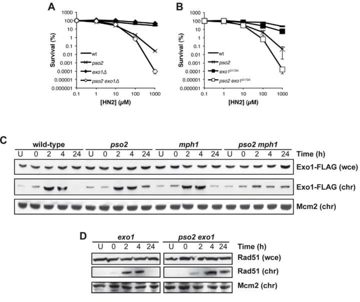
Exonuclease1 (Exo1) interacts with components of the MMR apparatus, including Msh2 [60], and like MutSa factors plays a redundant role with Pso2 during ICL repair [38]. To further explore this, we first asked whether the nuclease activity of Exo1 is required for its role in ICL repair. Anexo1disruptant exhibits no ICL sensitivity, but when combined with pso2 disruption we observed increased sensitivity as previously reported in a different genetic background [38] (Figure 5A). Mutation of aspartic acid 173 to alanine (D173A) in Exo1, that abolishes its exonuclease activityin vitro[61], produces a phenocopy of theexo1disruptant, where the single mutant has no sensitivity to HN2, but thepso2 exo1-D173A strain exhibits increased sensitivity, confirming that the nuclease activity of Exo1 is required for its role in ICL repair (Figure 5B). We next asked whether the Mph1-Mgm101-MutSa complex is involved in the targeting of Exo1 to chromatin during ICL repair. We followed the recruitment of Exo1-FLAG to chromatin after HN2 treatment for up to 24 hours. Exo1 was recruited to chromatin in wild type, pso2 and mph1 cells within 2 hours of ICL induction (Figure 5C). In contrast, in apso2 mph1 double disruptant, Exo1-FLAG recruitment was much reduced, suggesting that both pathways, involving Pso2 and Mph1-Mgm101-MutSa, respectively, are required for the efficient chromatin targeting of Exo1-FLAG to effect its role in ICL repair. Since, like Pso2, Exo1 is a 59-39exonuclease it is possible that these two factors are functionally redundant during ICL repair (see Discussion). As with pso2 mph1 and pso2 msh2 cells, Rad51 was recruited to chromatin with delayed kinetics inpso2 exo1 double disruptant compared to the exo1 single mutant, indicating that the initiation of recombination was delayed (Figure 5D). It is clear that the absence of Pso2 alone does impact
on the early phase of ICL repair, but the defect is compensated for to a significant extent by the activity of Exo1, since a delay in Rad51 recruitment at the whole chromatin level inpso2cells is not observed, at least at the whole chromatin level. It is, of course, possible that a loading defect would be observed if it was technically possible to examine individual ICL repair events in pso2cells. This absence of a delay in Rad51-chromatin association in the exo1 single disruptant indicates that the delay in Rad51 recruitment inpso2 exo1cells is not a result of the (very mild) DSB resection defects incurred by Exo1 loss [62], but is most likely to be the result of defective ICL processing.
The ICL repair pathway controlled by Mph1-Mgm101-MutSaalso involves the putative FANCJ- and FANCP-related factors Chl1 and Slx4
The existence of an ICL repair pathway involving Mph1 that is only revealed in the absence of Pso2, suggests that a simplified FA-related pathway might be operational in yeast. To this end, analysis of the yeast genome and proteome reveals two further factors that are putative FA homologs, notably the Chl1 helicase that has similarity with FANCJ [63] and Slx4/FANCP [64–66]. We therefore inactivated CHL1 and SLX4, alone and in combination withPSO2andMSH2(Figure 6). This revealed that bothchl1andslx4shows the same pattern of genetic interaction withpso2asmsh2,mph1andmgm101(Figure 6A and C). Moreover, disruption ofMSH2in apso2 chl1andpso2 slx4double disruptants does not further increase the strains sensitivity to HN2 (Figure 6B and D), indicating that Chl1 and Slx4 operate within the ICL repair pathway controlled by Mph1-Mgm101-MutSa. Together, our data suggest that a prototypical FA-related repair pathway may operate in budding yeast.
Discussion
Here we show that Mgm101 interacts with MutSaand Mph1, and collectively these factors control a key ICL repair pathway. Although Mph1 has significant similarity to FANCM, the disruption of MPH1 alone does not cause any increase in ICL sensitivity, in contrast to vertebratefancmmutants [16,17,26]. Co-disruption ofPSO2revealed an important role for Mph1 in ICL repair. Mgm101 has a well-established role in the transmission and repair of the mitochondrial genome [48,49,67], and we now show that Mgm101 also contributes to nuclear DNA repair. Cells disrupted forMGM101exhibit no increase in spontaneous forward mutation frequency (Figure S1E) or GCR rates (Figure 3A), suggesting that they undertake nuclear DNA replication with close to normal fidelity and efficiency. However, the interactions of Mgm101 with the nuclear repair factors Mph1 and MutSa promoted us to examine a role for Mgm101 in nuclear DNA metabolism. From our fractionation studies we were able to demonstrate that Mgm101 resides in both the nucleus and mitochondria, consistent with a role in DNA repair in both compartments.
Figure 3. Pso2 and Mph1-Mgm101-MutSaprotect against ICL-induced GCRs.(A) Left panel - schematic of the genomically-integrated (at theHXT13locus on chromosome V) substrate used to measure GCRs. Simultaneous loss of both theCAN1andURA3genes can only occurviaGCRs, producing cells able to form colonies able to grow on media containing canavanine and 5-FOA. See text for further details. Right panel - GCRs rates in wild type,pso2,mgm101, mph1and their respective double mutants, both spontaneously occurring and induced by a sub-lethal dose of HN2 (10 mM). Rates were determined as described in the Materials and Methods section. (B) Accumulation of DSBs in HN2-treated wild type,pso2,pso2 msh2,mgm101,pso2 mgm101andpso2 mgm101 msh2cells. Exponentially growing cells were treated with 100 mM HN2 for 2 hours at 28uC, or mock-treated (U) with water, and subsequently allowed to repair in minimal medium for 2, 4 and 24 hours. The U24 sample was mock-mock-treated allowed to repair for 24 hours. Samples were analysed on PFGE gels. (C) Rad53 phosphorylation following treatment with 100 mM HN2 and up to 2 hours recovery in wild type,pso2,msh2,mph1,pso2 msh2andpso2 mph1mutants.
It is well-established that several pathways contribute to the repair of ICLs, and these are, at least partly, utilised in a cell cycle phase-dependent manner [9,10,38,42]. Indeed, genetic and molecular evidence has been presented for a pathway involving the sequential action of NER followed by TLS, involving DNA polymerasef[6,10]. This pathway is thought to predominate in the G1-phase and early S-phase of the cell cycle, where a favoured HRR substrate (sister chromatid) is absent. Moreover, the sensitivity profile of rad52 deficient yeast cells though the cell cycle suggests that Rad52 is indeed dispensable in the primary ICL repair pathway operating in G1-phase and in quiescent (stationary
phase) cells [6,38]. Further, it is well-established that ICLs induce replication-associated DSBs [42,43], and that the repair of these DSBs is mainly dependent upon HRR, and hence Rad52 for their repair, in both yeast and mammalian cells [6,7,68]. Therefore, the fact that pso2 msh2 and pso2 mgm101 deficient cells accumulate ICL-associated DSBs argues that their repair by HR is blocked when both these pathways are deleted. This is corroborated by our observation that deletingRAD52inpso2 mgm101,pso2 mph1orpso2 msh2 strains does not lead to a further increase in sensitivity to ICL-inducing agents, and also by the delay in Rad51 chromatin recruitment we observed following ICL induction. Notably, a Figure 4. HRR and NHEJ mediated repair of ICL-associated DSBs is precluded in cells lacking Pso2 and components of the Mph1-Mgm101-MutSarepair pathways.(A–F) HN2 sensitivity of combinations ofpso2,msh2,rad52andyku70disruptants treated with HN2. All results are the mean of at least three independent experiments and the error bars show the standard error of the mean. (G) HN2-induced chromatin recruitment of Rad51 and Yku70 in wild type,pso2,msh2andmph1single and double disruptants. Mcm2 is shown as a loading control for chromatin associated protein. Chromatin bound material is labelled chr, and that from whole cell extract labelled wce.
doi:10.1371/journal.pgen.1002884.g004
rad52single mutant is less sensitive to ICLs thanpso2 mph1,pso2 msh2 and pso2 mgm101 double mutants, indicating that together these factors control ICL repair events in addition to HRR. In this regard, NHEJ factors appear to be utilised to repair ICL-associated DSBs in the absence of Pso2. Indeed, we have previously observed that rad52 yku70 double disruptant has a more severe DSB repair defective phenotype than arad52single mutant following HN2 treatment, consistent with a role for both HRR and NHEJ in the repair of ICL-associated DSBs [6].
We therefore suggest that the primary defect in cells lacking Pso2 and components of the Mph1-Mgm101-MutSa (and Exo1) pathway lie in processing an ICL repair intermediate that must be dealt with prior to the initiation of the recombinational/DSB repair phase of ICL repair (see Figure 7 for a model). Compelling data showing thatpso2mutants incise ICLs has been presented by several groups [34–36]. This suggests that a tethered cross-linked oligonucleotide is a possible substrate for degradation by exonuclease activity of Pso2, by analogy with the postulated role for hSNM1A [43]. We speculate that in the absence of Pso2 activity the same intermediate is recognised by MutSa, Mph1 and Mgm101. This leads to recruitment of Exo1, which operates with the same polarity (59-to-39) as Pso2 [69,70], to degrade the tethered oligonucleotide. In this regard, it is possible that the D-loop dissociation activity of Mph1 [23] controls commitment to the recombinational phase of repair until the ICL has been processed to provide a qualitatively useful substrate for HRR. Alternatively, or in addition, the ICL processing pathway that depends upon Mph1-Mgm101-MutSa might rely on the stable formation of a regressed fork for the recruitment of Exo1, another
potential function ascribed to Mph1 [21]. Given the increase in GCRs induced by the presence of ICLs in pso2 mgm101/msh2/ mph1cells, the inability to process this ICL repair intermediate is not only highly toxic, but also leads to drastic rearrangements of the genome, presumably through the use of low fidelity pathways to heal broken chromosomes [57]. Clearly the biochemical analysis of ICL intermediates as substrates for Pso2, Mph1, MutSa, Mgm101 and Exo1 factors is an important future area of study.
Recent reports suggest that human MMR factors might be important effectors of the FA repair pathway [71–73], reminis-cent of a role for MutSa collaborating with Mph1 during ICL repair in yeast. Our studies in yeast suggest that Pso2 is dominant over Mph1-Mgm101-MutSa and Exo1, as well as the FANCJ and FANCP homologs/orthologs Chl1 and Slx4, during ICL repair. However, it is possible that mammalian cells prioritise differently, sharing the burden between the FA pathway and SNM1A, since depletion of FA factors or SNM1A both produces pronounced ICL sensitisation in human cells. Indeed, a recent study indicated that breedingFANCD22/2mice withSNM12/2 produced enhanced perinatal mortality, consistent with redun-dancy between these pathways in mammals [74]. Moreover, evidence that hSNM1A recruitment to ICLs is controlled by Rad18, in an FA pathway-independent manner, has recently been presented [75]. Finally, it appears that a significant number of tumours somatically inactivate the FA pathway [76–78] or the MMR apparatus [79], which might help drive initial genomic instability. In such cases the inhibition of human SNM1 factors might be a means for selective sensitisation to ICL-inducing anticancer drugs.
Figure 6. The FANCJ- and FANCP-related factors Chl1 and Slx4, respectively, are both involved in the ICL repair pathway controlled by Mph1-Mgm101-MutSa.(A–D) HN2 sensitivity analysis of cells disrupted forPSO2andMSH2in combination with the FANCJ-related factorCHL1 and FANCP related factorSLX4. All results are the mean of at least three independent experiments and the error bars show the standard error of the mean.
doi:10.1371/journal.pgen.1002884.g006
Materials and Methods
Chemicals and enzymes
Analytical grade nitrogen mustard, L-canavanine, methyl methanesulfonate, cisplatin and hydrogen peroxide were pur-chased from Sigma-Aldrich (Poole, UK). Arthrobacter Luteus Zymolyase-20T was obtained from MP Biomedicals (Cambridge, UK). 5-fluoroorotic acid (5-FOA) was from Zymo Reseach (Irvine, CA, USA).
Yeast strains
Strains used in this study are listed in Table S1. Gene deletion and C-terminal tagging were carried out by PCR-based micro-homology targeted gene disruption and targeting strategies, using the pFA6a vector series and its derivatives [80–82]. Some deletions were also generated by synthetic genetic analysis methodology [83]. Deletions and tagging were confirmed by PCR analysis and restriction enzyme digestion.
Antibodies
For detection of FLAG-tagged Mgm101 protein, an affinity-purified rabbit antibody to the FLAG epitope tag was obtained from Sigma-Aldrich. Polyclonal anti-6xHIS antibodies (Abcam) was used to detect 6xHIS-tagged proteins. An anti-GFP polyclonal antibody from Roche was used to detect Mph1-GFP. A rabbit polyclonal, ChIP-grade histone H3 (Abcam) and mouse mono-clonal antibody to porin (Invitrogen) were used as controls for nuclear and mitochondrial localisation, respectively. Anti-Rad51 polyclonal antibodies were a kind gift of Patrick Sung. Immuno-detection of Rad53 was performed using a goat polyclonal antibody (Santa Cruz Biotechnology). Mouse anti-c-Myc (9E11, Abcam), mouse anti-HA (ab9110, Abcam), monoclonal mouse anti-FLAG (Sigma F1804) and goat polyclonal Msh2 (Santa Cruz Biotechnologies Inc, SC-26230) antibodies were employed in the gel filtration analysis.
Plasmids
MGM101 was amplified from yeast genomic DNA using primers containing cloning restriction sites and the nucleotide sequence for the FLAG epitope tag. Following amplification, the MGM101-FLAGconstruct was ligated into appropriately digested pYES2.0 yeast expression vector (Invitrogen, UK). Expression was induced by growth in glucose-free synthetic complete media containing 2% galactose and 1% raffinose as a carbon source.
Immunoprecipitation and co-immunoprecipitation Galactose-induced (5 hours) cell cultures were split into two separate aliquots, spun down, washed once in ddH2O, and
resuspended in lysis buffer (50 mM sodium phosphate, pH 7.4, 1 mM EDTA, 5% glycerol, supplemented with protease inhibitor cocktail and nuclease mix (both from Amersham Biosciences, Piscataway, NJ, USA), EDTA in lysis buffer was omitted in case of TALON Metal Affinity resin binding assay. Cells were disrupted by sonication and whole cell extracts were cleared by centrifuga-tion. Six milligrams of whole cell extracts were incubated with Anti-FLAG M2 Affinity Gel (Sigma-Aldrich, St. Louis, MO, USA) or TALON Metal Affinity Resin (Clontech, Palo Alta, CA, USA), and affinity purification of FLAG- and 6xHIS-tagged proteins was performed according to the protocols outlined in the manufactur-er’s manuals.
Liquid chromatography and MALDI-TOF/MS-based protein identification
Anti-FLAG M2 Affinity gel elute was subjected to the liquid chromatography (EASY -NLC, Bruker, Germany). A total of nine
potential peaks (Cutoff.400 intensity) were selected for further protein analysis. Proteins were trypsin digested automatically using a Proteineer DP protein digestion station (Bruker-Daltonics, Bremen, Germany). An aliquot of digestion solution was mixed with an aliquot of a-cyano-4-hydroxycinnamic acid (Bruker-Daltonics) in 33% aqueous acetonitrile and 0.25% trifluoroacetic Figure 7. A model for the two major ICL processing pathways in budding yeast, one controlled by Pso2 and the other by Mph1-Mgm101-MutSain collaboration with Exo1.See associated text for details.
acid. This mixture was deposited onto a 600mm AnchorChip prestructured MALDI probe (Bruker-Daltonics) and allowed to dry at room temperature. MALDI-MS data were obtained in an automated analysis loop using an Ultraflex time-of-flight (TOF) mass spectrometer (Bruker-Daltonics) equipped with a LIFT-MS/ MS device [84]. Spectra were acquired in the positive-ion mode at 50 Hz laser frequency, and 100 to 1000 individual spectra were averaged. Subsequently, selected precursor ions were subject to fragment ion analysis in the tandem time-of-flight (TOF/TOF) mode to obtain the corresponding MALDI-MS/MS spectra. Automated analysis of mass data was performed using the flexAnalysis software (Bruker-Daltonics). MALDI-MS and MALDI-MS/MS data were combined through the BioTools program (Bruker-Daltonics) to search a non-redundant protein database (NCBInr) using Mascot software (Matrix Science, London, UK). MALDI-MS and MALDI-MS/MS spectra as well as database search results were inspected in detail using flexAnalysis as well as in-house software.
Yeast cellular fractionation
Fractionation of yeast to isolate nuclear and mitochondrial fractions were performed as described [85,86], with minor modifications.
Gel filtration analysis
Approximately 60 mg of yeast whole cell extract prepared from a strain harbouring Msh6-HA, Mph1-FLAG and Mgm101-Myc constructs was loaded in a 200 ml volume onto a Superdex 200 column on an AKTA FPLC (Amersham Pharmacia Biotech) equilibrated with (25 mM HEPES pH 7.9, 10% glycerol, 150 mM NaCl, 1 mM EDTA and 1 mM DTT). The flow rate was set at 0.5 ml/min and fractions of 2 ml were collected following a void volume of 40 ml. The proteins from aliquots were subjected to SDS-PAGE on a 4–12% Bis-Tris gel (Invitrogen) and blotted onto Hybond-C (Amersham) membranes.
Chromatin binding assays
These were performed as described [86].
Nitrogen mustard, cisplatin, methyl methanesulfonate, ultraviolet light, and hydrogen peroxide sensitivity assays
Exponentially growing cells were diluted in serial in PBSA to a concentration of 26107cells/ml and incubated with HN2, CDDP, MMS or H2O2 at the stated doses for 1 hour at 30uC with
shaking, or exposed to 254 nm UV light at the doses shown. Samples were subsequently plated onto YPD plates and incubated at 30uC for 3 days before scoring survival.
Canavanine resistance forward mutation assays These were performed as previously described [87].
Pulsed-field gel electrophoresis analysis
This was performed as previously described [6], except plugs were analysed on a Beckmann TAFE machine.
Mitochondrial petite induction
Yeast cells were cultivated in YPD media until the stationary phase of growth was reached. The cells were then collected by centrifugation and washed with, and resuspended in, sterile water to the final concentration of 26107 cells/ml. The resulting cell suspension was subsequently treated with 10mM HN2 or 100mM
H2O2for 1 hour at 30uC. After treatment, cells were harvested by
centrifugation, and washed twice with, and resuspended in,
potassium phosphate buffer. Appropriately diluted cell suspension was plated in triplicate onto YPD, YPG or YPGE plates. Plates were incubated at 30uC for 2–3 days (YPD), 5 days (YPG, YPGE) before being scored.
Measurement of Gross Chromosomal Rearrangements Yeast cultures grown in SC media overnight were inoculated into fresh YPD media, where incubation continued until the cell suspension reached a density of 26107cells/ml. 261010of cells were collected by centrifugation, washed with, and resuspended in, potassium phosphate buffer to the final concentration of 26108 cells/ml. The cell suspension was treated with 10mM HN2 for
1 hour at 30uC. After treatment, cells were harvested, washed twice with, and resuspended in, potassium phosphate buffer. Cell suspension was diluted and plated in triplicate onto YPD plates to determine cell viability. For GCR measurement, the undiluted cell suspension (approximately 1010 of cells) was plated on 5-FOA plates supplemented with canavanine.
Supporting Information
Figure S1 (A) Mgm101-FLAG can be detected in both nuclear and mitochondrial fractions of fractionated yeast cells. Mitochon-drial porin and histone H3 were used for markers for the nuclear and mitochondrial fractions, respectively. (B–D) The rates of spontaneous, HN2- and H2O2-induced loss of functional
mito-chondria (petite formation) inpso2,msh2andmph1strains are not elevated above wt. Data is the average of at least three independent experiments, error bars show the standard error of the mean. (E) Spontaneous forward mutation frequencies of wt, pso2, msh2,mph1andmgm101strains measured by determining the number of canavanine resistant colonies arising per 108 survivors. See materials and methods for details. Data is the average of at least three independent experiments, error bars show the standard error of the mean.
(TIF)
Figure S2 (A–F) Cells disrupted forMGM101,MPH1orMSH2 alone or in combination with disruptions with PSO2, show no increase in sensitivity to H2O2or IR, whereasrad52cells are highly
IR sensitive. Data is the average of at least three independent experiments, error bars show the standard error of the mean. (G– I) Cells co-disrupted for PSO2and MSH2, MPH1orMGM101 are CDDP sensitive. (J–O) Cells disrupted for MGM101, MPH1 or MSH2alone or in combination with disruptions withPSO2, show no increase in sensitivity to UV or MMS.
(EPS)
Figure S3 (A) Cell cycle progression in wt,pso2, msh2andpso2 msh2 mutants, as determined by FACS analysis, following treatment with 100mM HN2. (B) Rad53 phosphorylation following treatment with 100mM HN2 and up to 24 hours recovery in wt,pso2,msh2andpso2 msh2mutants.
(TIF)
Figure S4 Co-disruption ofRAD52inpso2 mph1(A) orpso2 msh2 mgm101(B) cells does not lead to an increase in HN2 sensitivity. Data is the average of at least three independent experiments, error bars show the standard error of the mean. (C) HN2-induced chromatin recruitment of Rad51 in mgm101 and pso2 mgm101 single and double disruptants. Mcm2 is shown as a loading control for chromatin associated protein. Chromatin bound material is labelled chr, and that from whole cell extract labelled wce. (TIF)
Table S1 Genotypes and origin of yeast strain used in this study. (DOC)
Acknowledgments
We thank Ian Hickson and Leonard Wu for helpful insights throughout this work, and to Mika Abu for assistance with gel filtration. We are grateful to Richard Kolodner, Mike Liskay and Leonard Wu for yeast strains, and Patrick Sung for antibodies.
Author Contributions
Conceived and designed the experiments: TAW ZD MC PJM. Performed the experiments: TAW ZD SS MRB DV. Analyzed the data: PJM MC MRB TAW ZD. Contributed reagents/materials/analysis tools: MRB. Wrote the paper: MC PJM.
References
1. McHugh PJ, Spanswick VJ, Hartley JA (2001) Repair of DNA interstrand crosslinks: molecular mechanisms and clinical relevance. Lancet Oncol 2: 483– 490.
2. Dronkert ML, Kanaar R (2001) Repair of DNA interstrand cross-links. Mutat Res 486: 217–247.
3. Lehoczky P, McHugh PJ, Chovanec M (2007) DNA interstrand cross-link repair in Saccharomyces cerevisiae. FEMS Microbiol Rev 31: 109–133.
4. Ruhland A, Brendel M (1979) Mutagenesis by cytostatic alkylating agents in yeast strains of differing repair capacities. Genetics 92: 83–97.
5. Miller RD, Prakash L, Prakash S (1982) Genetic control of excision of Saccharomyces cerevisiae interstrand DNA cross-links induced by psoralen plus near-UV light. Mol Cell Biol 2: 939–948.
6. McHugh PJ, Sones WR, Hartley JA (2000) Repair of intermediate structures produced at DNA interstrand cross-links in Saccharomyces cerevisiae. Mol Cell Biol 20: 3425–3433.
7. Jachymczyk WJ, von Borstel RC, Mowat MR, Hastings PJ (1981) Repair of interstrand cross-links in DNA of Saccharomyces cerevisiae requires two systems for DNA repair: the RAD3 system and the RAD51 system. Mol Gen Genet 182: 196–205.
8. Zheng H, Wang X, Warren AJ, Legerski RJ, Nairn RS, et al. (2003) Nucleotide excision repair- and polymerase eta-mediated error-prone removal of mitomycin C interstrand cross-links. Mol Cell Biol 23: 754–761.
9. Niedernhofer LJ, Odijk H, Budzowska M, van Drunen E, Maas A, et al. (2004) The structure-specific endonuclease Ercc1-Xpf is required to resolve DNA interstrand cross-link-induced double-strand breaks. Mol Cell Biol 24: 5776– 5787.
10. Sarkar S, Davies AA, Ulrich HD, McHugh PJ (2006) DNA interstrand crosslink repair during G1 involves nucleotide excision repair and DNA polymerase zeta. Embo J 25: 1285–1294.
11. Raschle M, Knipscheer P, Enoiu M, Angelov T, Sun J, et al. (2008) Mechanism of replication-coupled DNA interstrand crosslink repair. Cell 134: 969–980. 12. Niedzwiedz W, Mosedale G, Johnson M, Ong CY, Pace P, et al. (2004) The
Fanconi anaemia gene FANCC promotes homologous recombination and error-prone DNA repair. Mol Cell 15: 607–620.
13. Knipscheer P, Raschle M, Smogorzewska A, Enoiu M, Ho TV, et al. (2009) The Fanconi anemia pathway promotes replication-dependent DNA interstrand cross-link repair. Science 326: 1698–1701.
14. Long DT, Raschle M, Joukov V, Walter JC (2011) Mechanism of RAD51-dependent DNA interstrand cross-link repair. Science 333: 84–87.
15. Joenje H, Patel KJ (2001) The emerging genetic and molecular basis of Fanconi anaemia. Nat Rev Genet 2: 446–457.
16. Meetei AR, Medhurst AL, Ling C, Xue Y, Singh TR, et al. (2005) A human ortholog of archaeal DNA repair protein Hef is defective in Fanconi anemia complementation group M. Nat Genet 37: 958–963.
17. Mosedale G, Niedzwiedz W, Alpi A, Perrina F, Pereira-Leal JB, et al. (2005) The vertebrate Hef ortholog is a component of the Fanconi anemia tumor-suppressor pathway. Nat Struct Mol Biol 12: 763–771.
18. Entian KD, Schuster T, Hegemann JH, Becher D, Feldmann H, et al. (1999) Functional analysis of 150 deletion mutants in Saccharomyces cerevisiae by a systematic approach. Mol Gen Genet 262: 683–702.
19. Scheller J, Schurer A, Rudolph C, Hettwer S, Kramer W (2000) MPH1, a yeast gene encoding a DEAH protein, plays a role in protection of the genome from spontaneous and chemically induced damage. Genetics 155: 1069–1081. 20. Schurer KA, Rudolph C, Ulrich HD, Kramer W (2004) Yeast MPH1 gene
functions in an error-free DNA damage bypass pathway that requires genes from Homologous recombination, but not from postreplicative repair. Genetics 166: 1673–1686.
21. Sun W, Nandi S, Osman F, Ahn JS, Jakovleska J, et al. (2008) The FANCM ortholog Fml1 promotes recombination at stalled replication forks and limits crossing over during DNA double-strand break repair. Mol Cell 32: 118–128. 22. Banerjee S, Smith S, Oum JH, Liaw HJ, Hwang JY, et al. (2008) Mph1p
promotes gross chromosomal rearrangement through partial inhibition of homologous recombination. J Cell Biol 181: 1083–1093.
23. Prakash R, Satory D, Dray E, Papusha A, Scheller J, et al. (2009) Yeast Mph1 helicase dissociates Rad51-made D-loops: implications for crossover control in mitotic recombination. Genes Dev 23: 67–79.
24. Prakash R, Krejci L, Van Komen S, Anke Schurer K, Kramer W, et al. (2005) Saccharomyces cerevisiae MPH1 gene, required for homologous recombination-mediated mutation avoidance, encodes a 39to 59DNA helicase. J Biol Chem 280: 7854–7860.
25. Gari K, Decaillet C, Stasiak AZ, Stasiak A, Constantinou A (2008) The Fanconi anemia protein FANCM can promote branch migration of Holliday junctions and replication forks. Mol Cell 29: 141–148.
26. Xue Y, Li Y, Guo R, Ling C, Wang W (2008) FANCM of the Fanconi anemia core complex is required for both monoubiquitination and DNA repair. Hum Mol Genet.
27. Rosado IV, Niedzwiedz W, Alpi AF, Patel KJ (2009) The Walker B motif in avian FANCM is required to limit sister chromatid exchanges but is dispensable for DNA crosslink repair. Nucleic Acids Res 37: 4360–4370.
28. Schwab RA, Blackford AN, Niedzwiedz W (2010) ATR activation and replication fork restart are defective in FANCM-deficient cells. Embo J 29: 806–818.
29. Collis SJ, Ciccia A, Deans AJ, Horejsi Z, Martin JS, et al. (2008) FANCM and FAAP24 function in ATR-mediated checkpoint signaling independently of the Fanconi anemia core complex. Mol Cell 32: 313–324.
30. Luke-Glaser S, Luke B, Grossi S, Constantinou A (2010) FANCM regulates DNA chain elongation and is stabilized by S-phase checkpoint signalling. Embo J 29: 795–805.
31. Henriques JA, Moustacchi E (1980) Isolation and characterization of pso mutants sensitive to photo-addition of psoralen derivatives in Saccharomyces cerevisiae. Genetics 95: 273–288.
32. Ruhland A, Haase E, Siede W, Brendel M (1981) Isolation of yeast mutants sensitive to the bifunctional alkylating agent nitrogen mustard. Mol Gen Genet 181: 346–351.
33. Ruhland A, Kircher M, Wilborn F, Brendel M (1981) A yeast mutant specifically sensitive to bifunctional alkylation. Mutat Res 91: 457–462.
34. Grossmann KF, Ward AM, Moses RE (2000) Saccharomyces cerevisiae lacking Snm1, Rev3 or Rad51 have a normal S-phase but arrest permanently in G2 after cisplatin treatment. Mutat Res 461: 1–13.
35. Magana-Schwencke N, Henriques JA, Chanet R, Moustacchi E (1982) The fate of 8-methoxypsoralen photoinduced crosslinks in nuclear and mitochondrial yeast DNA: comparison of wild-type and repair-deficient strains. Proc Natl Acad Sci U S A 79: 1722–1726.
36. Wilborn F, Brendel M (1989) Formation and stability of interstrand cross-links induced by cis- and trans-diamminedichloroplatinum (II) in the DNA of Saccharomyces cerevisiae strains differing in repair capacity. Curr Genet 16: 331–338.
37. Li X, Moses RE (2003) The beta-lactamase motif in Snm1 is required for repair of DNA double-strand breaks caused by interstrand crosslinks in S. cerevisiae. DNA Repair (Amst) 2: 121–129.
38. Barber LJ, Ward TA, Hartley JA, McHugh PJ (2005) DNA interstrand cross-link repair in the Saccharomyces cerevisiae cell cycle: overlapping roles for PSO2 (SNM1) with MutS factors and EXO1 during S phase. Mol Cell Biol 25: 2297– 2309.
39. De Silva IU, McHugh PJ, Clingen PH, Hartley JA (2000) Defining the roles of nucleotide excision repair and recombination in the repair of DNA interstrand cross-links in mammalian cells. Mol Cell Biol 20: 7980–7990.
40. Akkari YM, Bateman RL, Reifsteck CA, Olson SB, Grompe M (2000) DNA replication is required To elicit cellular responses to psoralen-induced DNA interstrand cross-links. Mol Cell Biol 20: 8283–8289.
41. Bessho T (2003) Induction of DNA replication-mediated double strand breaks by psoralen DNA interstrand cross-links. J Biol Chem 278: 5250–5254. 42. Hanada K, Budzowska M, Modesti M, Maas A, Wyman C, et al. (2006) The
structure-specific endonuclease Mus81-Eme1 promotes conversion of interstrand DNA crosslinks into double-strands breaks. Embo J 25: 4921–4932. 43. Wang AT, Sengerova B, Cattell E, Inagawa T, Hartley JM, et al. (2011) Human
SNM1A and XPF-ERCC1 collaborate to initiate DNA interstrand cross-link repair. Genes Dev 25: 1859–1870.
44. Zhang N, Lu X, Zhang X, Peterson CA, Legerski RJ (2002) hMutSbeta is required for the recognition and uncoupling of psoralen interstrand cross-links in vitro. Mol Cell Biol 22: 2388–2397.
45. Wu Q, Christensen LA, Legerski RJ, Vasquez KM (2005) Mismatch repair participates in error-free processing of DNA interstrand crosslinks in human cells. EMBO Rep 6: 551–557.
46. Lan L, Hayashi T, Rabeya RM, Nakajima S, Kanno S, et al. (2004) Functional and physical interactions between ERCC1 and MSH2 complexes for resistance to cis-diamminedichloroplatinum(II) in mammalian cells. DNA Repair (Amst) 3: 135–143.
47. Ho Y, Gruhler A, Heilbut A, Bader GD, Moore L, et al. (2002) Systematic identification of protein complexes in Saccharomyces cerevisiae by mass spectrometry. Nature 415: 180–183.
48. Chen XJ, Guan MX, Clark-Walker GD (1993) MGM101, a nuclear gene involved in maintenance of the mitochondrial genome in Saccharomyces cerevisiae. Nucleic Acids Res 21: 3473–3477.
for the repair of oxidatively damaged mitochondrial DNA. J Cell Biol 145: 291– 304.
50. Meeusen S, Nunnari J (2003) Evidence for a two membrane-spanning autonomous mitochondrial DNA replisome. J Cell Biol 163: 503–510. 51. Mbantenkhu M, Wang X, Nardozzi JD, Wilkens S, Hoffman E, et al. (2011)
Mgm101 is a Rad52-related protein required for mitochondrial DNA recombination. J Biol Chem.
52. Zuo X, Xue D, Li N, Clark-Walker GD (2007) A functional core of the mitochondrial genome maintenance protein Mgm101p in Saccharomyces cerevisiae determined with a temperature-conditional allele. FEMS Yeast Res 7: 131–140.
53. Drummond JT, Genschel J, Wolf E, Modrich P (1997) DHFR/MSH3 amplification in methotrexate-resistant cells alters the hMutSalpha/hMutSbeta ratio and reduces the efficiency of base-base mismatch repair. Proc Natl Acad Sci U S A 94: 10144–10149.
54. Contamine V, Picard M (2000) Maintenance and integrity of the mitochondrial genome: a plethora of nuclear genes in the budding yeast. Microbiol Mol Biol Rev 64: 281–315.
55. Reenan RA, Kolodner RD (1992) Isolation and characterization of two Saccharomyces cerevisiae genes encoding homologs of the bacterial HexA and MutS mismatch repair proteins. Genetics 132: 963–973.
56. Chen C, Kolodner RD (1999) Gross chromosomal rearrangements in Saccharomyces cerevisiae replication and recombination defective mutants. Nat Genet 23: 81–85.
57. Kolodner RD, Putnam CD, Myung K (2002) Maintenance of genome stability in Saccharomyces cerevisiae. Science 297: 552–557.
58. Myung K, Datta A, Chen C, Kolodner RD (2001) SGS1, the Saccharomyces cerevisiae homologue of BLM and WRN, suppresses genome instability and homeologous recombination. Nat Genet 27: 113–116.
59. Smith S, Hwang JY, Banerjee S, Majeed A, Gupta A, et al. (2004) Mutator genes for suppression of gross chromosomal rearrangements identified by a genome-wide screening in Saccharomyces cerevisiae. Proc Natl Acad Sci U S A 101: 9039–9044.
60. Tishkoff DX, Boerger AL, Bertrand P, Filosi N, Gaida GM, et al. (1997) Identification and characterization of Saccharomyces cerevisiae EXO1, a gene encoding an exonuclease that interacts with MSH2. Proc Natl Acad Sci U S A 94: 7487–7492.
61. Tran PT, Erdeniz N, Dudley S, Liskay RM (2002) Characterization of nuclease-dependent functions of Exo1p in Saccharomyces cerevisiae. DNA Repair (Amst) 1: 895–912.
62. Symington LS, Gautier J (2010) Double-Strand Break End Resection and Repair Pathway Choice. Annu Rev Genet.
63. White MF (2009) Structure, function and evolution of the XPD family of iron-sulfur-containing 59R39DNA helicases. Biochem Soc Trans 37: 547–551. 64. Stoepker C, Hain K, Schuster B, Hilhorst-Hofstee Y, Rooimans MA, et al.
(2011) SLX4, a coordinator of structure-specific endonucleases, is mutated in a new Fanconi anemia subtype. Nat Genet 43: 138–141.
65. Crossan GP, van der Weyden L, Rosado IV, Langevin F, Gaillard PH, et al. (2011) Disruption of mouse Slx4, a regulator of structure-specific nucleases, phenocopies Fanconi anemia. Nat Genet 43: 147–152.
66. Kim Y, Lach FP, Desetty R, Hanenberg H, Auerbach AD, et al. (2011) Mutations of the SLX4 gene in Fanconi anemia. Nat Genet 43: 142–146. 67. Zuo XM, Clark-Walker GD, Chen XJ (2002) The mitochondrial nucleoid
protein, Mgm101p, of Saccharomyces cerevisiae is involved in the maintenance of rho(+) and ori/rep-devoid petite genomes but is not required for hypersuppressive rho(2) mtDNA. Genetics 160: 1389–1400.
68. De Silva IU, McHugh PJ, Clingen PH, Hartley JA (2002) Defects in interstrand cross-link uncoupling do not account for the extreme sensitivity of ERCC1 and XPF cells to cisplatin. Nucleic Acids Res 30: 3848–3856.
69. Li X, Hejna J, Moses RE (2005) The yeast Snm1 protein is a DNA 59 -exonuclease. DNA Repair (Amst) 4: 163–170.
70. Hazrati A, Ramis-Castelltort M, Sarkar S, Barber LJ, Schofield CJ, et al. (2007) Human SNM1A rescues the DNA repair defects of yeast pso2 mutants. DNA Repair (Amst) in press.
71. Peng M, Litman R, Xie J, Sharma S, Brosh RM Jr, et al. (2007) The FANCJ/ MutLalpha interaction is required for correction of the cross-link response in FA-J cells. Embo J 26: 3238–3249.
72. Williams SA, Wilson JB, Clark AP, Mitson-Salazar A, Tomashevski A, et al. (2011) Functional and physical interaction between the mismatch repair and FA-BRCA pathways. Hum Mol Genet.
73. Huang M, Kennedy R, Ali AM, Moreau LA, Meetei AR, et al. (2011) Human MutS and FANCM complexes function as redundant DNA damage sensors in the Fanconi Anemia pathway. DNA Repair (Amst).
74. Hemphill AW, Bruun D, Thrun L, Akkari Y, Torimaru Y, et al. (2008) Mammalian SNM1 is required for genome stability. Mol Genet Metab 94: 38– 45.
75. Yang K, Moldovan GL, D’Andrea AD (2010) RAD18-dependent recruitment of SNM1A to DNA repair complexes by a ubiquitin-binding zinc finger. J Biol Chem 285: 19085–19091.
76. Marsit CJ, Liu M, Nelson HH, Posner M, Suzuki M, et al. (2004) Inactivation of the Fanconi anemia/BRCA pathway in lung and oral cancers: implications for treatment and survival. Oncogene 23: 1000–1004.
77. Wreesmann VB, Estilo C, Eisele DW, Singh B, Wang SJ (2007) Downregulation of Fanconi anemia genes in sporadic head and neck squamous cell carcinoma. ORL J Otorhinolaryngol Relat Spec 69: 218–225.
78. Taniguchi T, Tischkowitz M, Ameziane N, Hodgson SV, Mathew CG, et al. (2003) Disruption of the Fanconi anemia-BRCA pathway in cisplatin-sensitive ovarian tumors. Nat Med 9: 568–574.
79. Jiricny J (1998) Eukaryotic mismatch repair: an update. Mutat Res 409: 107– 121.
80. Wach A, Brachat A, Pohlmann R, Philippsen P (1994) New heterologous modules for classical or PCR-based gene disruptions in Saccharomyces cerevisiae. Yeast 10: 1793–1808.
81. Longtine MS, McKenzie A 3rd, Demarini DJ, Shah NG, Wach A, et al. (1998) Additional modules for versatile and economical PCR-based gene deletion and modification in Saccharomyces cerevisiae. Yeast 14: 953–961.
82. Goldstein AL, McCusker JH (1999) Three new dominant drug resistance cassettes for gene disruption in Saccharomyces cerevisiae. Yeast 15: 1541–1553. 83. Tong AH, Evangelista M, Parsons AB, Xu H, Bader GD, et al. (2001) Systematic genetic analysis with ordered arrays of yeast deletion mutants. Science 294: 2364–2368.
84. Suckau D, Resemann A, Schuerenberg M, Hufnagel P, Franzen J, et al. (2003) A novel MALDI LIFT-TOF/TOF mass spectrometer for proteomics. Anal Bioanal Chem 376: 952–965.
85. Zinser E, Daum G (1995) Isolation and biochemical characterization of organelles from the yeast, Saccharomyces cerevisiae. Yeast 11: 493–536. 86. Liang C, Stillman B (1997) Persistent initiation of DNA replication and
chromatin-bound MCM proteins during the cell cycle in cdc6 mutants. Genes Dev 11: 3375–3386.
87. McHugh PJ, Gill RD, Waters R, Hartley JA (1999) Excision repair of nitrogen mustard-DNA adducts in Saccharomyces cerevisiae. Nucleic Acids Res 27: 3259–3266.