Fibrillar Amyloid Plaque Formation Precedes
Microglial Activation
Christian K. E. Jung1☯, Kevin Keppler1☯, Sonja Steinbach1, Lidia Blazquez-Llorca1, Jochen Herms1,2*
1Department for Translational Brain Research, German Center for Neurodegenerative Diseases—site Munich (DZNE-M) and Center for Neuropathology and Prion Research (ZNP), Ludwig-Maximilians-University Munich, Munich, Germany,2Munich Cluster of Systems Neurology (SyNergy), Munich, Germany
☯These authors contributed equally to this work. *Jochen.Herms@med.uni-muenchen.de
Abstract
In Alzheimer’s disease (AD), hallmarkβ-amyloid deposits are characterized by the pres-ence of activated microglia around them. Despite an extensive characterization of the rela-tion of amyloid plaques with microglia, little is known about the initiarela-tion of this interacrela-tion. In this study, the detailed investigation of very small plaques in brain slices in AD transgenic mice of the line APP-PS1(dE9) revealed different levels of microglia recruitment. Analysing plaques with a diameter of up to 10μm we find that only the half are associated with clear morphologically activated microglia. Utilizingin vivoimaging of new appearing amyloid pla-ques in double-transgenic APP-PS1(dE9)xCX3CR1+/-mice further characterized the dy-namic of morphological microglia activation. We observed no correlation of morphological microglia activation and plaque volume or plaque lifetime. Taken together, our results dem-onstrate a very prominent variation in size as well as in lifetime of new plaques relative to the state of microglia reaction. These observations might question the existing view that am-yloid deposits by themselves are sufficient to attract and activate microgliain vivo.
Introduction
Microglia activation is a characteristic feature of Alzheimer’s disease (AD). Although microglia cells have been found to contact neurofibrillary tangles in the brain of Alzheimer’s patients ([1]), the association of microglia to amyloid plaques is a more frequent and distinguished event ([2]; [3]). Plaques are extracellular proteinaceous aggregates mainly composed of the β-amyloid peptide (Aβ). Proteolytically derived Aβmonomers successively aggregate into olig-omeric species and thence amyloid fibrils, ultimately forming the pathognomonic amyloid plaques which gradually grow in size ([4]). In the vicinity of amyloid plaques neurons are dam-aged, as apparent by dystrophic neurites, the progressive loss of dendritic spines as well as the loss of axons and neurons ([5]; [6]; [7]; [8]).
Microglia are the resident immune cells of the brain actively surveying the brain parenchy-ma with their fine processes. After brain injury or immunological challenges, microglia are
a11111
OPEN ACCESS
Citation:Jung CKE, Keppler K, Steinbach S, Blazquez-Llorca L, Herms J (2015) Fibrillar Amyloid Plaque Formation Precedes Microglial Activation. PLoS ONE 10(3): e0119768. doi:10.1371/journal. pone.0119768
Academic Editor:Joseph El Khoury, Massachusetts General Hospital and Harvard Medical School, UNITED STATES
Received:July 28, 2014
Accepted:January 16, 2015
Published:March 23, 2015
Copyright:© 2015 Jung et al. This is an open access article distributed under the terms of the
Creative Commons Attribution License, which permits unrestricted use, distribution, and reproduction in any medium, provided the original author and source are credited.
Data Availability Statement:All relevant data are within the paper and its Supporting Information files.
activated leading to the upregulated expression of immune-related molecules, including interleukin-1 (IL-1) and transforming growth factorβ([9]; [10]). Once activated, microglia prominently change their morphology: the ramified processes swell and withdraw, whilst their cell bodies enlarge ([11]). In the AD brain, activation of microglia and clustering around amy-loid deposits tend to occur early in the development of plaque pathology. Nevertheless, little is known about the initiating event of this interaction. Here, we specifically focus on very small newly-formed amyloid plaques and attempt to characterize the process by which microglia are attracted to these lesions.
Material and Methods
Mice
Mouse lines APP-PS1(dE9) ([12]), CX3CR1-GFP ([13]) and YFP-H ([14]) were purchased from The Jackson Laboratory, Bar Harbor, USA. Briefly, the transgenic APP-PS1 (dE9) mouse line expresses human APP with Swedish mutation and mutant human Presinilin1 (PS1 delta E9) both under the control of the mouse prion protein promoter resulting in abundant amyloid plaques in cortex and hippocampus[12]. The knockin of gfp to the Cx3cr1 locus in CX3CR1-GFP mice results in a CX3CR1-GFP-labeling of microglia[13]. In YFP-H mouse line the YFP is expressed under control of Thy1 promoter which leads to a sparse labeling of pyramidal neurons in cor-tex and hippocampus [14]. Mice were housed in standard cages with food and water ad libitum. The APP-PS1(dE9) line was crossed with CX3CR1-GFP. The resulting offspring [APP-PS1 (dE9)xCX3CR1+/-] was inbred to generate the [APP-PS1(dE9)xCX3CR1+/-], used for in vivo imaging experiments. For control experiments (S1 Fig.) [APP-PS1(dE9)xCX3CR1+/-] x [APP-PS1(dE9)xCX3CR1+/-] crossings resulted in [APP-PS1(dE9)xCX3CR1+/+], [APP-PS1(dE9) xCX3CR1+/-] and [APP-PS1(dE9)xCX3CR1-/-]. Furthermore APP-PS1(dE9) was crossed to the YFP-H line resulting in [APP-PS1(dE9)xYFP-H] mice, which were inbred. Mice of mixed gender were used for experiments at indicated age.
The studies were carried out in accordance with an animal protocol approved by the Ludwig-Maximilians-University Munich and the government of Upper Bavaria (Az. 55.2– 1–54–2531–188–09). The cranial window preparation andin vivoimaging were performed under anesthesia, and all efforts were made to minimize suffering of the animals.
Immunohistochemistry
Briefly, mice anesthetized with an intraperitoneal injection of ketamine/xylazine (0.14 mg ketamine / 0.01 mg xylazine per gram body weight; WDT/Bayer Health Care) were transcar-dially perfused with PBS and 4% PFA and brains were prepared. 100μm thick sections were
cut from postfixed brains. Free-floating sections were permeabilized with 2% Triton X-100 overnight and blocked with 10% normal goat serum and BSA. Microglia were immunohisto-chemically labeled using Iba1-antibody (Wako, 1:200; secondary antibody A-21244 by Invitro-gen, 1:500). Fibrillar Aβplaque staining was performed with 145μM Methoxy-X04 (provided
by Prof. Boris Schmidt from TU Darmstadt, Germany) in PBS for 30 min and subsequently washed with PBS. Slices of CX3CR1-/-and CX3CR1+/-mice were bleached before staining for Iba-1 and Methoxy-X04. Fluorescence images were acquired with a confocal laser scanning mi-croscope mounted on an inverted mimi-croscope support (LSM 510, Carl Zeiss). Images were ac-quired 0.22μm pixel size and frame distance of 2μm using a 40x/1.3 DIC oil objective. Images
were aquired from cortical or hippocampal tissue. Image stacks were acquired spanning a total depth of at least 10μm above and 10μm below each selected amyloid plaque.
Cranial window preparation and in vivo two-photon microscopy
Mice were anesthetized with an intraperitoneal injection of ketamine/xylazine (0.14 mg ketamine / 0.01 mg xylazine per gram body weight; WDT/Bayer Health Care). Additionally, a single dosage of dexamethasone (6μg/g body weight; Sigma) was intraperitoneally
adminis-tered immediately before surgery. Utilizing the open skull preparation a cranial window was placed above the somatosensory cortex. For repositioning during repetitive imaging a small metal bar, containing a hole for a screw, was glued next to the window. Directly after surgery mice received subcutaneously a single analgesic treatment with carprophen (7.5μg/g body
weight; Pfizer) and a single antibiotic treatment with cefotaxim (0.25 mg/g body weight; Pharmore).
Two-photonin vivoimaging was started after a 3–4 week recovery period post-surgery, uti-lizing a LSM 5MP setup (Carl Zeiss) equipped with a MaiTai laser (Spectra Physics) and a 20x water-immersion objective (1.0 NA) (Carl Zeiss). For amyloid plaque staining, methoxy-X04 (0.4 to 2.4 mg/kg; provided by Prof. Boris Schmidt from TU Darmstadt, Germany) was intra-peritoneally injected 24 hours before imaging ([15]). Methoxy-X04 is a derivate of Congo red, that specifically binds to amyloid deposits in post mortem tissue andin vivoand therefore pro-vides a fluorescent label [15]. Mice were anesthetized by Isofluoran. Imaging sessions lasted for no longer than 60 min and laser power was kept below 50 mW to avoid phototoxic effects. We imaged up to three cortical positions with each position spanning 425μm x 425μm x 201μm
in x-y-z (pixel distance in x/y: 0.42μm; pixel distance in z: 3μm). Care was taken to ensure
similar fluorescence levels in space and in time at every imaging session.
Data analysis
Analysis of confocal and multiphoton images was performed with unprocessed data. From multiphoton imaging data only amyloid plaques were selected for analysis when at all imaging sessions a total image stack with at least 10μm above and 10μm below the plaque was
re-corded. All interaction analysis of microglia with amyloid plaques was performed with 3D data sets. For plaque volume determination, smallest and largest diameter of each plaque was mea-sured from image projections using ZEN software (Zeiss). Assuming the plaque as a spheric el-ement, the volume was calculated using the corresponding mean diameter. For illustration purposes multiphoton images were deconvolved (AutoQuantX2, Media Cybernetics) and fil-tered with an edge-preserving algorithm, followed by a local contrast change (Imaris 5.0.1, Bit-plane). Confocal and multiphoton image stacks were aligned with Imaris 5.0.1, Bitplane.
Statistics: For histological ex vivo analysis a total of 188 small plaques from five animals were identified (54/55/24/23/32). All images were analysed independently and identically. Two photon in vivo imaging revealed a total of 21 new appearing plaques in four animals (2/4/9/6). All time series data points were analysed independently and identically.
Results
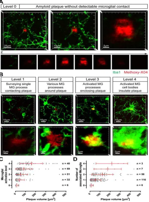
On brain slices of 12-month-old APP-PS1(dE9) mice, microglia cells were immunohistologi-cally stained and fibrillar amyloid deposits were labelled with Methoxy-XO4. Rarely, we ob-served small amyloid plaques that were devoid of any detectable microglial contact (Fig. 1A); in most cases, similar sized plaques were clearly contacted by microglia, partly exhibiting an activated appearance (Fig. 1B). To characterize the relationship between plaque size and micro-glial activation, we specifically imaged small amyloid plaques (which are supposed to corre-spond to novel plaques) in cortex or hippocampus. Therefore, plaques with a diameter of up to ~10μm (equates a volume of 524μm3) were chosen randomly, while the microglia channel
covering the total plaque volume with additional 10μm beneath and above each plaque.
Subse-quently, the microglial status around the plaque was classified into five levels: (0) Amyloid pla-que devoid of any detectable microglial contact; (1) Amyloid plapla-que contacted by a single surveying microglial process; (2) Amyloid plaque surrounded by various microglial processes;
Fig 1. Activation of microglia by small amyloid plaques.On brain slices of 12-month- old APP-PS1(dE9) mice microglial cells were immunohistochemically labelled for Iba1 (green) and amyloid plaques were stained with Methoxy-X04 (red). Small amyloid plaques were imaged and classified according to the microglial status.
AImage projections of an amyloid plaque lacking any microglial contact (level 0) at three magnifications. Below, the single x-y-planes clarifying that no microglial process touches the plaque.BExemplary images of the stages of plaque-associated microglial (MG) activation (level 1 to level 4). The upper lower magnification illustrates the microglial environment around the plaque, the higher magnification below highlights the individual plaque. Notice that, although each plaque has a similar size, microglial reaction is diverse.CSmall plaques were classified according to their microglial activation level and plotted against the plaque volume.D
The number of microglial cells associated to the individual plaques was plotted against the plaque volume. (The brains of five animals were analysed with a total of 188 small plaques. Red bars show mean with standard deviation.)
(3) Amyloid plaque enclosed by activated (explicitly swollen) microglial processes; (4) Amyloid plaque insulated from neuropil by activated microglial cell bodies (Fig. 1A-B). Whereas level 3 and level 4 exhibit distinct morphological signs of microglia activation, at level 2 the microglia start to recognize and orientate toward the plaque. At level 0 and level 1, shape and number of microglia processes at and nearby the amyloid plaque are indistinguishable from plaque-free brain areas. It should be noted that all plaques shown inFig. 1 A-Bare of comparable size. Hereunder, we will refer to activated microglia cells based on their morphological appearance (level 3 and level 4).
In total, 188 small amyloid plaques were investigated from the brains of five animals. Six very small plaques (volume between 0.9 and 33.5μm3) were lacking any microglial detectable
contact (level 0) (Fig. 1C). An additional 32 plaques were classified as level 1, with volumes reaching up to 168.3μm3while 40 plaques were classified as level 2 (volume between 3.6 and
143.8μm3). About 50% of small amyloid plaques were associated with clear morphological
signs of microglia activation (level 3 and 4;Fig. 1C). The recruitment and activation of micro-glial cells correlate weakly with plaque size (R2= 0.1040). For example 73% of plaques in level 3 are smaller than 100μm3, 12% are even smaller than 20μm3. Analyzing the number of
micro-glia, we found that most small amyloid plaques were associated with only one microglial cell (Fig. 1D). Only a very tiny quantity of small plaques were surrounded by three or four micro-glia, with the volume of these plaques ranging from 37.4μm3to 434.9μm3(Fig. 1D). These
re-sults indicate that small plaques present a variable recruitment and activation of microglial cells around them.
To confirm theex vivodata and explore the dynamic of microglia activation by amyloid pla-que formation we utilizedin vivotwo-photon microscopy. Therefore, we crossbred APP-PS1 (dE9) mice with CX3CR1 heterozygous knockout mice, which carry the EGFP gene at the CX3CR1 locus providing a specific fluorescent labelling of microglia in the brain. Although previous studies have shown an effect of heterozygous and homozygous knockout of CX3CR1 on amyloid plaque burden in Alzheimer-transgenic mice ([16]; [17]), in our hands plaque den-sity, plaque area, plaque size and microglia area were indistinguishable in cortex and hippo-campus of APP-PS1(dE9) and APP-PS1(dE9)xCX3CR1+/-(S1 Fig.).
Starting at an age of six months we repetitively imaged superficial volumes in the somato-sensory and the primary motor cortex of APP-PS1(dE9)xCX3CR1+/-mice to scan for newborn, Methoxy-X04 labelled amyloid plaques. To distinguish very small plaques from autofluores-cent spots we recorded additionally to the Methoxy-X04 emission (short pass 485 nm) nonspe-cific emission at 590–650 nm (S2 Fig.). New amyloid plaques were identified as Methoxy-X04 specifically-labelled structures that remained in place and typically grew in size over time (S3 Fig.). In four mice, we were able to detect 21 new appearing plaques in total and followed the microglial reaction on these plaques for up to 106 days (Fig. 2A). Interestingly, at the imag-ing time-point of first appearance out of the 21 new appearimag-ing plaques we could not observe a single plaques without any detectable microglial contact (level 0) (Fig. 2B). Except for one pla-que, at the first detection time point all plaques were classified as level 1 or level 2, lacking clear signs of microglial activation. Over time, most plaques induced a more serious morphological activation of microglia, typically terminating at level 4. Nevertheless, the time point when microglial activation was reached differed for individual plaques, ranging for level 4 between 7 and 42 days after first detection (Fig. 2B). Similarly, the plaque volume for microglial activation was different, with the smallest plaque inducing level 4 at a size of 10.3μm3and the largest at a
size of 104.8μm3(Fig. 2C). There was no correlation between plaque volume and plaque
plaques precedes the activation of microgial cells. Furthermore, the recruitment of microglia does not seem to be directly dependent on the lifetime or the initial size of the corresponding plaque. Although, eventually, all amyloid plaques are surrounded by activated microglial cells (level 3–4), there is substantial variability between individual plaques before reaching this level.
Fig 2.In vivomonitoring of amyloid plaque induced microglial activation.In the cortex of PS1(dE9) xCX3CR1+/-mice, individual new appearing plaques (red, Methoxy-X04 stained) and the subsequent microglia activation (green) were followed using two-photon microscopy.ATime series of image projections of a newly formed plaque (starting with day 1) and the corresponding microglial reaction. The upper row indicates the microglial environment, the lower row shows the plaque in higher magnification. Notice, at day 1 a microglial process is already contacting the plaque (level 1). At day 9 this plaque was classified as level 3 and level 4 was reached at day 29. Interestingly, at day 88 the plaque is mainly liberated from enclosing microglia, which at day 92 is reverted to level 4 again.BTime dependent development of microglial activation level for each plaque (colour coded) beginning with the time point of appearance. The box plots below indicate the distribution of initial time points when microglia activation was reached (level 3 and 4).CSize dependent development of microglial activation for individual plaques (colour coded). The box plots below indicate the distribution of initial volume when microglia activation was reached (level 3 and 4).DNo correlation between plaque lifetime and plaque volume when microglial activation level 3 or 4 are reached.
Discussion
In the brain of AD patients, the amyloid burden is accompanied by clustering of activated microglia around these amyloid plaques. The role of microglia in this pathology is controversial. Hypotheses range from clearance of Aβ-aggregates to potential negative in-flammatory effects and neuron elimination mediated by microglia ([18]; [19]; [20]). In this descriptive study, but the first of its kind, we aimed to analyse the initiation of microglial interaction with amyloid plaques in APP-PS1(dE9) transgenic mouse line. For very small plaques we distinguished five levels of increasing interaction in brain slices, four of which could be confirmed byin vivotwo-photon microscopy. We observed that the degree of microglia reaction showed a great variation with regards to size as well as lifetime of individu-al smindividu-all plaques.
In a more detailed description, we therefore herewith confirm the original finding by Meyer-Luehmann and colleagues that the formation of amyloid plaques precedes the recruit-ment of microglia ([21]). Typically, the plaque is touched by fine microglial processes before swollen processes and finally activated microglial cell bodies envelope the whole plaque. How-ever by the data it cannot be ruled out, that microglia might be involved in the formation of plaques by releasing certain factors into the microenvironment.
An important question is whether the amyloid deposit itself is sensed by the microglia and induces the subsequent microglial reactions. It has been shown that fibrillar Aβcan directly ac-tivate microglia IL-1 expression ([22]). Nonetheless, it is not clear why the here observed reac-tion of microglia to the individual small plaques is so diverse. After emergence of a plaque, we observed that the morphological activation of microglia takes between 7–42 days to start. The variation in initiation sizes and initiation time points after plaque formation of unequivocal microglial activation supports the hypothesis that secondary consequences of amyloid deposi-tion lead to the recruitment of microglia. This view is further supported by the fact that indeed sporadically small plaques without any detectable association to microglia have been found. One possible explanation could be that a developing amyloid plaque directly injures at some time point its surrounding neuropil, and the subsequent release of neuron-derived signalling molecules induces the recruitment of nearby microglial cells. Accordingly, neuritic dystrophies which accompany the development of amyloid plaques might represent the site of acute neuro-nal tissue damage (S4 Fig.). The different degree of neuropil alteration around a plaque over time could explain the different levels of microglial activation, but at a certain point all plaques are surrounded by activated microglia. However, we cannot rule out that neuronal injury might also be a consequence of microglia activation. Although the microglia in humans might respond differently to fibrillar Aβ, in AD brains microglial activation has been shown to prog-ress with the neuropathological stage of the disease as well as with the progprog-ression of individual amyloid plaques ([23], [24]). About one fifth of early, non-fibrillar amyloid plaques in AD brains were found not to be associated with microglia, again questioning whether Aβ aggre-gatesper secan recruit these cells ([23]). Interestingly, the late-stage dense core plaques showed less microglial association, paralleled by a reduced number of dystrophic neurites ([23]). Pre-sumably, at this final stage the immediate vicinity of the plaque lacks neuronal tissue that can be damaged.
Supporting Information
S1 Fig. Amyloid plaque deposition is comparable in APP-PS1(dE9)x xCX3CR1+/+ and APP-PS1(dE9)xCX3CR1+/-.Coronal brain sections of 12 months APP-PS1(dE9) xCX3CR1+/+(which are in fact APP-PS1(dE9)), APP-PS1(dE9)xCX3CR1+/-and APP-PS1 (dE9)xCX3CR1-/-were bleached and posteriorly immunohistochemically stained for micro-glia (Iba-1, green), amyloid plaques were labelled with Methoxy-X04 (blue). There was no sig-nificant difference in plaque density, plaque area, plaque size distribution or microglia area between CX3CR1+/+and CX3CR1+/-Alzheimer-transgenic animals in cortex and hippocam-pus. (We analysed 5 CX3CR1+/+-mice and 6 CX3CR1+/—mice with 5 sections per animal. Error bars indicate SEM.) Because of breading problems we were able to generate only one APP-PS1(dE9)xCX3CR1-/—mouse, which we separately included into the graphs with dashed lines. Although statistically not testable, plaque burden in this mouse seems to be highly re-duced by the CX3CR1-knockout. Data analysis was performed using Imaris 5.0.1, Bitplane. Surface rendering was performed for the various fluorescent channels resulting in the corre-sponding plaque or microglia area. Plaque density was determined by fluorescent
spot detection. (TIF)
S2 Fig. Detection of auto-fluorescent spots.Two-photon excitation of Methoxy-X04 labelled amyloid plaques was performed at 750 nm and the signal was detected using a short pass (SP) 485 nm filter. To exclude false positive fluorescent spots from analysis, we recorded additional-ly emission at 590–650 nm. These auto-fluorescent spots were found in the neuropil, but also within microglial cells.
(TIF)
S3 Fig. Growth of newborn plaques(A) The change in plaque volume was followed over the imaging period for each amyloid plaque analysed for microglia activation (Fig. 2). To compare plaques the volume was normalized to 1 for the time point of first appearance. (B) To estimate the growth rate data points of approximately weekly distance (+/- 1 day) were combined and plotted over 5 weeks. The slope of linear regression is 0.575 which results in a weekly growth rate of 4.025. (Error bars show SEM.)
(TIF)
S4 Fig. Amyloid plaques cause neuritic damage.In vivomicroscopic time series of image projections of a new appearing plaque (A) and a preexisting plaque (B) stained with Methoxy-X04 in a APP-PS1(dE9)xYFP-H mouse. In YFP-H mice the fluorescent reporter YFP is ex-pressed under control of a Thy-1 promotor providing to sparse labelling of neurons. While de-veloping the new (A) as well as the preexisting (B) plaque cause damage of the neuropil, exemplarily apparent as neuritic swellings around the plaques. Notice that in B the neuritic pa-thology is moderate at day 1, when the plaque has already a reasonable size, but strongly devel-ops over time.
(TIF)
Author Contributions
References
1. Sheng JG, Mrak RE, Griffin WS. Glial-neuronal interactions in Alzheimer disease: progressive associa-tion of IL-1alpha+ microglia and S100beta+ astrocytes with neurofibrillary tangle stages. J Neuropathol Exp Neurol. 1997; 56: 285–290. PMID:9056542
2. Dickson DW, Farlo J, Davies P, Crystal H, Fuld P, Yen SH. Alzheimer's disease. A double-labeling immunohistochemical study of senile plaques. Am J Pathol. 1988; 132: 86–101. PMID: 2456021
3. Itagaki S, McGeer PL, Akiyama H, Zhu S, Selkoe D. Relationship of microglia and astrocytes to amyloid deposits of Alzheimer disease. J Neuroimmunol. 1989; 24: 173–182. PMID:2808689
4. Burgold S, Bittner T, Dorostkar MM, Kieser D, Fuhrmann M, Mitteregger G, et al. In vivo multiphoton im-aging reveals gradual growth of newborn amyloid plaques over weeks. Acta Neuropathol. 2011; 121: 327–335. doi:10.1007/s00401-010-0787-6PMID:21136067
5. Bittner T, Burgold S, Dorostkar MM, Fuhrmann M, Wegenast-Braun BM, Schmidt B, et al. Amyloid pla-que formation precedes dendritic spine loss. Acta Neuropathol. 2012; 124: 797–807. doi:10.1007/ s00401-012-1047-8PMID:22993126
6. Trojanowski JQ, Shin RW, Schmidt ML, Lee VM. Relationship between plaques, tangles, and dystro-phic processes in Alzheimer's disease. Neurobiol Aging 1995; 16: 335–340; discussion 341–335. PMID:7566343
7. Tsai J, Grutzendler J, Duff K, Gan WB. Fibrillar amyloid deposition leads to local synaptic abnormalities and breakage of neuronal branches. Nat Neurosci. 2004; 7: 1181–1183. PMID:15475950
8. Shah P, Lal N, Leung E, Traul DE, Gonzalo-Ruiz A, Geula C. Neuronal and axonal loss are selectively linked to fibrillar amyloid-{beta} within plaques of the aged primate cerebral cortex. Am J Pathol. 2010; 177: 325–333. doi:10.2353/ajpath.2010.090937PMID:20489158
9. Cunningham C. Microglia and neurodegeneration: the role of systemic inflammation. Glia 2013; 61: 71–90. doi:10.1002/glia.22350PMID:22674585
10. Dheen ST, Kaur C, Ling EA. Microglial activation and its implications in the brain diseases. Curr Med Chem. 2007; 14: 1189–1197. PMID:17504139
11. Stence N, Waite M, Dailey ME. Dynamics of microglial activation: a confocal time-lapse analysis in hip-pocampal slices. Glia 2001; 33: 256–266. PMID:11241743
12. Jankowsky JL, Fadale DJ, Anderson J, Xu GM, Gonzales V, Jenkins NA, et al. Mutant presenilins spe-cifically elevate the levels of the 42 residue beta-amyloid peptide in vivo: evidence for augmentation of a 42-specific gamma secretase. Hum Mol Genet. 2004; 13: 159–170. PMID:14645205
13. Jung S, Aliberti J, Graemmel P, Sunshine MJ, Kreutzberg GW, Sher A, et al. Analysis of fractalkine re-ceptor CX(3)CR1 function by targeted deletion and green fluorescent protein reporter gene insertion. Mol Cell Biol. 2000; 20: 4106–4114. PMID:10805752
14. Feng G, Mellor RH, Bernstein M, Keller-Peck C, Nguyen QT, Wallace M, et al. Imaging neuronal sub-sets in transgenic mice expressing multiple spectral variants of GFP. Neuron 2000; 28: 41–51. PMID: 11086982
15. Klunk WE, Bacskai BJ, Mathis CA, Kajdasz ST, McLellan ME, Frosch MP, et al. Imaging Abeta plaques in living transgenic mice with multiphoton microscopy and methoxy-X04, a systemically administered Congo red derivative. J Neuropathol Exp Neurol. 2002; 61: 797–805. PMID:12230326
16. Lee S, Varvel NH, Konerth ME, Xu G, Cardona AE, Ransohoff RM, et al. CX3CR1 deficiency alters microglial activation and reduces beta-amyloid deposition in two Alzheimer's disease mouse models. Am J Pathol. 2010; 177: 2549–2562. doi:10.2353/ajpath.2010.100265PMID:20864679
17. Liu Z, Condello C, Schain A, Harb R, Grutzendler J. CX3CR1 in microglia regulates brain amyloid depo-sition through selective protofibrillar amyloid-beta phagocytosis. J Neurosci. 2010; 30: 17091–17101. doi:10.1523/JNEUROSCI.4403-10.2010PMID:21159979
18. Akiyama H, Mori H, Saido T, Kondo H, Ikeda K, McGeer PL. Occurrence of the diffuse amyloid beta-protein (Abeta) deposits with numerous Abeta-containing glial cells in the cerebral cortex of patients with Alzheimer's disease. Glia 1999; 25: 324–331. PMID:10028915
19. Fuhrmann M, Bittner T, Jung CK, Burgold S, Page RM, Mitteregger G, et al. Microglial Cx3cr1 knockout prevents neuron loss in a mouse model of Alzheimer's disease. Nat Neurosci. 2010; 13: 411–413. doi: 10.1038/nn.2511PMID:20305648
20. Wyss-Coray T. Tgf-Beta pathway as a potential target in neurodegeneration and Alzheimer's. Curr Alz-heimer Res. 2006; 3: 191–195. PMID:16842094
22. Halle A, Hornung V, Petzold GC, Stewart CR, Monks BG, Reinheckel T, et al. The NALP3 inflamma-some is involved in the innate immune response to amyloid-beta. Nat Immunol. 2008; 9: 857–865. doi: 10.1038/ni.1636PMID:18604209
23. Griffin WS, Sheng JG, Roberts GW, Mrak RE. Interleukin-1 expression in different plaque types in Alz-heimer's disease: significance in plaque evolution. J Neuropathol. 1995; Exp Neurol 54: 276–281. PMID:7876895