Case 10754
Postmolar gestational trophoblastic neoplasia
Ines Leite , Teresa Margarida Cunha , Ana Félix 1 2 3
Genital (Female) Imaging Section:
2013, Apr. 3 Published:
22 year(s), female Patient:
Authors' Institution
1- Serviço de Imagiologia, Hospital Santa Maria, EPE, Lisboa, Portugal
2- Serviço de Radiologia, Instituto Português de Oncologia de Lisboa Francisco Gentil, EPE, Lisboa, Portugal
3- Serviço de Anatomia Patológica, Instituto Português de Oncologia de Lisboa Francisco Gentil, EPE, Lisboa, Portugal
Clinical History
A 9-week pregnant patient presented with massive vaginal bleeding and hyperemesis. A full-term pregnancy was reported 2 years before. Pelvic ultrasonography detected molar pregnancy and a hystological diagnosis of complete hydatiform mole (HM) was attained. However, the symptoms relapsed after uterine evacuation and HCG levels remained persistently elevated (50% rise over 4 weeks).
Imaging Findings
The diagnosis of complete HM was attained on histology (Fig.1, 2).
However, gestational trophoblastic neoplasia (GTN) diagnosis was suspected after uterine evacuation, and both metastatic workup and risk factor evaluation were undertaken.
invaded the myometrium. It showed isointensity compared to myometrium on T1-weighted MR images, except for some probably hemorrhagic high-signal intensity foci (Fig.3A). Intratumoral molar-like cystic components were easily detected on T2-weighted MR images (Fig.3B, 4A, 5A). In addition, the mass showed avid gadolinium uptake (Fig.3B, 4B). Parametrial and adnexal
involvement were excluded.
Chest Computed Tomography (CT) demonstrated multiple (n=15) pulmonary metastases, randomly distributed (Fig.6).
Brain involvement was excluded by CT examination and hCG measurement in cerebrospinal fluid. High-risk metastatic disease (score=7) was established with GTN scoring system[1]. The patient received EMACO (etoposide, actinomycin-D, methotrexate, vincristine and cyclophosphamide) combination therapy and still remains disease-free, 3 years after diagnosis.
Discussion
Postmolar GTN is characterised by persistent trophoblastic proliferation and occurs in the form of invasive mole or choriocarcinoma.
It usually presents with vaginal bleeding and follows either complete (15-20%) or partial moles (<5%). Rising or plateauing hCG levels, histological confirmation of choriocarcinoma or evidence of metastases establish the diagnosis [1].
Invasive mole is a benign tumour defined as HM persistence associated with myometrial invasion [2]. It follows HM in 10% of cases and is infrequently associated with other gestations [2]. Lung and vagina metastases were described in 15% of cases; vaginal vault and parametrium extension have also been reported [3, 4].
Choriocarcinoma is composed of only trophoblastic cells; absence of chorionic villi and evidence of extensive necrotic and hemorrhagic areas are main features [5]. It has high propensity for
metastases, with lungs (75%) and vagina (50%) being most commonly involved [4]. Choriocarcinomas arise in 5% of cases after molar evacuation; however, only 50% of choriocarcinomas follow HM [2, 5].
Both entities present as echogenic and hypervascular masses on ultrasonography. MR imaging has also a limited role in their differentiation, but it is excellent for local staging, providing myometrial and parametrial invasion assessment [5]. Invasive mole commonly appears more aggressive than choriocarcinoma, since the well-enhanced mass usually presents molar-like cystic structures, has ill-defined contours and can deeply invade the myometrium. On the contrary, choriocarcinoma tends to have well-defined nodular margins, as myometrial invasion occurs through venous sinuses. In both these situations, the masses are iso- or hyperintense on T1-weighted images and have variable signal intensity on T2-weighted images, depending on hemorrhagic component and age. Necrotic areas presenting a peripheral enhancing rim and low intratumoral vascularisation are very characteristic [2, 5].
Before treatment, patients are classified into low and high risk groups, according to FIGO score. The overall cure rate is currently >90%, but high-risk GTN requires intensive and combined chemotherapy with poorer outcome [1, 3]. HCG postreatment levels should be obtained monthly during the first year because disease recurrence is reported in up to 3% of cases [1].
invasive mole with pulmonary metastases, is based on the identification of molar-like cystic
structures after uterine evacuation and on the fact that choriocarcinomas only represent 10% to 30% of persistent GTN following a complete hydatiform mole[5].
Final Diagnosis
Postmolar gestational trophoblastic neoplasia
Differential Diagnosis List
Retained products of conception, Endometritis
Figures
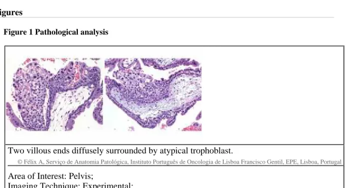
Figure 1 Pathological analysis
Two villous ends diffusely surrounded by atypical trophoblast.
© Félix A, Serviço de Anatomia Patológica, Instituto Português de Oncologia de Lisboa Francisco Gentil, EPE, Lisboa, Portugal
Area of Interest: Pelvis;
Imaging Technique: Experimental;
Procedure: Diagnostic procedure;
Special Focus: Pathology;
Chorionic villi with myxoid stroma and large central cisterns, and hyperplastic trophoblast.
© Félix A, Serviço de Anatomia Patológica, Instituto Português de Oncologia de Lisboa Francisco Gentil, EPE, Lisboa, Portugal
Area of Interest: Pelvis;
Imaging Technique: Experimental;
Procedure: Diagnostic procedure;
Special Focus: Pathology;
Figure 3 Axial T1WI (A) and T2WI (B)
The mass was isointense on T1WI (A) and presented high-signal intensity foci, probably
haemorrhagic. The assessment of deep myometrial extension and the exclusion of
parametrial involvement were better depicted on T2WI (B).
© Cunha TM, Serviço de Radiologia, Instituto Português de Oncologia de Lisboa Francisco Gentil, EPE, Lisboa, Portugal
Area of Interest: Pelvis;
Imaging Technique: MR;
Procedure: Diagnostic procedure;
Special Focus: Pathology;
Partial disruption of junctional zone was evident in the fundic region (A). The endometrial
cavity was filled with molarlike structures (A), which showed contrast enhancement and
were suggestive of invasive mole origin (B).
© Cunha TM, Serviço de Radiologia, Instituto Português de Oncologia de Lisboa Francisco Gentil, EPE, Lisboa, Portugal
Area of Interest: Pelvis;
Imaging Technique: MR;
Procedure: Diagnostic procedure;
Special Focus: Pathology;
Figure 5 Para-sagittal T2WI (A) and fat-suppressed postcontrast T1WI (B)
The mass has an aggressive appearance and invades the deep myometrium, which is also
demonstrated in the para-sagittal images (A, B).
© Cunha TM, Serviço de Radiologia, Instituto Português de Oncologia de Lisboa Francisco Gentil, EPE, Lisboa, Portugal
Area of Interest: Pelvis;
Imaging Technique: MR;
Procedure: Diagnostic procedure;
Special Focus: Pathology;
Figure 6 Axial CT of the chest (A, B)
Scattered well-defined pulmonary nodules highly suggestive of metastases (A, B).
Area of Interest: Pelvis;
Imaging Technique: MR;
Procedure: Diagnostic procedure;
Special Focus: Pathology;
MeSH
[C13] Female Genital Diseases and Pregnancy Complications
References
[1] Lurain J (2011) Gestational trophoblastic disease II: classification and management of gestational trophoblastic neoplasia Am J Obstet Gynecol 204(1):11-8
[2] Jung SE, Byun JY, Lee JM (2001) MR imaging of maternal disease in pregnancy AJR 177(6):1293-300
[3] Lurain JR (2010) Gestational trophoblastic disease I: epidemiology, pathology, clinical presentation and diagnosis of gestational trophoblastic disease, and management of hydatiform mole Am J Obstet Gynecol 203(6):531-9
[4] Green CL, Angtuaco TL, Shah HR (1996) Gestational trophoblastic disease: a spectrum of radiologic diagnosis Radiographics 16(6):1371-84
[5] Hammond C, Soper J editors (1990) Gestational Trophoblastic disease in Danforth's Obstetrics and Gynecology 6th ed Lippincott Philadelphia, USA