Case 10653
Unusual ovarian tumor in female teenager
Joana Ip , Teresa Margarida Cunha , Zita Seabra , Ana Félix1 1 2 1 Instituto Português de Oncologia de Lisboa Francisco Gentil, Lisbon
1
Hospital de Santo António dos Capuchos-CHLC, Lisbon
2
Instituto Portugues Oncologia de Lisboa
Genital (Female) Imaging Section:
2013, Jan. 25 Published:
13 year(s), female Patient:
Authors' Institution
Instituto Portugues de Oncologia de Lisboa Francisco Gentil, Lisbon, Portugal
Email:joana_fs@hotmail.com
Clinical History
A 13-year-old female, presented to her general practitioner with diffuse abdominal pain. An abdominal ultrasound was performed showing a large mass in the right adnexal area. She was otherwise healthy and all laboratory tests were within normal values but delta-4-androstenedione which was 6.73ng/dl (normal values: 0.46-3.39 ng/dl).
Imaging Findings
Abdominal ultrasound reported a solid inhomogeneous mass occupying the right adnexal area. Transvaginal ultrasound was not indicated since the patient had no prior sexual relations.
measuring 7x6, 5x7cm with well-defined contours. The exam showed hypointense and isointense areas on the T1-weighted images. The T2-weighted sequences demonstrated intermediate-to-high signal intensity and a 14mm-cyst in the periphery. A small amount of ascites was noticed in the right ovarian fossa. The left ovary and the uterus were unremarkable. There were no pathologic lymph nodes.
After surgical resection pathology confirmed the diagnosis of a poorly differentiated Sertoli-Leydig Cell Tumor (SLCT) with retiform areas.
The patient was treated with chemotherapy (bleomycin, ethoposide and cisplatinum) and maintains SLCT 4-year-remission.
Discussion
Sertoli-Leydig cell tumours are rare (0, 5%-1% of all ovarian tumours) and usually occur unilaterally in females under 30 years (mean 25 years) [1-3]. They are derived from the sex cord-stromal structures of the ovary, four subtypes are defined: well-differentiated,
intermediately-differentiated, poorly-differentiated and retiform. These tumours can cause
hyperandrogenism resulting in virilization in one-third to one-half of the patients [1, 4]. In our case the patient presented mildly elevated levels of testosterone derivates without virilization and abdominal pain. Regardless the initial size the majority of these are localized tumours (stage I) with favorable outcome [1, 3, 4].
On CT and MRI, SLCT are well-defined, enhancing solid masses with variable-sized intratumoral cysts. Calcifications are rare and enhancement varies according to the solid component. T1W images are usually isointense and T2W images are partially isointense or slightly hyperintense on the solid component and hyperintense in the cystic content [1, 3, 5]. On transvaginal ultrasound they appear as large isoechoic/hypoechoic tumours with cystic component often due to internal
haemorrhage or necrosis [2]. Our MRI depicted a large mass with a peripheral small cyst, with high intensity signal on T2W and low intensity on T1W.
In the differential diagnosis several tumour types arising in young patients and sharing a similar morphology should be considered.
Granulosa cell tumour is the most common sex cord-stromal tumour representing 70% of all sex cord-stromal tumours. Adult-type is more frequent affecting postmenopausal women. Presenting similar radiological features, the juvenile type may resemble to SLCT, often as large multiloculated tumour. The symptoms are related to elevated level of estrogens [1-2] and can present as isosexual pseudoprecocity. Our patient did not present these symptoms and MRI showed large tumour with small cyst rather than a large-cystic-tumour.
Fibromas and thecomas are solid masses with low signal intensity on T2-weighted images because they have abundant collagen. These tumours often have variable extent of calcification or
Sclerosing stromal cell tumour (SSCT) also a very rare tumour is usually a large mass with cystic and solid components usually without hormonal activity. On CT and MRI they show typical early peripheral enhancement with centripetal progression [2]. Since we did not have contrast-enhanced sequences we should consider a SSCT except for the levels of androstenedione.
Final Diagnosis
Sertoli-Leydig Cell Tumour
Differential Diagnosis List
Granulosa Cell Tumour (adult and juvenile type), Fibroma, Techoma, Sclerosing Stromal Cell Tumour
Figures
Figure 1 Axial T1-weighted image
The image shows large mass with heterogeneous low signal intensity on the right adnexa.
© Cunha TM, Radiology Department. Instituto Português de Oncologia de Lisboa Francisco Gentil, Portugal
Area of Interest: Genital / Reproductive system female;
Imaging Technique: MR;
Procedure: Diagnostic procedure;
Special Focus: Neoplasia;
The image shows a well-delineated right ovary mass with intermediate-to-high signal
intensity and a peripheral anteriorly located 14mm-cyst. A small amount of ascites is
detected.
© Cunha TM, Radiology Department, Instituto Português de Oncologia de Lisboa Francisco Gentil, Portugal
Area of Interest: Genital / Reproductive system female;
Imaging Technique: MR;
Procedure: Diagnostic procedure;
Special Focus: Neoplasia;
The image shows a well delineated right ovary mass with intermediate-to-high signal
intensity. A small amount of ascites is detected.
© Cunha TM, Radiology Department, Instituto Português de Oncologia de Lisboa Francisco Gentil, Portugal
Area of Interest: Genital / Reproductive system female;
Imaging Technique: MR;
The image shows a well delineated right ovary mass with intermediate-to-high signal
intensity.
© Cunha TM, Radiology Department, Instituto Português de Oncologia de Lisboa Francisco Gentil, Portugal
Area of Interest: Genital / Reproductive system female;
Imaging Technique: MR;
Procedure: Diagnostic procedure;
Special Focus: Neoplasia;
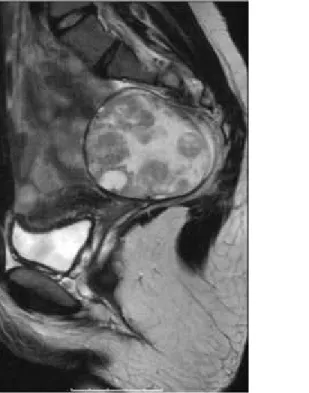
Figure 3 Sagittal T2-weighted images
The image shows the mass with intermediate-to-high signal intensity.
© Cunha TM, Radiology Department, Instituto Português de Oncologia de Lisboa Francisco Gentil, Portugal
Area of Interest: Genital / Reproductive system female;
Imaging Technique: MR;
The image shows the mass with intermediate-to-high signal intensity and a peripheral
anteriorly located 14mm-cyst. Minimal amount of fluid is also seen.
© Cunha TM, Radiology Department, Instituto Português de Oncologia de Lisboa Francisco Gentil, Portugal
Area of Interest: Genital / Reproductive system female;
Imaging Technique: MR;
Procedure: Diagnostic procedure;
Special Focus: Neoplasia;
Figure 4 Coronal T2-weighted image
The image shows the mass with intermediate-to-high signal intensity and a peripheral
anteriorly located 14mm-cyst. Minimal amount of fluid is also seen.
© Cunha TM, Radiology Department, Instituto Português de Oncologia de Lisboa Francisco Gentil, Portugal
Imaging Technique: MR;
Procedure: Diagnostic procedure;
Special Focus: Neoplasia;
Figure 5 SCLT with retiform pattern
Tumour area with microcystic spaces surrounded by edematous stroma.
© Félix A, Pathology Department, Instituto Português de Oncologia de Lisboa Francisco Gentil, Lisbon, Portugal
Area of Interest: Genital / Reproductive system female;
Imaging Technique: MR;
Procedure: Surgery;
Special Focus: Neoplasia;
Figure 6 Poorly differentiated
Tumour shows nodule of poorly differentiated cells admixed with gonadal stroma. Mitotic
activity is present.
© Félix A, Pathology Department, Instituto Português de Oncologia de Lisboa Francisco Gentil, Lisbon, Portugal
Area of Interest: Genital / Reproductive system female;
Imaging Technique: MR;
Special Focus: Neoplasia;
MeSH
[C13.371.270.750] Ovarian Neoplasms
Tumors or cancer of the OVARY. These neoplasms can be benign or malignant. They are classified according to the tissue of origin, such as the surface epithelium, the stromal endocrine cells, and the totipotent germ cells.
References
[1] Shanbhogue AK, Shanbhogue DK, Prasad SR, Surabh VR, Fasih N, Menias CO (2010) Clinical Syndromes associated with ovarian neoplasms Radiographics 30 (4): 903-19
[2] Jung SE, Rha SE, Lee JM, Park SY, Oh SN, Cho KS, Lee EJ, Byun JY, Hahn ST (2005) CT and MRI Findings of Sex Cord-Stromal Tumor of the Ovary AJR Am J Roentgenol 185 (1): 207-15
[3] Jung SE, Lee JM, Rha SE, Byun SY, Jung JI, Hahn ST (2002) CT and MR Imaging of Ovarian Tumors with Emphasis on Differential Diagnosis Radiographics 22: 1305-1325
[4] Tanaka YO, Tsunoda H, Kitagawa Y, Ueno T, Yoshikawa H, Saida Y (2004) Functioning Ovarian Tumors: Direct and Indirect Findings at MR Imaging Radiographics 24: S147-S166
[5] Iyer VR, Lee S (2010) MRI, CT, and PET/CT for Ovarian Cancer Detection and Adnexal Lesion Characterization AJR Am J Roentgenol 194 (2): 311-21
Citation
Joana Ip , Teresa Margarida Cunha , Zita Seabra , Ana Félix1 1 2 1 Instituto Português de Oncologia de Lisboa Francisco Gentil, Lisbon
1
Hospital de Santo António dos Capuchos-CHLC, Lisbon (2013, Jan. 25)
2