Case 10870
Sclerosing stromal tumor of the ovary
Ana Loureiro ; Teresa Margarida Cunha ; Ana Félix 1 1 2
Genital (Female) Imaging Section:
2013, May. 10 Published:
14 year(s), female Patient:
Authors' Institution
Instituto Português de Oncologia de Lisboa Francisco Gentil, Department of Radiology, Lisbon,
1
Portugal
Instituto Português de Oncologia de Lisboa Francisco Gentil, Department of Pathology, Lisbon,
2
Portugal
Clinical History
A 14-year-old girl without relevant clinical history presented with pyelonephritis.
On the renal and pelvic US performed for the evaluation of pyelonephritis, a solid mass in the left adnexal area was found.
The patient had normal levels of CA-125, CEA, -FP, and -hCG.
Imaging Findings
A pelvic MRI was performed and documented a well-defined solid ovoid mass with 8cm larger dimension, on the left adnexal area. This mass had homogeneous low-signal intensity on
T1-weighted images (T1WI). The T2-weighted images (T2WI) showed central high-signal intensity areas with a peripheral rim of low-signal intensity. Contrast-enhanced MRI revealed early avid contrast enhancement of the periphery. There was a small amount of ascites on the Douglas recess. The uterus and the right ovary didn't show any abnormalities.
The patient underwent surgical excision of the left adnexa.
measuring 8 x 7 x 5cm, of the left ovary, soft in consistency and slightly lobulated, compatible with a sclerosing stromal tumour of the ovary.
Discussion
Sclerosing stromal tumours (SST) are an extremely rare sub-type of sex-cord stromal ovarian tumours, and were described for the first time by Chalvardjian and Scully in 1973 [1].
This tumour occurs predominantly in women younger than 30-year-old, different from other types of stromal tumours [1, 2].
The most common presenting symptom is pelvic pain. It is often hormonally inactive although there are few reports of irregular menses, genital bleeding or virilization, more common during
pregnancy.
Histologically, this tumour is characterized by cellular pseudolobules, interlobular fibrosis, marked vascularity and a dual cell population (collagen-producing spindle cells and lipid containing round or ovoid cells). The heterogeneity due to the variation in cellular size and shape are helpful features in the differential diagnosis of SST, and contrasts with the relative homogeneity of thecoma and fibromas [3-6].
Sonographically SSTs are described as unilateral, multilocular cystic or solid masses with multiple small cysts or clefts. Color Doppler imaging shows increased vascularity in the periphery and between the cysts. A small amount of ascites may be present [3, 4].
On T2WI shows hyperintense cystic areas and hypointense nodules located inside the hyperintense stroma, corresponding to the pseudolobulation that characterizes histologically the SST, and a thin rim of low-signal intensity in the periphery. T1WI may show the same thin low-signal intensity outer rim that corresponds to the compressed ovarian cortex and a low-signal intensity central area. At dynamic contrast-enhanced imaging, early and strong peripheral enhancement with centripetal progression is characteristic. Lack of enhancement of the central area, even on delayed images, correspond to collagenous acellular areas [2, 5, 6].
The differential diagnosis should include in the pediatric and young adult population: Sertoli-Leydig cell tumour, granulosa cell tumour, cystadenofibroma; and in adults: thecoma/fibroma, malignant epithelial ovarian tumours and metastases.
Sertoly-Leydig cell tumour presents as a solid, well-defined, enhancing mass that can have some cystic/necrotic components, in a young woman with virilization. Granulosa cell tumour most commonly present as a multilocular cystic/solid mass with cystic components, low-signal on T2WI, in a patient with a thickened endometrial stripe due to tumour estrogens secretion.
Cystadenofibrona can present as a complex unilocular/multilocular cystic mass with papillary solid projections.
Fibromas/thecomas usually appear with low-signal intensity on T2WI, and have a progressive enhancement.
Ovarian metastases and malignant epithelial tumours are more common in older patients and usually don't show the progressive centripetal enhancement [6].
Final Diagnosis
Sclerosing stromal tumour of the ovary.
Differential Diagnosis List
Sertoli-Leydig Cell Tumour , Ovarian Fibroma / Thecoma, Granulosa Cell Tumour ,
Dysgerminoma, Ovarian Carcinoma, Krukenberg Tumour, Metastases to the Ovary, Massive Ovarian Edema
Figures
Figure 1 T2-weighted MR images show a left ovarian mass.
Axial T2-weighted MRI shows an heterogeneous mass, well-circumscribed, with regular
contours, revealing a low-signal intensity outer rim and central intermediate-signal intensity
nodules alternate with high-signal intensity cystic areas. This pseudolobular pattern
characterizes this tumor.
© Cunha TM, Department of Radiology, Istituto Português de Oncologia de Lisboa Francisco Gentil, Lisbon, Portugal
Area of Interest: Genital / Reproductive system female;
Imaging Technique: MR;
Coronal T2-weighted MR image shows the pseudolobular pattern that characterizes this
tumor: intermediate-signal intensity nodules that alternate with the high-signal intensity
cystic areas.
© Cunha TM, Department of Radiology, Istituto Português de Oncologia de Lisboa Francisco Gentil, Lisbon, Portugal
Area of Interest: Genital / Reproductive system female;
Imaging Technique: MR;
Procedure: Diagnostic procedure;
Special Focus: Neoplasia;
Sagittal T2-weighted MR image shows the pseudolobulation of the tumor.
© Cunha TM, Department of Radiology, Istituto Português de Oncologia de Lisboa Francisco Gentil, Lisbon, Portugal
Procedure: Diagnostic procedure;
Special Focus: Neoplasia;
Figure 2 Axial T1-weighted MR images.
Non-enhanced T1-weighted MR image reveals a mass with homogeneous low-signal
intensity.
© Cunha TM, Department of Radiology, Istituto Português de Oncologia de Lisboa Francisco Gentil, Lisbon, Portugal
Area of Interest: Genital / Reproductive system female;
Imaging Technique: MR;
Procedure: Diagnostic procedure;
Special Focus: Neoplasia;
Fat-suppressed image, after gadolinium enhancement, shows avid enhancement of the
periphery of the mass.
© Cunha TM, Department of Radiology, Istituto Português de Oncologia de Lisboa Francisco Gentil, Lisbon, Portugal
Area of Interest: Genital / Reproductive system female;
Imaging Technique: MR;
Procedure: Diagnostic procedure;
Special Focus: Neoplasia;
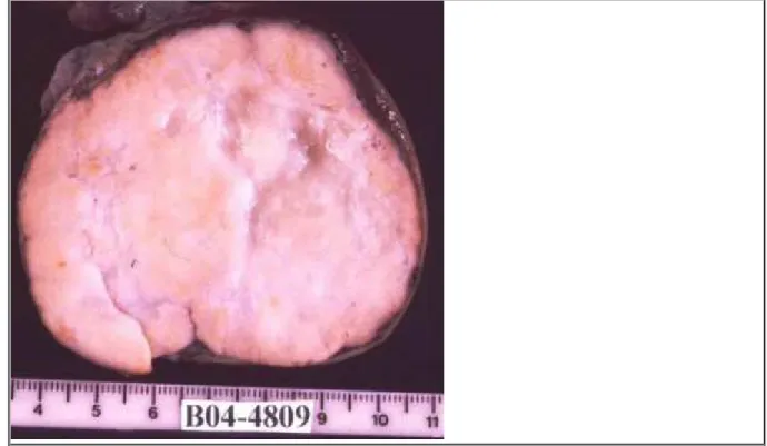
Yellowish tone solid mass, soft in consistency and slightly lobulated, with a central
oedematous area.
© Félix A, Department of Pathology, Istituto Português de Oncologia de Lisboa Francisco Gentil, Lisbon, Portugal
Area of Interest: Genital / Reproductive system female;
Imaging Technique: Experimental;
Procedure: Diagnostic procedure;
Special Focus: Neoplasia;
Figure 4 Microscopic examination.
Pseudolobulation is created by cellular and fibrous areas (H&E 1x).
Area of Interest: Genital / Reproductive system female;
Imaging Technique: Experimental;
Procedure: Diagnostic procedure;
Special Focus: Neoplasia;
Interface between cellular and fibrous areas (H&E 10x).
© Félix A, Department of Pathology, Istituto Português de Oncologia de Lisboa Francisco Gentil, Lisbon, Portugal
Area of Interest: Genital / Reproductive system female;
Imaging Technique: Experimental;
Procedure: Diagnostic procedure;
Special Focus: Neoplasia;
Cellular area with large polyhedral and spindle tumor cells (H&E 40x).
© Félix A, Department of Pathology, Istituto Português de Oncologia de Lisboa Francisco Gentil, Lisbon, Portugal
Area of Interest: Genital / Reproductive system female;
Imaging Technique: Experimental;
MeSH
[A05.360.319.114.630] Ovary
The reproductive organ (GONADS) in female animals. In vertebrates, the ovary contains two functional parts: the OVARIAN FOLLICLE for the production of female germ cells
(OOGENESIS); and the endocrine cells (GRANULOSA CELLS, THECA CELLS, and LUTEAL CELLS) for the production of ESTROGENS and PROGESTERONE.
[C13.371] Genital Diseases, Female
[C13.371.056] Adnexal Diseases
Diseases of the uterine appendages (ADNEXA UTERI) including diseases involving the OVARY, the FALLOPIAN TUBES, and ligaments of the uterus (BROAD LIGAMENT; ROUND
LIGAMENT).
[C13.371.056.630] Ovarian Diseases
Disorders of ovarian function. They have varied pathology depending on the phase of the reproductive life from fetal stage to adulthood.
[C13.371.056.630.705] Ovarian Neoplasms
Tumors or cancer of the OVARY. These neoplasms can be benign or malignant. They are classified according to the tissue of origin, such as the surface epithelium, the stromal endocrine cells, and the totipotent germ cells.
[C13.371.270] Genital Neoplasms, Female
[E01.370.350.500] Magnetic Resonance Imaging
Non-invasive method of demonstrating internal anatomy based on the principle that atomic nuclei in a strong magnetic field absorb pulses of radiofrequency energy and emit them as radiowaves which can be reconstructed into computerized images. The concept includes proton spin tomographic techniques.
References
[1] Chalvardjian A, et al (1973) Sclerosing stromal tumors of the ovary Cancer Mar; (3):664-70
[2] Matsubayashi R, et al (1999) Sclerosing stromal tumor of the ovary: radiologic findings Eur Radiol. 9(7):1335-8
[3] Fox H, et al (2003) Obstetrical and Gynecological Pathology 5th Ed. 842-3
[4] Lee MS, et al (2001) Ovarian sclerosing stromal tumors: gray scale and color Doppler sonographic findings J Ultrasound Med Apr; (4):413-7
[5] Hricak H, et al (2007) Diagnostic Imaging Gynecology 1th Ed. 7-40 - 7-42
[6] Kim JY, et al (2003) Sclerosing stromal tumor of the ovary: MR-pathologic correlation in three cases Korean J Radiol Jul-Sep; 4(3):194-9