biomolecules
ISSN 2218-273X www.mdpi.com/journal/biomolecules/ Review
The Interplay between Alpha-Synuclein Clearance and Spreading
Tomás Lopes da Fonseca 1,2,†, Anna Villar-Piqué 1,† and Tiago Fleming Outeiro 1,2,3,*
1 Department of Neurodegeneration and Restorative Research, Center for Nanoscale Microscopy
and Molecular Physiology of the Brain, University Medical Center Göttingen, Göttingen 37073, Germany; E-Mails: [email protected] (T.L.F.); [email protected] (A.V.-P.)
2 Instituto de Fisiologia, Faculty of Medicine, University of Lisbon, Lisboa 1649-028, Portugal 3 CEDOC, Faculdade de Ciências Médicas, Universidade Nova de Lisboa, Lisboa 1150, Portugal
† These authors contributed equally to this work.
* Author to whom correspondence should be addressed; E-Mail: [email protected].
Academic Editor: Stephan N. Witt
Received: 17 March 2015 / Accepted: 7 April 2015 / Published: 14 April 2015
Abstract: Parkinson’s Disease (PD) is a complex neurodegenerative disorder classically characterized by movement impairment. Pathologically, the most striking features of PD are the loss of dopaminergic neurons and the presence of intraneuronal protein inclusions primarily composed of alpha-synuclein (-syn) that are known as Lewy bodies and Lewy neurites in surviving neurons. Though the mechanisms underlying the progression of PD pathology are unclear, accumulating evidence suggests a prion-like spreading of -syn pathology. The intracellular homeostasis of -syn requires the proper degradation of the protein by three mechanisms: chaperone-mediated autophagy, macroautophagy and ubiquitin-proteasome. Impairment of these pathways might drive the system towards an alternative clearance mechanism that could involve its release from the cell. This increased release to the extracellular space could be the basis for -syn propagation to different brain areas and, ultimately, for the spreading of pathology and disease progression. Here, we review the interplay between -syn degradation pathways and its intercellular spreading. The understanding of this interplay is indispensable for obtaining a better knowledge of the molecular basis of PD and, consequently, for the design of novel avenues for therapeutic intervention.
1. Introduction
1.1. Parkinson’s Disease
Parkinson’s disease (PD) is the most common neurodegenerative disorder with movement impairment. At the clinical level, the disease is classically characterized by resting tremor, bradykinesia, postural instability, and muscular rigidity [1]. Although PD was initially classified as a movement disorder, it is now well accepted that non-motor symptoms precede and follow motor disabilities. Thus, PD is currently regarded as a disorder affecting the whole-brain [2]. In fact, hyposmia is one of the prevalent symptoms in early stages of the disease, with several olfactory-related brain areas found severely affected in PD patients [3–8].
Pathologically, PD is characterized by the degeneration of dopaminergic neurons in the substantia nigra pars compacta, and the accumulation of intracellular, proteinaceous inclusions in the surviving neurons, known as Lewy bodies (LBs) and Lewy neurites (LNs) [1].
According to Braak’s staging theory, the evolution of LB pathology is thought to initiate in either the lower brainstem or in the olfactory bulb. Then, as the disease progresses, LB pathology appears in other brain areas, including the cerebral cortex, impacting different neural networks and leading to non-motor symptoms such as depression, cognitive decline and hallucination episodes [4,9–11]. Defining the responsible mechanism for PD pathology progression has challenged the scientific community for almost 200 years. During the last decade, the “prion-like” hypothesis has gained particular emphasis based on findings from independent experiments in PD patients. In patients that received grafts of normal fetal mesencephalic brain tissue, a time-dependent accumulation of LBs and reduced immunostaining for dopamine transporter was observed [12–16].
Only a minority of PD cases have been linked to genetic factors (less than 10%) with the majority being considered sporadic, of unknown origin. Until now, more than 20 genes have been associated with PD [17], but this number is expected to increase as new studies including larger number of patients are conducted. The first gene linked to familial forms of PD was SNCA, encoding for the protein alpha-synuclein (-syn). Thus far, mutations, as well as duplication or triplications of the SNCA gene, are associated with familial forms of PD [18–25]. Additionally, recent studies also revealed that polymorphisms in the SNCA gene lead to increased risk for developing PD [26–29]. Thus, for all of the above, -syn is regarded as one of the major culprits in both genetic and idiopathic forms of PD.
1.2. Alpha-Synuclein
the protein due to abundance of glutamates and aspartates. These negative residues appear to have an important effect in modulating -syn aggregation [35] and, in fact, this domain is essential for the formation of calcium-mediated annular oligomers via direct binding [36,37]. In addition, the C-terminus of -syn has been suggested to play a role on the protein’s chaperone-like activity [38].
For years, -syn was regarded as a natively unfolded, monomeric protein. Recently, some studies proposed it may occur as a natively folded helical tetramer [39–41], but this subject remains highly controversial [42]. Nevertheless, the possibility that -syn occurs naturally in a folded state brought new perspectives into the process of its oligomerization and aggregation, one of the major mysteries in the field [17]. Major efforts have been placed in understanding which form of -syn acts as the toxic species but consensus has not been reached. Currently, it is generally accepted that smaller oligomeric species are more toxic than larger aggregated forms [43–45]. However, some authors still claim that the final mature aggregates are the most dangerous for cell homeostasis [46,47].
-syn is prone to several types of posttranslational modifications (PTMs). Ubiquitination [48–51], sumoylation [52–54] and N–terminal acetylation [55–57] have been described. In addition, -syn can be phosphorylated [58] in two serines (S129 and S87) and three tyrosines (Y125, Y133 and Y135). It is estimated that approximately 90% of the -syn present in LBs is phosphorylated in S129 [59]. Several kinases, including CKs, PLKs and GRKs, can phosphorylate -syn on S129 [58,60–65]. Interestingly, some of those enzymes were found up-regulated in PD brains [63] or present in LBs [61,66]. S129 phosphorylation can inhibit -syn fibrillization [67,68], and a similar effect was observed for S87 [69]. Unfortunately, the full functional relevance of phosphorylation in both physiological and pathological contexts is elusive [70–72].
In contrast, nitration of tyrosine residues in -syn (Y39, Y125, Y133, Y136) is known to produce toxic effects. In particular, nitrated -syn is present in LBs [73] and nitrated -syn oligomers promote mitochondrial impairment and cell death in mammalian cell culture [74]. Furthermore, administration of nitrated -syn in the substantia nigra of rats induces severe dopaminergic neuronal cell death, and the down-regulation of striatal dopamine and dopamine receptor D2 [75]. Nitration on Y39 blocks -syn fibril formation and reduces monomer degradation via the ubiquitin proteasome system [76].
Finally, truncated -syn is found in LBs and in animal models and it is thought that some familial -syn mutations might promote its truncation [48,77,78]. In vitro, C-terminally truncated -syn enhances fibril assembly and induces fibril formation of full-length -syn [79,80]. Nevertheless, the opposite effect was reported upon calpain1 or neurosin mediated truncation [81–84]. Interestingly, these two proteases cleave -syn near the NAC domain highlighting the importance of this region on -syn aggregation. 2. Protein Degradation Systems
lysosome [88]. Therefore, and considering the purpose of this review, we will focus specifically on the pathways culminating in the lysosomal compartment.
Autophagy (meaning “self-eating”, in Greek) consists in the process of decomposition and degradation of cellular components and organelles via the lysosomal compartment. Autophagy itself serves two main purposes: the clearance of deleterious intracellular components, and the recycling of macromolecules from functional pre-existing organelles and proteins to guarantee proteome renewal [89].
Depending on the cargo delivery method, autophagy can be divided in three main types: chaperone-mediated autophagy (CMA), macroautophagy and microautophagy. To our knowledge, there is presently no evidence linking -syn to microautophagy. Thus, here we focus only on CMA and macroautophagy.
2.1. -Syn and CMA—A Symbiotic Relation with a “Do not Disturb” Sign
CMA is a particular cellular mechanism for protein degradation linked to the lysosome. Unlike other degradation systems, CMA is based on the recognition of a specific amino acid sequence (KFERQ) [90]. This motif is found in nearly 30% of cytoplasmic proteins, including -syn, and in some compartment-associated proteins [90]. Protein misfolding, partial folding or PTMs, such as phosphorylation and acetylation, can promote or inhibit the CMA pathway [91]. Mechanistically, CMA relies on the proper identification and binding of cytosolic Hsc70 (cHsc-70) to the target substrate [92]. This complex is later directed to the lysosomal membrane where it interacts with the lysosome-associated membrane protein type 2a (LAMP2A) [93,94]. The final step of this translocation process requires the lysosome-associated Hsc70 (lHsc70) that targets the substrate to degradation (Figure 1A) [95].
The reciprocal interaction between -syn and CMA protein degradation has been of high interest in the last decade. Using in vitro purified lysosomes from liver, it was initially described that -syn can be actively degraded via CMA and, more interestingly, that two -syn familial mutations, A30P and A53T, impair CMA degradation (Figure 1D). Both -syn mutants exhibited higher affinity to the LAMP2A receptor, blocking CMA at the LAMP2A level which, ultimately, leads to a full impairment of the pathway [85]. Later on, it was shown that -syn can be degraded through this pathway, in several cell culture models [96]. In mice, increase of both LAMP2A and Hsc70 levels was observed upon overexpression of wild-type (wt) -syn [97]. In a comprehensive study on -syn PTMs, a slight inhibition of CMA was observed upon nitration and oxidation (Figure 1B). Furthermore this pathway is stalled when -syn is phosphorylated (Figure 1C) or exposed to dopamine. Interestingly, just the latter can also completely block the CMA degradation machinery (Figure 1D) [98]. The previous report also proved that CMA is only capable of degrading monomers and dimers of -syn, but not oligomers [98].
In addition to the direct effect of -syn on CMA, this interplay may also impact other degradation pathways. In particular, the A53T -syn mutation can block CMA leading to an activation of macroautophagy and an increase of toxicity cultured cells [99].
Recently, microRNAs (miRNAs) gained attention also in the context of -syn degradation via CMA. Interestingly, miRNAs have been linked to PD [104], and several miRNAs targeting the CMA pathway (LAMP2A or Hsc70) have been described. The majority was found to impair -syn degradation and potentially alter the aggregation state of the protein [105,106]. In an initial report, four miRNAs were found to reduce LAMP2A levels while three others directly down-regulated Hsc70. In all cases an increase in -syn was observed. Moreover, these 7 miRNAs were found to be up-regulated in the substantia nigra from PD patients [106], correlating with a decrease in both CMA related protein levels. A more recent study identified another miRNA, miRNA320a, although it had been previously discarded [105,106]. This particular miRNA320a has been linked to a panoply of diseases ranging from fibromyalgia [107] to Waldenström macroglobulinemia [108] and more recently to the Ebola virus [109].
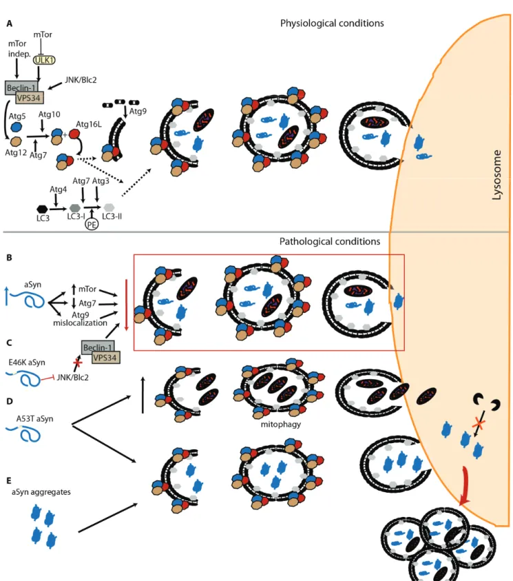
Figure 1. -syn and the CMA. (A) Under physiological conditions, Hsc70 recognizes the KFERQ domain of -syn (1) and targets the protein towards the lysosome (2). At the lysosomal membrane, -syn interacts with LAMP-2A and promotes its oligomerization (3) leading to the entrance of the protein into the lysosome (4). Once inside the lysosome, -syn is degraded by proteases (5); (B) PTMs such as oxidation and nitration slightly inhibit this pathway and reduce -syn degradation; (C) Phosphorylation of -syn on S129 impairs its degradation via CMA. However, while the phosphorylated form of protein does not block this pathway (D), dopamine-modified -syn and some familial mutations (A30P and A53T) that are also not degraded via CMA, can block this pathway and prevent the degradation of other CMA substrates.
2.2. Macroautophagy of -Syn—The Last Resource to Avoid Protein Aggregation
Macroautophagy, commonly referred to as “autophagy”, is the most scrutinized and well known of the three autophagic mechanisms. This “content-blind” pathway relies on the formation of de novo double membrane-bound vesicles to sequester intracellular components, including whole organelles, towards the lysosome [110,111]. This membrane formation mainly relies on the autophagy related protein (Atg) 9, both in yeast and humans [112–115]. Macroautophagy is found constitutively active but further activation via the mTOR pathway, the mammalian target of rapamycin [116], or the PI3kinase/beclin/vsp34 pathway, also known as mTOR-independent pathway, is possible [117]. Autophagosome formation requires two ubiquitination steps highly regulated by Atg proteins [118–120]. Initially Atg12 is conjugated with Atg5, a process involving Atg7 and Atg10 [121,122]. The Atg12-Atg5 complex is later targeted to the autophagosome with Atg16 [123,124]. The second ubiquitination step requires Atg8 (also known by LC3). LC3 is C-terminally cleaved by Atg4 to form LC3-I [125,126], which is then conjugated to the lipid phosphatidylethanolamine (PE) by Atg7 and Atg3 to generate LC3-II [122,127]. Interestingly, the Atg12-Atg5 complex originated from first ubiquitination seems to be necessary for the LC3 processing and localization at the autophagosome membrane (Figure 2A) [128,129].
Depending on the cargo being degraded, alternative players can be involved in the task. In the case of protein aggregates, an alternative has been reported and is usually referred to as aggrephagy. Mechanistically, aggrephagy can be divided in two pathways: HDAC6, or BAG-3 mediated. The first requires the addition of lysine (K) 63-poliubiquitination chains to the substrate. This can be achieved with the involvement of the PD-related protein Parkin [130]. HDAC6 can then recognize the substrate and translocate it, in a microtubule-dependent manner, towards the aggregosome [130,131]. Once in the aggregosome, the aggregate degradation requires the intervention of p62 and NBR1 that can work separately or possibly in a duet, since they can interact [132]. Importantly, both proteins interact with PE-LC3, an essential component of the autophagosome membrane [133–135]. Both p62 and NBR1 also interact with K63-poliubiquitinated chains suggesting that this PTM could be essential for HDAC6-mediated aggrephagy [135–137].
-syn degradation via macroautophagy has been mostly studied using specific inhibitors and enhancers of this pathway. Both in cell culture or in vivo, wt and mutated -syn can be degraded through macroautophagy, a process that may be at least partly modulated by Beclin-1 [142–144]. Furthermore, blockade of this pathway generally leads to the accumulation of high molecular weight species of -syn, although different macroautophagy inhibitors reveal distinct outcomes [88,96]. Another line of evidence indicates that macroautophagy inhibitors affect exclusively mutant A53T -syn, while other studies describe different behaviors that vary according to the selected inhibitor [102,145]. In yeast, an organism lacking CMA, our group recently demonstrated that -syn phosphorylation on S129 can modulate the protein degradation via macroautophagy and, ultimately, inclusion formation [68]. In yeast, it was also described that the interplay between phosphorylation and sumoylation has a direct impact on the autophagic degradation of -syn [54]. In general, it is believed that macroautophagy is crucial for the lysosomal degradation of oligomeric and aggregated -syn, since CMA is unable to handle large protein species [98,146]. Once inside the lysosome, -syn is mainly degraded by Cathepsin D (CTSD) [147]. In cells, overexpression of an inactive mutant of this aspartyl protease leads to increased -syn levels [148] while CTSD knockout mice exhibited accumulation of higher molecular weight -syn species [149].
Interestingly, -syn can also have a direct impact on this lysosomal degradation pathway. -syn overexpression can inhibit macroautophagy via an interaction with Rab1a that culminates in a mislocalization of Atg9 [150]. Furthermore, an increase in mTor and decrease in Atg7 levels has been observed in both Dementia with LB (DLB) patients and -syn transgenic mice (Figure 2B) [151]. The same study also reported the presence of enlarged autophagosomes and lysosomes, as observed in cells overexpressing -syn [142]. In a recent work, -syn aggregates were able to resist macroautophagy leading to a failure of the pathway and accumulation of autophagosomes (Figure 2E) [47].
In addition to the effects of wt -syn on autophagy, mutations associated with familial forms of PD also seem to differentially affect this protein degradation pathway. In the E46K -syn mutant, an impairment of autophagy is observed, via inactivation of the JNK1/Blc2 pathway (Figure 2C) [152]. While the A53T mutation seems to promote increased mitophagy [153] and the accumulation of autophagosomes due to impaired degradation (Figure 2D) [154].
In summary, and despite conflicting evidence, it seems unquestionable that there is a strong interplay between -syn and autophagy pathways, and that this may enable therapeutic interventions.
3. Prion-Like Spreading of -Syn Pathology
For the last decade, it has been postulated that neurodegeneration-related proteins, which are usually present in an abnormal aggregated state, can intercellularly transmit their abnormal conformation to homotypic native proteins. Here, we focus on the concept of spreading in the context of the overall cellular protein homeostasis. In this regard, protein degradation is a key mechanism that undoubtedly influences -syn propagation, although its precise modulatory effect is still unclear.
3.1. Insight from Pathological Studies
of the prion-like spreading of -syn pathology. Although Braak’s PD staging could be merely due to the differential disease susceptibility among distinct brain regions, the stereotypic pattern and location of the zones affected in each stage suggests a spatial-temporal dissemination of the -syn pathology, which indeed correlates with cognitive decline [155]. From the first regions affected, the olfactory bulb and brainstem, -syn pathology spreads caudo-rostrally to susceptible midbrain regions and to subcortical and cortical areas as PD progresses [4]. The initial sites where -syn lesions appear led Braak and co-workers to postulate the theory that indeed sporadic -syn pathology is initiated by an exogenous pathogenic agent [156,157], although experimental evidence for such agent is still lacking.
The discovery of LBs in young neurons that were grafted into the brains of PD patients boosted the idea of the prion-like spreading of pathology in the brain [15,16]. The grafted cells were obtained from human embryos and the transplantation was performed only 11–16 years before the pathological examination of the brains, upon death of the patients. Since this period of time is extremely short for -syn pathology to spontaneously develop under normal conditions [15,16,158], these findings suggested that -syn pathology could be transmitted from diseased to healthy cells. Among the distinct agents and factors that could be responsible for transmitting the pathology, the misfolded/aggregated state of -syn was a strong contender [159], establishing the basis for the current hypothesis of the prion-like spreading of -syn pathology. The -syn that accumulated in the grafted neurons displayed the same characteristics of LBs and LNs in the host tissue [160], suggesting a common gradual formation mechanism. In addition, those cells were also described to suffer a degenerative process and loss of dopaminergic phenotype [161,162]. The presence of LBs in young grafted neurons has been repeatedly confirmed in experiments in rodents overexpressing human -syn. In both mice [163] and rats [164,165], the time-dependent transfer of -syn from the host to the graft was demonstrated by the presence of human -syn puncta in the transplanted cells. In rats, the inclusions formed were described to consist of a core of human -syn surrounded by endogenous rat -syn, clearly suggesting a seeding mechanism [164].
Another relevant finding was the discovery of -syn in human body fluids, such as cerebrospinal fluid (CSF) and blood plasma [166,167]. Taking into account that -syn is traditionally considered a cytoplasmic protein, its extracellular presence could be associated to passive release caused by cell death and neurodegeneration. However, the similar levels of monomeric -syn found in the CSF and plasma of PD and control individuals hinted at a physiological secretion process [166,167]. Unfortunately, there is still no consensus on the role of extracellular -syn and whether this is related to pathological conditions. In an attempt to clarify this issue, some studies suggest the occurrence of increased levels of the -syn oligomeric fraction in the CSF of patients with synucleinopathies [168,169].
Overall, these studies indicate that, on one side, there is a fraction of extracellular -syn present in physiological conditions and, on the other, that there might be an increase in extracellular -syn oligomeric species in diseased individuals. This needs to be further confirmed but requires the development of novel and better tools, such as antibodies that recognize specific types of -syn species and/or conformers.
3.2. The Proof of Concept: in Vivo -Syn Propagation Experiments
human A53T -syn triggered an early onset of the pathology with expected clinical and biochemical hallmarks, when injected into the brains of young asymptomatic mice [170,171]. The same result was obtained when using recombinant human -syn fibrils assembled in vitro [170]. This finding was crucial to demonstrate that -syn pathology can be accelerated and spread through the brain after inoculation with pathological material. The acceleration of the pathology was interpreted as a seeding effect of the exogenous material over the otherwise slow and age-dependent aggregation of the endogenously expressed -syn.
Subsequent experiments provided additional insight into the process. Injecting recombinant mouse -syn fibrils in the dorsal striatum of young wt mice induced pathological -syn transformation leading to motor deficits [172]. Similar induction of -syn pathology was described in another study comparing the effect of recombinant human -syn and of insoluble brain fractions extracted from DLB patients [173]. -Syn accumulation was described in several brain regions distant from the injection site, demonstrating the propagation capacity of the injected material [172,173]. In addition to the biochemical hallmarks, the exogenous induction of -syn pathology also includes a progressive nigrostriatal neurodegeneration in the recipient animals as demonstrated by the injection of PD brain extracts into the brains of wt mice [174]. Data derived from experiments performed with recombinant protein or with brain-derived pathological samples should be interpreted in a complementary way. On one hand, using brain homogenates demonstrates that biological protein material is able to induce homotypic pathology in another organism, a primary characteristic of prion diseases. On the other hand, experiments using recombinant -syn fibrils enable the confirmation that -syn is the major component for pathology induction. Interestingly, it was demonstrated that mouse -syn fibrils displayed a higher efficiency in inducing a synucleinopathy in wt mice than human -syn fibrils, suggesting the existence of a species barrier effect [173,175]. Finally, similar experiments were performed in monkeys injected with LB-fractions derived from sporadic PD brains, demonstrating that the induction of a synucleinopathy also occurs in non-human primates and that, in the context of propagation of -syn pathology, the monkey-human species barrier is, at best, rather soft [174].
Despite existing discrepancies regarding the methodologies used in different works [176], and the number of open questions that need to be answered [177], the recent studies demonstrate that synucleinopathies can be induced by external agents, which raises the issue of whether these neurological disorders can also have an infectious origin, even if they do not appear to be contagious.
3.3. Mechanisms of -Syn Cell-to-Cell Propagation
Although the prion-like spreading of -syn pathology hypothesis is becoming widely accepted, little is still known about the molecular mechanisms underlying this process. It is possible that the mechanisms of -syn propagation are probably dependent on the cell type, their physiological state, and the type of -syn species transmitted. The various possible mechanisms have been extensively reviewed by many in the field [17,178].
ongoing neurodegenerative process. However, the lack of a clear increase in -syn CSF levels between diseased and control individuals [179] limits the likelihood of this process as the main mechanism of release.
Given the membrane-binding capacity of -syn, another possibility is that secretion might take place through an unconventional ER/Golgi-independent secretion pathway [180], or exosome-mediated pathway [181,182]. Indeed, the role of exosomes on the spreading of -syn pathology is gaining attention, and it was recently shown that exosomes isolated from plasma of PD patients contain higher levels of -syn when compared to exosomes from control individuals [183,184].It is also possible that exosomal encapsulation of -syn might confer partial protection against extracellular protein degradation mechanisms. However, the precise role of exosomes in the spreading of -syn pathology needs to be further detailed.
Other putative mechanisms involved in cell-to-cell transfer of -syn include endocytosis. This was confirmed in experiments using dynasore, a potent endocytosis inhibitor that dramatically reduces -syn uptake [163]. Nevertheless, no -syn-specific endocytic receptor has been described thus far. Alternative intercellular propagation pathways, such as transmission through axonal transport [185], or via a trans-synaptic mechanism [186], have been proposed. Importantly, the intracellular trafficking of -syn was found to be both anterograde and retrograde [187].
Another outstanding issue in the field relates to the factor(s) initiating the transfer and spreading of -syn, since -syn is primarily a cytoplasmic protein of which only a small fraction appears to be secreted under physiological conditions [180]. Stress conditions, promoting disturbances of cellular proteostasis, such as impairment of lysosomal and proteasomal function, and protein misfolding, are associated with an increase of -syn release from cells [187].
Several factors enhance -syn secretion, including dopamine, which specifically promotes secretion of -syn aggregates [188], or the PD-associated Leucine-rich repeat kinase 2 (LRRK2), found not only to promote -syn release but also the cell-to-cell transmission process [189]. Importantly, mutations in the SNCA gene associated with familial forms of PD also increase -syn secretion from SH-SY5Y cells [190,191].
Nevertheless, despite recent progress, it seems that we are only at the tip of the iceberg concerning the molecular underpinnings of -syn spreading, and much more research needs to be carried out to enable a full understanding of the propagation mechanisms and the dissemination of human synucleinopathies throughout the nervous system.
3.4. What Species of -Syn are Transmitted and Spread the Pathology?
-Syn, as most amyloidogenic proteins, can be manipulated in vitro to generate distinct types of aggregates. This can be achieved by modulating the self-assembly conditions , by the inclusion of co-factors, such as metal ions or lipids , or due to the presence of mutations [190,193–200]. Besides structural differences, a prominent hallmark of protein strains is their seeding and self-propagation capacity. In this sense, some initial studies about the intercellular transmission of -syn already showed that distinct oligomeric species might display distinct seeding and transmission efficiencies [201,202]. In one of the pioneering studies, variation of the ionic strength of the assembly buffer resulted in the formation of two distinct -syn conformers that displayed distinct physico-chemical properties, self-propagation ability and toxicity [203]. Using a different number of iterative seeding cycles, another group described the generation of two recombinant -syn species that, despite minor structural variation, exhibited striking differences in toxicity and in the cross-seeding ability over tau protein aggregation in cultured neurons. The differential tau cross-seeding efficiency of both strains was also confirmed in a mouse model of tau pathology [175]. The putative existence of -syn strains might have a relevant impact in the spreading of the pathology, since one of the key features of a strain can be its self-propagation ability [203]. A provocative hypothesis could be that -syn strains with a more aggressive propagation activity are the ones involved in pathology, but isolation of -syn strains from various types of diseased brains is necessary to explore this possibility.
4. Interplay between -Syn Clearance Mechanisms and Spreading
4.1. Lysosomal Activity and the Generation of Spreading-Competent -Syn Species
aggregates were secreted in Rab11a-related exosomes, larger aggregates were released in a passive method via membrane shredding [212].
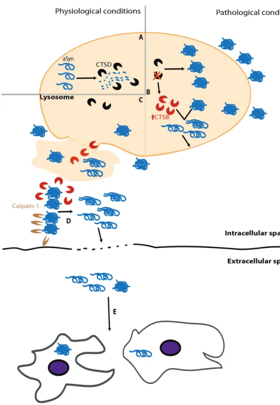
Impairment of -syn clearance caused by lysosomal dysfunction can be due to enzymatic failure. A relevant player of this paradigm is glucocerebrosidade (GCase). This lysosomal hydrolase is associated with Gaucher’s disease, the most common lysosomal storage disease (LSD) [213]. It has been shown that GCase deficiency leads to neuroinflammation and -syn accumulation [214] and that this depletion can also promote -syn intercellular transmission [215]. Interestingly, -syn can inhibit GCase activity, thereby enhancing -syn aggregation [216]. Among lysosomal enzymes, CTSD plays a prominent role as it is the major enzyme processing -syn in the lysosome (Figure 3A) [147]. Despite the growing number of studies devoted to study the role of CTSD in PD and other synucleinopathies, its activity in diseased brains has been poorly explored. Initial studies revealed similar activity levels of CTSD in the brain and CSF of PD and control individuals [217,218]. However, an exhaustive histochemical analysis of individual neurons in the susbtantia nigra of PD revealed a decrease in CSTD staining, compared to age-matched controls, particularly in cells containing -syn inclusions [219]. Although the functional interpretation of this finding is not trivial, experiments with CTSD deficient animal models provided evidence that impairment of CTSD function impairs -syn clearance, leading to its aggregation in neurons (Figure 3B) [149,220]. Importantly, CTSD deficiency entails other deleterious consequences, including reduction of proteasome activity, which in turn also enhances -syn accumulation, and up-regulation of other cathepsins as a compensatory cellular mechanism [220]. Although the increased levels of other lysosomal enzymes might, in principle, alleviate -syn aggregation, this does not seem to work and might result in a two-edged sword. In this regard, increased cathepsin B (CTSB) levels, resulting from CTSD deficiency, display a detrimental activity over -syn aggregates, generating fragments with high seeding efficiency that eventually cause the rupture of lysosome, promoting an abrupt alteration of the whole cell homeostasis leading to cell death (Figure 3B) [221]. Considering that extracellular -syn aggregates can, upon internalization, induce lysosome rupture [222], the putative cleavage of -syn by CTSB would create a damaging situation where the cytoplasm would be deficient in lysosomes and burdened with -syn seeds and CTSB molecules, which can be active in the cytosol potentially amplifying the amount of -syn seeds (Figure 3C) [223]. The eventual cell death caused by lysosomal rupture and release of the cytoplasmic content into the extracellular space might induce a pro-inflammatory response [222], as well as the spreading of -syn seeds that could trigger a similar process in neighboring cells, thereby propagating -syn pathology (Figure 3C–E).
Mouse models of -syn aggregation with decreased Calpain1 activity present a reduced number of -syn inclusions and alleviated nigral degeneration and synaptic defects. In addition, Calpain1 inhibition also reduces the levels of the astrocytic marker GFAP [225,226], suggesting a possible reduction of the inflammatory response. Moreover, antibodies blocking Calpain1-induced -syn truncation prevent the intercellular propagation of -syn and neurodegeneration in a mouse model of PD [227]. One possibility is that this effect occurs at the extracellular space, where Calpain1 is secreted along with -syn species [227]. Considering that -syn accumulation is a known enhancer of Calpain1 secretion [228], one can speculate that this vicious cycle contributes to the spreading of -syn pathology and inflammation. In agreement with this hypothesis, a relevant relationship between -syn and Ca2+ has also been put forward [229]. Ca2+ homeostasis might be strongly affected by -syn, as it was demonstrated that transgenic mice overexpressing -syn present long-lasting Ca2+ transients [230]. Ca2+ dysregulation has also been reported to be caused by exogenous -syn [231], which promotes Ca2+ influx and, eventually, Calpain activation. Therefore,
it seems that -syn accumulation is an enhancer of Calpain1 activity and vice-versa, accounting for numerous detrimental effects, including -syn pathology and a massive dysregulation of Ca2+ homeostasis.
Although a comprehensive examination of these data is still necessary, this may suggest a novel interpretation of the calpain-cathepsin hypothesis that was put forward in the field of Alzheimer’s disease [232,233]—in the case of PD, the final outcome would not only be cell necrosis but also the spreading of -syn pathology.
4.2. Macrosecretion and Lysosome-Mediated Exocytosis
A recent wave of discoveries brought a new meaning to the interaction between the autophagy and secretory pathways, which has been highly neglected in the PD field thus far.
On one hand, the macroautophagy machinery can regulate an unconventional (ER-to-Golgi independent) pathway of extracellular protein secretion, henceforth simply designated as macrosecretion. As previously mentioned, parts of the membranes forming the phagosome originate from the ER [234]. It is believed that the same process is at the basis of macrosecretion [235]. Although little is known about the molecular mechanisms involved, Atg5, GRASP55, and Rab8a were found to be crucial players [236]. Thus, autophagy inhibition also leads to macrosecretion blockade, possibly leading to intracellular protein accumulation [237].
No research has been done so far on the role of macrosecretion in either PD or other synucleinopathies but this pathway has already been linked to the intracellular accumulation of Abeta peptide upon autophagy impairment [238]. Thus, it would be very interesting to understand (1) what cellular inputs push the system towards one of the directions; (2) if -syn can be secreted through this pathway; (3) if -syn can also affect macrosecretion; and (4) if inflammation in PD is associated with the release of cytokines via macrosecretion (Figure 4A,B).
Lysosome exocytosis is beneficial in LSDs [239] but its possible role in PD and other synucleinopathies has not been analysed. Lysosome exocytosis, an “unconventional secretion pathway”, shares several resemblances with synaptic vesicle release, including a Ca2+ dependency [240].
-syn in this pathway. Since it has been postulated that -syn has an important role in neurotransmitter release [244,245], the question is whether it can also modulate lysosome exocytosis, and whether -syn can be released to the extracellular environment via lysosome secretion upon autophagy impairment (Figure 4C,D).
Figure 4. Macrosecretion and lysosome-mediated exocytosis. (A) Since autophagosomes can be secreted to the extracellular space, it is possible that -syn release occurs via this pathway, both in normal conditions and upon autophagosome accumulation due to lysosome impairment; (B) -syn can alter the levels of important players in macroautophagy and eventually play a role in macrosecretion; (C) Lysosomes can fuse with the plasma membrane and release their content, a mechanism named lysosome exocytosis. The process is similar to the one observed in neurotransmitter release, -syn may play a role; (D) Alternatively, impairment of protein degradation might promote the accumulation of high molecular weight species of -syn inside the lysosome. One can speculate that, as observed in LSDs, the lysosomes might fuse with the plasma membrane and release -syn oligomers and aggregates into the extracellular media and start the propagation of pathology.
4.3. Extracellular -Syn Species
type, physiological state and -syn species. One important issue, yet unresolved, is the fate and/or the physiological role of the -syn species that are present in the extracellular space. The clearance of extracellular -syn species can be achieved through proteolysis by extracellular proteases or uptake by surrounding cells. The first possibility has been demonstrated in cellular models, and distinct enzymes have been found to cleave extracellular -syn. These include neurosin [246], plasmin [247] and matrix metalloproteases [248] but the outcome of their activity on -syn-induced toxicity is still debatable. The role of these extracellular proteases exceeds the scope of this review and readers are referred to other reviews [249].
The internalization of -syn species by neighboring cells can be also regarded as a clearing mechanism, particularly when microglia are involved [250], although in this case, it may also entail opposing effects. As explained above, the internalization of pathological forms of -syn can seed the pathogenic conversion of soluble -syn in the receiving cell, and this might be one of the fundamental steps for -syn propagation and spreading of pathology (Figure 3E) [178]. This effect has been well described in surrounding neurons where new -syn inclusions are formed [185] and in astrocytes where -syn can trigger aggregation and an inflammatory response [251].
4.4. The Putative Role of Glial Cells in the Spreading of -Syn Pathology
PD, as other neurodegenerative disorders, is characterized by neuroinflammation in specific brain regions [252,253]. This process includes the enrolment of various types of brain cells such as microglia and astrocytes [254–256] and the accumulation of different molecules involved in the neuroinflammatory response [257]. However, little is known about the precise relationship between -syn pathology and neuroinflammation, and its role in disease progression and pathology propagation [258,259].
Although -syn is mainly a neuronal protein [260], -syn inclusions can occur in astrocytes and oligodendrocytes, in a manner that correlates with nigral degeneration [261,262]. The analysis of PD brains showed -syn inclusions in astrocytes with a particular distribution pattern suggesting that misfolded or abnormal -syn molecules may escape from damaged terminal axons and then be integrated by neighboring astrocytes [263], in agreement with the current hypothesis of prion-like spreading of -syn pathology [251]. Likewise, -syn can be taken up by oligodendrocytes [264] and induce the formation of cytoplasmic inclusions [265]. In addition to inclusion formation, -syn accumulation might be a relevant inducer of the local inflammatory response, as suggested by the addition of exogenous -syn in cultured astrocytes and by the analysis of MSA brains, where astrocyte activation was greater in the vicinity of -syn inclusions [265].
studies revealed that TLR2 receptors seem to be specific for oligomeric species of -syn [271].Indeed, -syn conformation of appears to be crucial and, since monomeric extracellular -syn enhances microglial phagocytosis, aggregated species inhibit this activity [272]. While these observations are still hard to reconcile, one can speculate that perhaps aggregated species of -syn might, at some point, hinder the intracellular clearance systems, therefore causing glial cells to also contribute to the spreading process. 5. Conclusions
The activity of protein clearance mechanisms depends on the physiological state of the cell and its surrounding environment. They play a crucial role in clearing misfolded protein species that may appear and have deleterious consequences for the cell. The kinetics of protein aggregation, following a sigmoidal process, affords biological systems an opportunity to interfere with protein aggregation in the early rate-limiting step, thus, protecting the cell from potentially dangerous, aggregation-prone species. However, for reasons that are currently still unclear, some proteins may escape the cellular quality control systems, misfold, and aggregate, leading to a variety of detrimental consequences, including cell death. In some instances, cellular efforts to counteract the protein aggregation process, through the activation of protein degradation pathways, might become two-edged swords, enhancing protein aggregation and the generation of species that can be transmitted to neighboring cells.
Although the majority of studies performed thus far have focused on the role of autophagy-lysosomal pathway on -syn propagation, it will also be important to assess the role of the UPS in this process, since the impairment of this pathway might precipitate a cascade of events culminating in increased extracellular release and, thereby, spreading of pathology. Ultimately, we posit that the comprehension of the synergistic and compensatory mechanisms of these pathways might lead to the identification of novel targets and strategies for modulating the spreading of pathology in PD and other synucleinopathies. Acknowledgments
Tomás Lopes da Fonseca is supported by Fundação para a Ciência e Tecnologia (SFRH/BD/74881/2010). Anna Villar-Piqué is supported by the Dorothea Schlözer Programme of the Georg August University Göttingen. Tiago Fleming Outeiro is supported by the DFG Center for Nanoscale Microscopy and Molecular Physiology of the Brain. We apologize to those whose work could not be directly cited due to space limitations.
Author Contributions
All authors wrote the manuscript. Conflicts of Interest
References
1. Marti, M.J.; Tolosa, E.; Campdelacreu, J. Clinical overview of the synucleinopathies. Mov. Disord. 2003, 18, S21–S27.
2. Chaudhuri, K.R.; Naidu, Y. Early Parkinson’s disease and non-motor issues. J. Neurol. 2008, 255, 33–38.
3. Harding, A.J.; Stimson, E.; Henderson, J.M.; Halliday, G.M. Clinical correlates of selective pathology in the amygdala of patients with Parkinson’s disease. Brain 2002, 125, 2431–2445. 4. Braak, H.; del Tredici, K.; Rub, U.; de Vos, R.A.; Jansen Steur, E.N.; Braak, E. Staging of brain
pathology related to sporadic Parkinson’s disease. Neurobiol. Aging 2003, 24, 197–211.
5. Baba, T.; Kikuchi, A.; Hirayama, K.; Nishio, Y.; Hosokai, Y.; Kanno, S.; Hasegawa, T.; Sugeno, N.; Konno, M.; Suzuki, K.; et al. Severe olfactory dysfunction is a prodromal symptom of dementia associated with Parkinson’s disease: A 3 year longitudinal study. Brain 2012, 135, 161–169. 6. Moessnang, C.; Frank, G.; Bogdahn, U.; Winkler, J.; Greenlee, M.W.; Klucken, J. Altered activation
patterns within the olfactory network in Parkinson’s disease. Cereb. Cortex 2011, 21, 1246–1253. 7. Wang, J.; You, H.; Liu, J.F.; Ni, D.F.; Zhang, Z.X.; Guan, J. Association of olfactory bulb volume and olfactory sulcus depth with olfactory function in patients with Parkinson disease. Am. J. Neuroradiol. 2011, 32, 677–681.
8. Ansari, K.A.; Johnson, A. Olfactory function in patients with Parkinson’s disease. J. Chronic Dis. 1975, 28, 493–497.
9. Simuni, T.; Sethi, K. Nonmotor manifestations of Parkinson’s disease. Ann. Neurol. 2008, 64, S65–S80.
10. Park, A.; Stacy, M. Non-motor symptoms in Parkinson’s disease. J. Neurol. 2009, 256, 293–298. 11. Gallagher, D.A.; Lees, A.J.; Schrag, A. What are the most important nonmotor symptoms in
patients with Parkinson’s disease and are we missing them? Mov. Disord. 2010, 25, 2493–2500. 12. Olanow, C.W.; Goetz, C.G.; Kordower, J.H.; Stoessl, A.J.; Sossi, V.; Brin, M.F.; Shannon, K.M.;
Nauert, G.M.; Perl, D.P.; Godbold, J.; et al. A double-blind controlled trial of bilateral fetal nigral transplantation in Parkinson’s disease. Ann. Neurol. 2003, 54, 403–414.
13. Lindvall, O.; Sawle, G.; Widner, H.; Rothwell, J.C.; Bjorklund, A.; Brooks, D.; Brundin, P.; Frackowiak, R.; Marsden, C.D.; Odin, P.; et al. Evidence for long-term survival and function of dopaminergic grafts in progressive Parkinson’s disease. Ann. Neurol. 1994, 35, 172–180.
14. Kordower, J.H.; Freeman, T.B.; Snow, B.J.; Vingerhoets, F.J.; Mufson, E.J.; Sanberg, P.R.; Hauser, R.A.; Smith, D.A.; Nauert, G.M.; Perl, D.P.; et al. Neuropathological evidence of graft survival and striatal reinnervation after the transplantation of fetal mesencephalic tissue in a patient with Parkinson’s disease. N. Engl. J. Med. 1995, 332, 1118–1124.
15. Li, J.Y.; Englund, E.; Holton, J.L.; Soulet, D.; Hagell, P.; Lees, A.J.; Lashley, T.; Quinn, N.P.; Rehncrona, S.; Bjorklund, A.; et al. Lewy bodies in grafted neurons in subjects with Parkinson’s disease suggest host-to-graft disease propagation. Nat. Med. 2008, 14, 501–503.
16. Kordower, J.H.; Chu, Y.; Hauser, R.A.; Freeman, T.B.; Olanow, C.W. Lewy body-like pathology in long-term embryonic nigral transplants in Parkinson’s disease. Nat. Med. 2008, 14, 504–506. 17. Wales, P.; Pinho, R.; Lazaro, D.F.; Outeiro, T.F. Limelight on alpha-synuclein: Pathological and
18. Polymeropoulos, M.H.; Lavedan, C.; Leroy, E.; Ide, S.E.; Dehejia, A.; Dutra, A.; Pike, B.; Root, H.; Rubenstein, J.; Boyer, R.; et al. Mutation in the alpha-synuclein gene identified in families with Parkinson’s disease. Science 1997, 276, 2045–2047.
19. Kruger, R.; Kuhn, W.; Muller, T.; Woitalla, D.; Graeber, M.; Kosel, S.; Przuntek, H.; Epplen, J.T.; Schols, L.; Riess, O. Ala30pro mutation in the gene encoding alpha-synuclein in Parkinson’s disease. Nat. Genet. 1998, 18, 106–108.
20. Zarranz, J.J.; Alegre, J.; Gomez-Esteban, J.C.; Lezcano, E.; Ros, R.; Ampuero, I.; Vidal, L.; Hoenicka, J.; Rodriguez, O.; Atares, B.; et al. The new mutation, E46K, of alpha-synuclein causes parkinson and Lewy body dementia. Ann. Neurol. 2004, 55, 164–173.
21. Appel-Cresswell, S.; Vilarino-Guell, C.; Encarnacion, M.; Sherman, H.; Yu, I.; Shah, B.; Weir, D.; Thompson, C.; Szu-Tu, C.; Trinh, J.; et al. Alpha-synuclein p.H50Q, a novel pathogenic mutation for Parkinson’s disease. Mov. Disord. 2013, 28, 811–813.
22. Lesage, S.; Anheim, M.; Letournel, F.; Bousset, L.; Honore, A.; Rozas, N.; Pieri, L.; Madiona, K.; Durr, A.; Melki, R.; et al. G51D alpha-synuclein mutation causes a novel parkinsonian-pyramidal syndrome. Ann. Neurol. 2013, 73, 459–471.
23. Pasanen, P.; Myllykangas, L.; Siitonen, M.; Raunio, A.; Kaakkola, S.; Lyytinen, J.; Tienari, P.J.; Poyhonen, M.; Paetau, A. A novel alpha-synuclein mutation A53E associated with atypical multiple system atrophy and Parkinson’s disease-type pathology. Neurobiol. Aging 2014, 35, 2180.e1–2180.e5.
24. Singleton, A.B.; Farrer, M.; Johnson, J.; Singleton, A.; Hague, S.; Kachergus, J.; Hulihan, M.; Peuralinna, T.; Dutra, A.; Nussbaum, R.; et al. Alpha-synuclein locus triplication causes Parkinson’s disease. Science 2003, 302, 841.
25. Chartier-Harlin, M.C.; Kachergus, J.; Roumier, C.; Mouroux, V.; Douay, X.; Lincoln, S.; Levecque, C.; Larvor, L.; Andrieux, J.; Hulihan, M.; et al. Alpha-synuclein locus duplication as a cause of familial Parkinson’s disease. Lancet 2004, 364, 1167–1169.
26. Satake, W.; Nakabayashi, Y.; Mizuta, I.; Hirota, Y.; Ito, C.; Kubo, M.; Kawaguchi, T.; Tsunoda, T.; Watanabe, M.; Takeda, A.; et al. Genome-wide association study identifies common variants at four loci as genetic risk factors for Parkinson’s disease. Nat. Genet. 2009, 41, 1303–1307.
27. Simon-Sanchez, J.; Schulte, C.; Bras, J.M.; Sharma, M.; Gibbs, J.R.; Berg, D.; Paisan-Ruiz, C.; Lichtner, P.; Scholz, S.W.; Hernandez, D.G.; et al. Genome-wide association study reveals genetic risk underlying Parkinson’s disease. Nat. Genet. 2009, 41, 1308–1312.
28. Edwards, T.L.; Scott, W.K.; Almonte, C.; Burt, A.; Powell, E.H.; Beecham, G.W.; Wang, L.; Zuchner, S.; Konidari, I.; Wang, G.; et al. Genome-wide association study confirms snps in SNCA and the mapt region as common risk factors for Parkinson disease. Ann. Hum. Genet. 2010, 74, 97–109. 29. Nalls, M.A.; Pankratz, N.; Lill, C.M.; Do, C.B.; Hernandez, D.G.; Saad, M.; DeStefano, A.L.;
Kara, E.; Bras, J.; Sharma, M.; et al. Large-scale meta-analysis of genome-wide association data identifies six new risk loci for Parkinson’s disease. Nat. Genet. 2014, 46, 989–993.
30. Maroteaux, L.; Campanelli, J.T.; Scheller, R.H. Synuclein: A neuron-specific protein localized to the nucleus and presynaptic nerve terminal. J. Neurosci. 1988, 8, 2804–2815.
32. Vamvaca, K.; Volles, M.J.; Lansbury, P.T., Jr. The first N-terminal amino acids of alpha-synuclein are essential for alpha-helical structure formation in vitro and membrane binding in yeast. J. Mol. Biol. 2009, 389, 413–424.
33. Bartels, T.; Ahlstrom, L.S.; Leftin, A.; Kamp, F.; Haass, C.; Brown, M.F.; Beyer, K. The N-terminus of the intrinsically disordered protein alpha-synuclein triggers membrane binding and helix folding. Biophys. J. 2010, 99, 2116–2124.
34. Giasson, B.I.; Murray, I.V.; Trojanowski, J.Q.; Lee, V.M. A hydrophobic stretch of 12 amino acid residues in the middle of alpha-synuclein is essential for filament assembly. J. Biol. Chem. 2001, 276, 2380–2386.
35. Izawa, Y.; Tateno, H.; Kameda, H.; Hirakawa, K.; Hato, K.; Yagi, H.; Hongo, K.; Mizobata, T.; Kawata, Y. Role of C-terminal negative charges and tyrosine residues in fibril formation of alpha-synuclein. Brain Behav. 2012, 2, 595–605.
36. Nielsen, M.S.; Vorum, H.; Lindersson, E.; Jensen, P.H. Ca2+ binding to alpha-synuclein regulates ligand binding and oligomerization. J. Biol. Chem. 2001, 276, 22680–22684.
37. Lowe, R.; Pountney, D.L.; Jensen, P.H.; Gai, W.P.; Voelcker, N.H. Calcium(II) selectively induces alpha-synuclein annular oligomers via interaction with the C-terminal domain. Protein Sci. 2004, 13, 3245–3252.
38. Souza, J.M.; Giasson, B.I.; Lee, V.M.; Ischiropoulos, H. Chaperone-like activity of synucleins. FEBS Lett. 2000, 474, 116–119.
39. Bartels, T.; Choi, J.G.; Selkoe, D.J. Alpha-synuclein occurs physiologically as a helically folded tetramer that resists aggregation. Nature 2011, 477, 107–110.
40. Wang, W.; Perovic, I.; Chittuluru, J.; Kaganovich, A.; Nguyen, L.T.; Liao, J.; Auclair, J.R.; Johnson, D.; Landeru, A.; Simorellis, A.K.; et al. A soluble alpha-synuclein construct forms a dynamic tetramer. Proc. Natl. Acad. Sci. USA 2011, 108, 17797–17802.
41. Dettmer, U.; Newman, A.J.; Luth, E.S.; Bartels, T.; Selkoe, D. In vivo cross-linking reveals principally oligomeric forms of alpha-synuclein and beta-synuclein in neurons and non-neural cells. J. Biol. Chem. 2013, 288, 6371–6385.
42. Fauvet, B.; Mbefo, M.K.; Fares, M.B.; Desobry, C.; Michael, S.; Ardah, M.T.; Tsika, E.; Coune, P.; Prudent, M.; Lion, N.; et al. Alpha-synuclein in central nervous system and from erythrocytes, mammalian cells, and escherichia coli exists predominantly as disordered monomer. J. Biol. Chem. 2012, 287, 15345–15364.
43. Outeiro, T.F.; Putcha, P.; Tetzlaff, J.E.; Spoelgen, R.; Koker, M.; Carvalho, F.; Hyman, B.T.; McLean, P.J. Formation of toxic oligomeric alpha-synuclein species in living cells. PLOS ONE 2008, 3, e1867.
44. Winner, B.; Jappelli, R.; Maji, S.K.; Desplats, P.A.; Boyer, L.; Aigner, S.; Hetzer, C.; Loher, T.; Vilar, M.; Campioni, S.; et al. In vivo demonstration that alpha-synuclein oligomers are toxic. Proc. Natl. Acad. Sci. USA 2011, 108, 4194–4199.
46. El-Agnaf, O.M.; Jakes, R.; Curran, M.D.; Middleton, D.; Ingenito, R.; Bianchi, E.; Pessi, A.; Neill, D.; Wallace, A. Aggregates from mutant and wild-type alpha-synuclein proteins and NAC peptide induce apoptotic cell death in human neuroblastoma cells by formation of beta-sheet and amyloid-like filaments. FEBS Lett. 1998, 440, 71–75.
47. Tanik, S.A.; Schultheiss, C.E.; Volpicelli-Daley, L.A.; Brunden, K.R.; Lee, V.M. Lewy body-like alpha-synuclein aggregates resist degradation and impair macroautophagy. J. Biol. Chem. 2013, 288, 15194–15210.
48. Anderson, J.P.; Walker, D.E.; Goldstein, J.M.; de Laat, R.; Banducci, K.; Caccavello, R.J.; Barbour, R.; Huang, J.; Kling, K.; Lee, M.; et al. Phosphorylation of Ser-129 is the dominant pathological modification of alpha-synuclein in familial and sporadic Lewy body disease. J. Biol. Chem. 2006, 281, 29739–29752.
49. Tofaris, G.K.; Razzaq, A.; Ghetti, B.; Lilley, K.S.; Spillantini, M.G. Ubiquitination of alpha-synuclein in lewy bodies is a pathological event not associated with impairment of proteasome function. J. Biol. Chem. 2003, 278, 44405–44411.
50. Rott, R.; Szargel, R.; Haskin, J.; Shani, V.; Shainskaya, A.; Manov, I.; Liani, E.; Avraham, E.; Engelender, S. Monoubiquitylation of alpha-synuclein by seven in absentia homolog (SIAH) promotes its aggregation in dopaminergic cells. J. Biol. Chem. 2008, 283, 3316–3328.
51. Shin, Y.; Klucken, J.; Patterson, C.; Hyman, B.T.; McLean, P.J. The co-chaperone carboxyl terminus of Hsp70-interacting protein (CHIP) mediates alpha-synuclein degradation decisions between proteasomal and lysosomal pathways. J. Biol. Chem. 2005, 280, 23727–23734.
52. Krumova, P.; Meulmeester, E.; Garrido, M.; Tirard, M.; Hsiao, H.H.; Bossis, G.; Urlaub, H.; Zweckstetter, M.; Kugler, S.; Melchior, F.; et al. Sumoylation inhibits alpha-synuclein aggregation and toxicity. J. Cell Biol. 2011, 194, 49–60.
53. Kim, Y.M.; Jang, W.H.; Quezado, M.M.; Oh, Y.; Chung, K.C.; Junn, E.; Mouradian, M.M. Proteasome inhibition induces alpha-synuclein sumoylation and aggregate formation. J. Neurol. Sci. 2011, 307, 157–161.
54. Shahpasandzadeh, H.; Popova, B.; Kleinknecht, A.; Fraser, P.E.; Outeiro, T.F.; Braus, G.H. Interplay between sumoylation and phosphorylation for protection against alpha-synuclein inclusions. J. Biol. Chem. 2014, 289, 31224–31240.
55. Dikiy, I.; Eliezer, D. N-terminal acetylation stabilizes N-terminal helicity in lipid- and micelle-bound alpha-synuclein and increases its affinity for physiological membranes. J. Biol. Chem. 2014, 289, 3652–3665.
56. Maltsev, A.S.; Ying, J.; Bax, A. Impact of N-terminal acetylation of alpha-synuclein on its random coil and lipid binding properties. Biochemistry 2012, 51, 5004–5013.
57. Bartels, T.; Kim, N.C.; Luth, E.S.; Selkoe, D.J. N-alpha-acetylation of alpha-synuclein increases its helical folding propensity, gm1 binding specificity and resistance to aggregation. PLOS ONE 2014, 9, e103727.
59. Fujiwara, H.; Hasegawa, M.; Dohmae, N.; Kawashima, A.; Masliah, E.; Goldberg, M.S.; Shen, J.; Takio, K.; Iwatsubo, T. Alpha-synuclein is phosphorylated in synucleinopathy lesions. Nat. Cell Biol. 2002, 4, 160–164.
60. Pronin, A.N.; Morris, A.J.; Surguchov, A.; Benovic, J.L. Synucleins are a novel class of substrates for G protein-coupled receptor kinases. J. Biol. Chem. 2000, 275, 26515–26522.
61. Arawaka, S.; Wada, M.; Goto, S.; Karube, H.; Sakamoto, M.; Ren, C.H.; Koyama, S.; Nagasawa, H.; Kimura, H.; Kawanami, T.; et al. The role of G-protein-coupled receptor kinase 5 in pathogenesis of sporadic Parkinson’s disease. J. Neurosci. 2006, 26, 9227–9238.
62. Inglis, K.J.; Chereau, D.; Brigham, E.F.; Chiou, S.S.; Schobel, S.; Frigon, N.L.; Yu, M.; Caccavello, R.J.; Nelson, S.; Motter, R.; et al. Polo-like kinase 2 (PLK2) phosphorylates alpha-synuclein at serine 129 in central nervous system. J. Biol. Chem. 2009, 284, 2598–2602. 63. Mbefo, M.K.; Paleologou, K.E.; Boucharaba, A.; Oueslati, A.; Schell, H.; Fournier, M.;
Olschewski, D.; Yin, G.; Zweckstetter, M.; Masliah, E.; et al. Phosphorylation of synucleins by members of the polo-like kinase family. J. Biol. Chem. 2010, 285, 2807–2822.
64. Ishii, A.; Nonaka, T.; Taniguchi, S.; Saito, T.; Arai, T.; Mann, D.; Iwatsubo, T.; Hisanaga, S.; Goedert, M.; Hasegawa, M. Casein kinase 2 is the major enzyme in brain that phosphorylates ser129 of human alpha-synuclein: Implication for alpha-synucleinopathies. FEBS Lett. 2007, 581, 4711–4717.
65. Basso, E.; Antas, P.; Marijanovic, Z.; Goncalves, S.; Tenreiro, S.; Outeiro, T.F. PLK2 modulates alpha-synuclein aggregation in yeast and mammalian cells. Mol. Neurobiol. 2013, 48, 854–862. 66. Ryu, M.Y.; Kim, D.W.; Arima, K.; Mouradian, M.M.; Kim, S.U.; Lee, G. Localization of CKII
beta subunits in lewy bodies of Parkinson’s disease. J. Neurol. Sci. 2008, 266, 9–12.
67. Chen, L.; Feany, M.B. Alpha-synuclein phosphorylation controls neurotoxicity and inclusion formation in a drosophila model of Parkinson disease. Nat. Neurosci. 2005, 8, 657–663.
68. Tenreiro, S.; Reimao-Pinto, M.M.; Antas, P.; Rino, J.; Wawrzycka, D.; Macedo, D.; Rosado-Ramos, R.; Amen, T.; Waiss, M.; Magalhaes, F.; et al. Phosphorylation modulates clearance of alpha-synuclein inclusions in a yeast model of Parkinson’s disease. PLOS Genet. 2014, 10, e1004302.
69. Paleologou, K.E.; Oueslati, A.; Shakked, G.; Rospigliosi, C.C.; Kim, H.Y.; Lamberto, G.R.; Fernandez, C.O.; Schmid, A.; Chegini, F.; Gai, W.P.; et al. Phosphorylation at S87 is enhanced in synucleinopathies, inhibits alpha-synuclein oligomerization, and influences synuclein-membrane interactions. J. Neurosci. 2010, 30, 3184–3198.
70. Oueslati, A.; Fournier, M.; Lashuel, H.A. Role of post-translational modifications in modulating the structure, function and toxicity of alpha-synuclein: Implications for Parkinson’s disease pathogenesis and therapies. Prog. Brain Res. 2010, 183, 115–145.
71. Sato, H.; Kato, T.; Arawaka, S. The role of Ser129 phosphorylation of alpha-synuclein in neurodegeneration of Parkinson’s disease: A review of in vivo models. Rev. Neurosci. 2013, 24, 115–123.
73. Giasson, B.I.; Duda, J.E.; Murray, I.V.; Chen, Q.; Souza, J.M.; Hurtig, H.I.; Ischiropoulos, H.; Trojanowski, J.Q.; Lee, V.M. Oxidative damage linked to neurodegeneration by selective alpha-synuclein nitration in synucleinopathy lesions. Science 2000, 290, 985–989.
74. Liu, Y.; Qiang, M.; Wei, Y.; He, R. A novel molecular mechanism for nitrated -synuclein-induced cell death. J. Mol. Cell Biol. 2011, 3, 239–249.
75. Yu, Z.; Xu, X.; Xiang, Z.; Zhou, J.; Zhang, Z.; Hu, C.; He, C. Nitrated alpha-synuclein induces the loss of dopaminergic neurons in the substantia nigra of rats. PLOS ONE 2010, 5, e9956.
76. Hodara, R.; Norris, E.H.; Giasson, B.I.; Mishizen-Eberz, A.J.; Lynch, D.R.; Lee, V.M.; Ischiropoulos, H. Functional consequences of alpha-synuclein tyrosine nitration: Diminished binding to lipid vesicles and increased fibril formation. J. Biol. Chem. 2004, 279, 47746–47753.
77. Li, W.; West, N.; Colla, E.; Pletnikova, O.; Troncoso, J.C.; Marsh, L.; Dawson, T.M.; Jakala, P.; Hartmann, T.; Price, D.L.; et al. Aggregation promoting C-terminal truncation of alpha-synuclein is a normal cellular process and is enhanced by the familial Parkinson’s disease-linked mutations. Proc. Natl. Acad. Sci. USA 2005, 102, 2162–2167.
78. Baba, M.; Nakajo, S.; Tu, P.H.; Tomita, T.; Nakaya, K.; Lee, V.M.; Trojanowski, J.Q.; Iwatsubo, T. Aggregation of alpha-synuclein in lewy bodies of sporadic Parkinson’s disease and dementia with lewy bodies. Am. J. Pathol. 1998, 152, 879–884.
79. Murray, I.V.; Giasson, B.I.; Quinn, S.M.; Koppaka, V.; Axelsen, P.H.; Ischiropoulos, H.; Trojanowski, J.Q.; Lee, V.M. Role of alpha-synuclein carboxy-terminus on fibril formation in vitro. Biochemistry 2003, 42, 8530–8540.
80. Ulusoy, A.; Febbraro, F.; Jensen, P.H.; Kirik, D.; Romero-Ramos, M. Co-expression of C-terminal truncated alpha-synuclein enhances full-length alpha-synuclein-induced pathology. Eur. J. Neurosci. 2010, 32, 409–422.
81. Mishizen-Eberz, A.J.; Guttmann, R.P.; Giasson, B.I.; Day, G.A., 3rd; Hodara, R.; Ischiropoulos, H.; Lee, V.M.; Trojanowski, J.Q.; Lynch, D.R. Distinct cleavage patterns of normal and pathologic forms of alpha-synuclein by calpain I in vitro. J. Neurochem. 2003, 86, 836–847.
82. Mishizen-Eberz, A.J.; Norris, E.H.; Giasson, B.I.; Hodara, R.; Ischiropoulos, H.; Lee, V.M.; Trojanowski, J.Q.; Lynch, D.R. Cleavage of alpha-synuclein by calpain: Potential role in degradation of fibrillized and nitrated species of alpha-synuclein. Biochemistry 2005, 44, 7818–7829.
83. Iwata, A.; Maruyama, M.; Akagi, T.; Hashikawa, T.; Kanazawa, I.; Tsuji, S.; Nukina, N. Alpha-synuclein degradation by serine protease neurosin: Implication for pathogenesis of synucleinopathies. Hum. Mol. Genet. 2003, 12, 2625–2635.
84. Kasai, T.; Tokuda, T.; Yamaguchi, N.; Watanabe, Y.; Kametani, F.; Nakagawa, M.; Mizuno, T. Cleavage of normal and pathological forms of alpha-synuclein by neurosin in vitro. Neurosci. Lett. 2008, 436, 52–56.
85. Cuervo, A.; Stefanis, L.; Fredenburg, R.; Lansbury, P. Impaired degradation of mutant -synuclein by chaperone-mediated autophagy. Science 2004, 305, 1292–1295.
86. Liu, C.W.; Corboy, M.J.; DeMartino, G.N.; Thomas, P.J. Endoproteolytic activity of the proteasome. Science 2003, 299, 408–411.
88. Lee, H.J.; Khoshaghideh, F.; Patel, S.; Lee, S.J. Clearance of alpha-synuclein oligomeric intermediates via the lysosomal degradation pathway. J. Neurosci. 2004, 24, 1888–1896.
89. Mizushima, N.; Klionsky, D.J. Protein turnover via autophagy: Implications for metabolism. Annu. Rev. Nutr. 2007, 27, 19–40.
90. Dice, J.F. Peptide sequences that target cytosolic proteins for lysosomal proteolysis. Trends Biochem. Sci. 1990, 15, 305–309.
91. Kiffin, R.; Christian, C.; Knecht, E.; Cuervo, A.M. Activation of chaperone-mediated autophagy during oxidative stress. Mol. Biol. Cell 2004, 15, 4829–4840.
92. Chiang, H.L.; Terlecky, S.R.; Plant, C.P.; Dice, J.F. A role for a 70-kilodalton heat shock protein in lysosomal degradation of intracellular proteins. Science 1989, 246, 382–385.
93. Eskelinen, E.L.; Cuervo, A.M.; Taylor, M.R.; Nishino, I.; Blum, J.S.; Dice, J.F.; Sandoval, I.V.; Lippincott-Schwartz, J.; August, J.T.; Saftig, P. Unifying nomenclature for the isoforms of the lysosomal membrane protein LAMP-2. Traffic 2005, 6, 1058–1061.
94. Cuervo, A.M.; Dice, J.F. A receptor for the selective uptake and degradation of proteins by lysosomes. Science 1996, 273, 501–503.
95. Agarraberes, F.A.; Terlecky, S.R.; Dice, J.F. An intralysosomal Hsp70 is required for a selective pathway of lysosomal protein degradation. J. Cell Biol. 1997, 137, 825–834.
96. Vogiatzi, T.; Xilouri, M.; Vekrellis, K.; Stefanis, L. Wild type alpha-synuclein is degraded by chaperone-mediated autophagy and macroautophagy in neuronal cells. J. Biol. Chem. 2008, 283, 23542–23556.
97. Mak, S.K.; McCormack, A.L.; Manning-Bog, A.B.; Cuervo, A.M.; di Monte, D.A. Lysosomal degradation of alpha-synuclein in vivo. J. Biol. Chem. 2010, 285, 13621–13629.
98. Martinez-Vicente, M.; Talloczy, Z. Dopamine-modified -synuclein blocks chaperone-mediated autophagy. J. Clin. Invest. 2008, 118, 777–788.
99. Xilouri, M.; Vogiatzi, T.; Vekrellis, K.; Park, D.; Stefanis, L. Abberant alpha-synuclein confers toxicity to neurons in part through inhibition of chaperone-mediated autophagy. PLOS ONE 2009, 4, e5515.
100. Xilouri, M.; Brekk, O.R.; Landeck, N.; Pitychoutis, P.M.; Papasilekas, T.; Papadopoulou-Daifoti, Z.; Kirik, D.; Stefanis, L. Boosting chaperone-mediated autophagy in vivo mitigates alpha-synuclein-induced neurodegeneration. Brain 2013, 136, 2130–2146.
101. Cuervo, A.M.; Dice, J.F. Age-related decline in chaperone-mediated autophagy. J. Biol. Chem. 2000, 275, 31505–31513.
102. Alvarez-Erviti, L.; Rodriguez-Oroz, M.C.; Cooper, J.M.; Caballero, C.; Ferrer, I.; Obeso, J.A.; Schapira, A.H. Chaperone-mediated autophagy markers in Parkinson disease brains. Arch. Neurol. 2010, 67, 1464–1472.
103. Eskelinen, E.L.; Schmidt, C.K.; Neu, S.; Willenborg, M.; Fuertes, G.; Salvador, N.; Tanaka, Y.; Lullmann-Rauch, R.; Hartmann, D.; Heeren, J.; et al. Disturbed cholesterol traffic but normal proteolytic function in LAMP-1/LAMP-2 double-deficient fibroblasts. Mol. Biol. Cell 2004, 15, 3132–3145.
105. Li, G.; Yang, H.; Zhu, D.; Huang, H.; Liu, G.; Lun, P. Targeted suppression of chaperone-mediated autophagy by miR-320A promotes alpha-synuclein aggregation. Int. J. Mol. Sci. 2014, 15, 15845–15857.
106. Alvarez-Erviti, L.; Seow, Y.; Schapira, A.H.; Rodriguez-Oroz, M.C.; Obeso, J.A.; Cooper, J.M. Influence of microRNA deregulation on chaperone-mediated autophagy and alpha-synuclein pathology in Parkinson’s disease. Cell Death Dis. 2013, 4, e545.
107. Bjersing, J.L.; Bokarewa, M.I.; Mannerkorpi, K. Profile of circulating microRNAs in fibromyalgia and their relation to symptom severity: An exploratory study. Rheumatol. Int. 2015, 35, 635–642. 108. Kubiczkova-Besse, L.; Sedlarikova, L.; Kryukov, F.; Nekvindova, J.; Radova, L.; Almasi, M.;
Pelcova, J.; Minarik, J.; Pika, T.; Pikalova, Z.; et al. Combination of serum microRNA-320A and microRNA-320B as a marker for waldenstrom macroglobulinemia. Am. J. Hematol. 2015, 90, E51–E52.
109. Sheng, M.; Zhong, Y.; Chen, Y.; Du, J.; Ju, X.; Zhao, C.; Zhang, G.; Zhang, L.; Liu, K.; Yang, N.; et al. Hsa-miR-1246, Hsa-miR-320a and Hsa-miR-196B-5P inhibitors can reduce the cytotoxicity of ebola virus glycoprotein in vitro. Sci. China Life Sci. 2014, 57, 959–972.
110. Noda, T.; Suzuki, K.; Ohsumi, Y. Yeast autophagosomes: De novo formation of a membrane structure. Trends Cell Biol. 2002, 12, 231–235.
111. Kraft, C.; Martens, S. Mechanisms and regulation of autophagosome formation. Curr. Opin. Cell Biol. 2012, 24, 496–501.
112. Reggiori, F.; Tucker, K.A.; Stromhaug, P.E.; Klionsky, D.J. The Atg1-Atg13 complex regulates Atg9 and Atg23 retrieval transport from the pre-autophagosomal structure. Dev. Cell 2004, 6, 79–90. 113. Reggiori, F.; Shintani, T.; Nair, U.; Klionsky, D.J. Atg9 cycles between mitochondria and the
pre-autophagosomal structure in yeasts. Autophagy 2005, 1, 101–109.
114. Orsi, A.; Razi, M.; Dooley, H.C.; Robinson, D.; Weston, A.E.; Collinson, L.M.; Tooze, S.A. Dynamic and transient interactions of Atg9 with autophagosomes, but not membrane integration, are required for autophagy. Mol. Biol. Cell 2012, 23, 1860–1873.
115. Young, A.R.; Chan, E.Y.; Hu, X.W.; Kochl, R.; Crawshaw, S.G.; High, S.; Hailey, D.W.; Lippincott-Schwartz, J.; Tooze, S.A. Starvation and ULK1-dependent cycling of mammalian Atg9 between the TGN and endosomes. J. Cell Sci. 2006, 119, 3888–3900.
116. Cardenas, M.E.; Cutler, N.S.; Lorenz, M.C.; di Como, C.J.; Heitman, J. The TOR signaling cascade regulates gene expression in response to nutrients. Genes Dev. 1999, 13, 3271–3279.
117. Zeng, X.; Overmeyer, J.H.; Maltese, W.A. Functional specificity of the mammalian Beclin-Vps34 PI 3-kinase complex in macroautophagy versus endocytosis and lysosomal enzyme trafficking. J. Cell Sci. 2006, 119, 259–270.
118. Thumm, M.; Egner, R.; Koch, B.; Schlumpberger, M.; Straub, M.; Veenhuis, M.; Wolf, D.H. Isolation of autophagocytosis mutants of saccharomyces cerevisiae. FEBS Lett. 1994, 349, 275–280. 119. Tsukada, M.; Ohsumi, Y. Isolation and characterization of autophagy-defective mutants of
saccharomyces cerevisiae. FEBS Lett. 1993, 333, 169–174.
121. Mizushima, N.; Noda, T.; Yoshimori, T.; Tanaka, Y.; Ishii, T.; George, M.D.; Klionsky, D.J.; Ohsumi, M.; Ohsumi, Y. A protein conjugation system essential for autophagy. Nature 1998, 395, 395–398.
122. Tanida, I.; Tanida-Miyake, E.; Ueno, T.; Kominami, E. The human homolog of saccharomyces cerevisiae Apg7p is a protein-activating enzyme for multiple substrates including human Apg12p, GATE-16, GABARAP, and MAP-LC3. J. Biol. Chem. 2001, 276, 1701–1706.
123. Mizushima, N.; Noda, T.; Ohsumi, Y. Apg16p is required for the function of the Apg12p-Apg5p conjugate in the yeast autophagy pathway. EMBO J. 1999, 18, 3888–3896.
124. Romanov, J.; Walczak, M.; Ibiricu, I.; Schuchner, S.; Ogris, E.; Kraft, C.; Martens, S. Mechanism and functions of membrane binding by the Atg5-Atg12/Atg16 complex during autophagosome formation. EMBO J. 2012, 31, 4304–4317.
125. Tanida, I.; Sou, Y.S.; Ezaki, J.; Minematsu-Ikeguchi, N.; Ueno, T.; Kominami, E. HsAtg4b/Hsapg4b/ autophagin-1 cleaves the carboxyl termini of three human Atg8 homologues and delipidates microtubule-associated protein light chain 3- and GABAA receptor-associated protein-phospholipid conjugates. J. Biol. Chem. 2004, 279, 36268–36276.
126. Kabeya, Y.; Mizushima, N.; Ueno, T.; Yamamoto, A.; Kirisako, T.; Noda, T.; Kominami, E.; Ohsumi, Y.; Yoshimori, T. LC3, a mammalian homologue of yeast Apg8p, is localized in autophagosome membranes after processing. EMBO J. 2000, 19, 5720–5728.
127. Tanida, I.; Tanida-Miyake, E.; Komatsu, M.; Ueno, T.; Kominami, E. Human Apg3p/Aut1p homologue is an authentic E2 enzyme for multiple substrates, GATE-16, GABARAP, and MAP-LC3, and facilitates the conjugation of Hapg12p to Hapg5p. J. Biol. Chem. 2002, 277, 13739–13744. 128. Otomo, C.; Metlagel, Z.; Takaesu, G.; Otomo, T. Structure of the human Atg12~Atg5 conjugate
required for LC3 lipidation in autophagy. Nat. Struct. Mol. Biol. 2013, 20, 59–66.
129. Fujita, N.; Itoh, T.; Omori, H.; Fukuda, M.; Noda, T.; Yoshimori, T. The Atg16l complex specifies the site of LC3 lipidation for membrane biogenesis in autophagy. Mol. Biol. Cell 2008, 19, 2092–2100. 130. Olzmann, J.A.; Li, L.; Chudaev, M.V.; Chen, J.; Perez, F.A.; Palmiter, R.D.; Chin, L.S.
Parkin-mediated K63-linked polyubiquitination targets misfolded DJ-1 to aggresomes via binding to HDAC6. J. Cell Biol. 2007, 178, 1025–1038.
131. Kawaguchi, Y.; Kovacs, J.J.; McLaurin, A.; Vance, J.M.; Ito, A.; Yao, T.P. The deacetylase HDAC6 regulates aggresome formation and cell viability in response to misfolded protein stress. Cell 2003, 115, 727–738.
132. Lamark, T.; Perander, M.; Outzen, H.; Kristiansen, K.; Overvatn, A.; Michaelsen, E.; Bjorkoy, G.; Johansen, T. Interaction codes within the family of mammalian Phox and Bem1p domain-containing proteins. J. Biol. Chem. 2003, 278, 34568–34581.
133. Noda, N.N.; Kumeta, H.; Nakatogawa, H.; Satoo, K.; Adachi, W.; Ishii, J.; Fujioka, Y.; Ohsumi, Y.; Inagaki, F. Structural basis of target recognition by Atg8/LC3 during selective autophagy. Genes Cells Devoted Mol. Cell. Mech. 2008, 13, 1211–1218.