Detection of mutator phenotype in Brazilian patients with acute
and chronic myeloid leukemia
Flávio Monteiro Ayres
1,4, Euza Guimarães Momotuk
2, Celso da Cunha Bastos
3and Aparecido Divino da Cruz
41
Universidade Federal de Goiás, Instituto de Ciências Biológicas,
Programa de Pós-Graduação em Biologia (Genética), Goiânia, GO, Brazil.
2
Universidade Federal de Goiás, Instituto de Ciências Biológicas, Departamento de Biologia Geral,
Goiânia, GO, Brazil.
3
Universidade Federal de Goiás, Faculdade de Medicina, Departamento de Clínica Médica,
Goiânia, GO, Brazil.
4
Universidade Católica de Goiás, Departamento de Biologia, Núcleo de Pesquisas Replicon,
Goiânia, GO, Brazil.
Abstract
The multisteps of tumorigenesis involve the classic chromosomal instability and the mutator phenotype pathways featured by a predisposition to acquire mutations in tumor suppressor genes and oncogenes. Expansion and contraction of microsatellite sequences due to a deficient mismatch repair system are a marker of the mutator phenotype. Controversial results regarding the extent of microsatellite instability (MSI) have been reported in the development and progression of myeloid malignancies. Here, we investigated MSI and loss of heterozygosity (LOH) frequencies at the microsatelliteloci BAT-26, D7S486, D8S135, ANK1, IFNA, TP53 and bcr of 19 Brazilian patients with acute (AML) and chronic myeloid leukemia (CML). One AML patient and one CML patient were categorized as having a high degree of microsatellite instability (MSI-H), corresponding to 10.5% (2/19) of all patients. LOH atloci BAT-26 and TP53 was present in 30% of the patients with AML alone. Despite the small sample size, our results suggest that the mutator phenotype, as verified by MSI frequency, could play a role in the leukemogenesis of a small subset of patients with myeloid leukemia.
Key words: loss of heterozygosity, microsatellite instability, mismatch repair, mutator phenotype, myeloid leukemia. Received: February 15, 2003; Accepted: May 16, 2004.
Introduction
Leukemia is a proliferative disorder of the leukopoietic cells. The complex origin and nature of this disorder allow its classification into acute and chronic myeloid or lymphoid types (Harris et al., 2000). The multistep pathways of tumorigenesis involve the classic chromosomal instability pathway featured by chromo-somal loss and LOH of tumor suppressor genes. In another possible pathway, a multistep mutator phenotype leads to the accumulation of mutations in tumor suppressor genes and oncogenes, due to a deficiency in the DNA mismatch repair system [MMR] (Peltomaki, 2001). Patients with such a predisposition to acquire mutations are likely to have
cancer early in life, more than one primary tumor, and a family history of cancer (Ben-Yehudaet al., 1996).
Cells harboring a proficient MMR system can correct nucleotide mismatches during DNA replication or recom-bination and trigger apoptosis following serious DNA dam-age (Aquilina and Bignami, 2001). Deficiency to repair nucleotide mismatches causing loops in the template DNA strand during replication is associated to deletions of nu-cleotides in microsatellite loci. On the other hand, defi-ciency to repair the new DNA strand is related to the length expansion of microsatelliteloci(Naidoo and Chetty, 1999). Microsatellites are highly polymorphic markers composed by short tracts of nucleotide repeats, dispersed throughout the human genome and prone to mutations by replication slippage. Increased frequency of somatic variation in the length of these markers, known as microsatellite instability
www.sbg.org.br
Send correspondence to Flávio Monteiro Ayres. Unidade Uni-versitária de Ciências Exatas e Tecnológicas, Universidade Esta-dual de Goiás, Br 153 km 98, 75000-000 Anápolis, GO, Brazil. E-mail: [email protected].
(MSI), is a clear evidence of mutator phenotype (Aquilina and Bignami, 2001).
Controversial results regarding the extent of MSI have been reported in the development and progression of myeloid malignancies. This disagreement may be due to the number of patients studied, panel of markers chosen, MSI assessment criteria, source of non-tumoral control sample or methodological differences (Wadaet al., 1994; Pabstet al., 1996; Moriet al., 1997; Tasakaet al., 1997; Boyeret al., 1998; Auneret al., 1999; Rimszaet al., 2000; Das-Guptaet al., 2001; Ribeiroet al., 2002). In this con-text, considering the scarcity of reports regarding such fre-quencies in Brazil, we aimed to investigate microsatellite alterations and the role of the mutator phenotype in a group of Brazilian patients with myeloid leukemia.
Materials and Methods
Patients and samples
Peripheral blood samples from 10 patients with acute and nine patients with chronic myeloid leukemia were col-lected in heparin at the Hematology Service ofHospital das Clínicas (Federal University of Goiás, Brazil). Addi-tionally, exfoliated cells from the oral mucosa of every pa-tient were obtained by gently scraping the inside lining of the mouth with a spatula. Patients were not selected based on stage, type nor therapy of the disease. Samples were col-lected from the patients following informed consent, ac-cording to the guidelines approved by the National Health Council of the Brazilian Ministry of Health.
DNA isolation
DNA samples were obtained from peripheral blood cells of all 19 patients and from bone marrow cells of two patients. DNA was isolated using conventional phenol-chloroform extraction and ethanol precipitation methods
(Sambrooket al., 1989). Control DNA was extracted from the exfoliated buccal cells, using RapidPrep Micro Genomic DNA isolation Kit for Cells and Tissues (Amersham Biotech Inc., Piscataway, NJ) according to the manufacturer’s instructions.
Microsatellite analysis
DNA amplification by polymerase chain reaction (PCR) was performed in a final volume of 25µL containing
100 ng of genomic DNA, 50 mmol/L KCl, 10 mmol/L Tris HCl (pH 8.3), 1.5 mmol/L MgCl, 0.2 mmol/L each dNTP, 0.4µmol/L of each sense and antisense primers for
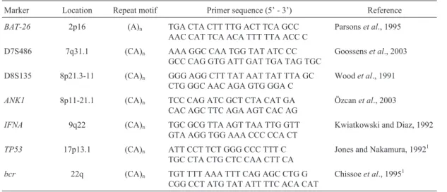
individ-uallocus, and 1 U of Taq Polymerase (Amersham Biotech Inc.). A panel of seven microsatellite markers was used in this study (see Table 1). The touchdown thermal protocol included denaturation at 92 °C for 1 min, annealing at 58-48 °C (decreasing 1 °C/cycle for 10 cycles, followed by 15 cycles at 48 °C) for 1 min, and extension at 72 °C for 1 min. PCR products were visualized using an 8% polyacrylamide gel stained with 0.5µg/mL ethidium bro-mide. Gels were analyzed using a VDS system (Amersham Biosciences Inc., Umeå, Sweden) and the TotalLab soft-ware version 1.0 (Nonlinear Dynamics Ltd., Newcastle Upon Tyne, UK) to determine band sizes (bp) and densi-ties. Decrease in the density of any given heterozygous al-lele was measured as arbitrary units of pixel concentration. The fractional allelic loss (FAL) was calculated using the formula FAL = CL/CI, where CL is the number of chromo-some arms lost and CI is the overall number of informative chromosome arms.
Statistical analysis
Means of MSI or LOH were analyzed using Fisher’s exact test with an overall significance level of 0.05 (95% CI) for comparisons between the two means.
Table 1- Microsatellite markers.
Marker Location Repeat motif Primer sequence (5’ - 3’) Reference
BAT-26 2p16 (A)n TGA CTA CTT TTG ACT TCA GCC
AAC CAT TCA ACA TTT TTA ACC C
Parsonset al., 1995
D7S486 7q31.1 (CA)n AAA GGC CAA TGG TAT ATC CC
GCC CAG GTG ATT GAT TGA TAG TGC
Goossenset al., 2003
D8S135 8p21.3-11 (CA)n GGG AGG CTT TAT AAT TAT TTA GC
CTG GGC AAC AGA GTG GGA C
Woodet al., 1991
ANK1 8p11-21.1 (CA)n TCC CAG ATC GCT CTA CAT GA
CAC AGC TTC AGA AGT CAC AG
Özcanet al., 2003
IFNA 9q22 (CA)n TGC GCG TTA AGT TAA TTG GTT
GTA AGG TGG AAA CCC CCA CT
Kwiatkowski and Diaz, 1992
TP53 17p13.1 (CA)n ATT CCT TCT GGG CCC TTT C
TGC CTA CTG CTC CAA CTT CA
Jones and Nakamura, 19921
bcr 22q (CA)n TGT TTT AAA TTT CAG AGC CTG G
CGG CCT ATG TAT ATT TTC ACA CAT
Chissoeet al., 19951
Results
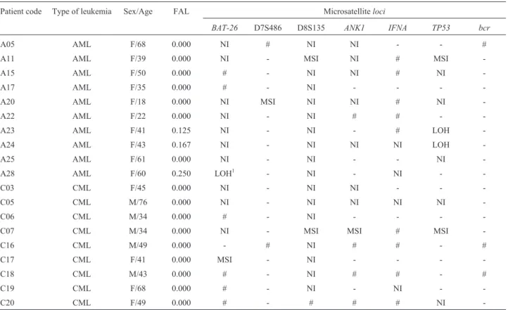
MSI and LOH were investigated for seven microsatellite markers scattered over six different chromo-somes of AML and CML patients. MSI was identified as a variation in the length of microsatellite alleles in the leuke-mic sample, as compared to the corresponding constitu-tional material from buccal cells. Seven out of 109 paired PCR amplifications exhibited instability at theloci BAT-26,
D7S486, D8S135,ANK1andTP53(Figure 1). Instabilities
were verified in two patients with AML and two with CML, corresponding to approximately 21% (4/19) of patients with MSI . Two of these four patients had a high degree of MSI (MSI-H≥30% of assessedloci), suggesting the occur-rence of the mutator phenotype. The other two patients with MSI had a low degree of MSI (MSI-L≤30% of assessed loci), suggesting the occurrence of another pathway of leukemogenesis rather than the mutator phenotype. Table 2 shows details of the results for each patient.
The criterion for LOH identification was the absence or a decrease to less than 50% in the intensity of one hetero-zygous band. The authors were aware of LOH misclassification due to technical artifacts, such as preferential amplification or amplification failure due to DNA degradation and low amount of template. However, DNA preparations of good quality were available, and only
heterozygous markers were considered informative. By using this criterion, markersbcrand D7S486 were hetero-zygous in most patients and, consequently, highly informa-tive for LOH. Amplification of 31 paired sets of informative alleles from AML patients permitted the detec-tion of two LOH events for markerTP53and another two for markerBAT-26(Figure 1). LOH events were found in three patients with AML, corresponding to 30% (3/10) of the AML patients. However, no LOH was detected in the 29 paired sets of informative alleles from CML patients (Table 2). Background data and statistic details for MSI and LOH are presented in Table 3.
Discussion
We studied 19 patients with myeloid leukemia and found 10.5% (2/19) of them with MSI–L and 10.5% (2/19) with MSI-H, which suggested the occurrence of the mutator phenotype in two of our patients (A11 and C07). It is noteworthy that MSI and LOH were not associated in any patient, corroborating the hypothesis that these two path-ways of tumorigenesis do not occur together (Ponz de Leon
et al., 1999). Additionally, it has been suggested that MSI is an adverse prognostic factor for leukemia and lymphoma patients, associated with short-term relapse and resistance to chemotherapy (Indraccoloet al., 1999). The association
Table 2- Analysis of MSI and LOH in AML and CML patients.
Patient code Type of leukemia Sex/Age FAL Microsatelliteloci
BAT-26 D7S486 D8S135 ANK1 IFNA TP53 bcr
A05 AML F/68 0.000 NI # NI NI - - #
A11 AML F/39 0.000 NI - MSI NI # MSI
-A15 AML F/50 0.000 # - NI NI # NI
-A17 AML F/35 0.000 # - NI - - -
-A20 AML F/18 0.000 NI MSI NI NI # NI
-A22 AML F/22 0.000 NI - NI # # -
-A23 AML F/41 0.125 NI - NI - # LOH
-A24 AML F/43 0.167 NI - NI NI NI LOH
-A25 AML F/61 0.000 NI - NI - - NI
-A28 AML F/60 0.250 LOH1 - NI - NI -
-C03 CML F/45 0.000 NI - NI NI - -
-C05 CML M/76 0.000 NI - NI NI NI NI
-C06 CML M/34 0.000 # - NI - - -
-C07 CML M/34 0.000 NI - MSI MSI # MSI
-C16 CML M/49 0.000 - # NI # # - #
C17 CML F/41 0.000 MSI - NI - - -
-C18 CML M/43 0.000 # - NI # # - #
C19 CML F/68 0.000 # - NI - NI -
-C20 CML F/49 0.000 # - # # # NI
of mutator phenotype andTP53molecular inactivation, as reported by Ben-Yehudaet al. (1996) and Zhuet al. (1999), might be an alternative pathway for de novo
leukemogenesis in elderly people or for an accentuated level of genomic instability in therapy-related leukemia pa-tients. Both hypotheses are thought to indicate a rather neg-ative prognosis. Moreover, an increased incidence of adverse cytogenetic abnormalities has been found in AML
patients with MSI, and even a patient with favorable cytogenetic findings, but with MSI, was reported to have had early relapse of AML (Das-Guptaet al., 2001). In con-trast, in CML patients, MSI has been identified as an un-common event (Mori et al., 1997; Auner et al., 1999; Ribeiroet al., 2002), although there is one former report suggesting that MSI may occur as a late event in the evolu-tion of CML toward blast crisis (Wadaet al., 1994).
In conformity with these previous findings, which correlate MSI and mutator phenotype with a more aggres-sive progression of AML, the prognosis of our AML patient (A11) with MSI-H was poor. The patient was initially diag-nosed with myelodysplastic syndrome, which progressed to AML. Remission was successfully induced by chemo-therapy, but the patient relapsed 17 months later and died during the second remission induction. On the other hand, our findings of MSI-H in CML were associated with a good prognosis. The CML patient C07 was treated with busulfan for six years, when the disease converted to the accelerated phase. The busulfan treatment was then replaced by hydroxyurea, and the disease has been under control for the last eight years. Cytogenetically this patient was found to be positive for the Ph chromosome, and bone marrow trans-plantation could not be done due to the lack of a histocompatible donor.
In the present study, the mean age of AML patients was 44 years (ranging from 18 to 68 years), and of CML pa-tients 49 years (range: 34 to 76 years). Those of our papa-tients who had MSI-H were 34 and 39 years old, while those who had MSI-L were 18 and 41 years old. It is of interest that our AML patients with MSI were younger than those studied
Table 3- MSI and LOH frequencies in AML and CML reported elsewhere.
Type of leukemia (Nr. of cases)
Nr. ofloci MSI and LOH frequencies p value1 Reference
AML (14) 5 7.1% of the patients had MSI 0.31 Ribeiroet al., (2002)
CML (29) 0% of MSI for all patients 0.05
AML (52) 11 10% of the patients had MSI 0.32 Das-Guptaet al., (2001)
AML (132) 3 0% of MSI for all patients < 0.01 Rimszaet al., (2000)
CML (48) 12 0% of MSI for all patients 0.01 Auneret al., (1999)
AML (39) 14 0% of MSI for all patients 0.04 Boyeret al., (1998)
0% of LOH at thelocus BAT-26 0.03
CML (30) 82 0% of MSI for all patients 0.04 Moriet al.,(1997)
3.3% of LOH at thelocus TP53 0.78
0% of LOH at thelocus D7S486 1.00
AML (17) 69 5.9% of MSI at thelocus TP53 0.48 Tasakaet al., (1997)
35% of the patients had MSI in (CA)nmotifs 0.33
AML (36) 22 5.6% of the patients had MSI in (CA)nmotifs 0.16 Pabstet al., (1996)
5.5% of LOH at 2p 0.20
CML (21) 14% of the patients had MSI in (CA)nmotifs 0.31
CML (39) 5 35.9% of the patients had MSI 0.35 Wadaet al., (1994)
1p value for Fisher’s exact test with an overall significance level of 0.05 (95% CI) for the two means comparison.
by Das-Guptaet al. (2001), who reported that MSI in AML was restricted to elderly patients. It was suggested that ex-posure to environmental mutagens over a long period of time could lead to an increased frequency of MSI in elderly AML patients, due to defective MMR (Zhuet al., 1999; Das-Guptaet al., 2001). However, our findings of MSI-H were not associated with any history of chronical exposure to environmental mutagens.
All three new alleles found in the AML patients had greater lengths than those of the respective control sample, while CML patients had two larger and two smaller new al-leles in the tumoral tissue, as compared to their correspond-ing wild-type controls (Figure 1). These new alleles with an increased length are an evidence of enhanced MMR defi-ciency in the new strand, immediately post DNA replica-tion of leukemia cells (Naidoo and Chetty, 1999). This expansion of repetitive sequences may be part of a potential third tumorigenesis pathway characterized by epigenetic regulation. The epigenetic mechanisms regarding hematopoietic malignancies have been described as being likely to inactivate tumor suppressor genes by methylation of their promoter regions (Chimet al., 2002).
The absence of amplification of theBAT-26 locusof patient A28 suggested that a double allele deletion had oc-curred. Also, polymorphisms or mutations in the primer-binding site of one or both alleles could result in allele am-plification failure or even the finding of null alleles. The possibility of amplification failure due to sample artifact was excluded after experimental repetitions and successful amplification of other sixlociusing the same DNA sample. Although theBAT-26 locusis an intronic site of the MMR genehMSH2(Rimszaet al., 2000), the lack of MSI in that patient may be due to some enzymatic overlap to repair DNA mismatches (Aquilina and Bignami, 2001). LOH of
TP53was found in our study with FAL values of 0.125 and 0.167 for patients A23 and A24, respectively. Once leuke-mia patients are highly expected to have chromosomal ab-errations (Willman, 1999), the frequency of LOH in our study could be an underestimate, because only sevenloci
were surveyed to screen six different chromosomes. Addi-tionally, contaminating normal cells could have obscured LOH assessment (Moriet al., 1997).
The MSI frequencies found in the present study were statistically different (p < 0.05, Fisher’s exact test) from those found by Mori et al. (1997), Boyer et al. (1998), Auneret al. (1999) and Rimszaet al. (2000), who reported absence of MSI in myeloid leukemias. However, there was no statistical difference from some frequencies reported earlier by Wadaet al. (1994), Pabstet al. (1996), Tasakaet al. (1997), Das-Gupta et al. (2001) and Ribeiro et al. (2002), whose findings demonstrated MSI in patients with myeloid leukemia. A statistical difference was also ob-served between the LOH frequency reported by Boyeret al. (1998) for the markerBAT-26in AML patients and the fre-quency found in this study. However, there was no
statisti-cal difference between our LOH frequencies and those re-ported forlociD7S486 andTP53(Moriet al., 1997) and also for some regions of the short arm of chromosome 2 de-scribed by Pabstet al. (1996).
In this and in most of the studies focusing on hemato-logical malignancies, MSI was not a general phenomenon, supporting that MSI is not suitable for differential diagno-sis among different types of leukemia. Moreover, because tumor samples at diagnosis were not available, the roles of MSI and the mutator phenotype in the onset of myeloid leu-kemia in our patients were not clarified. Nonetheless, our findings regarding the mutator phenotype in myeloid leu-kemia suggest that this phenotype could play a role, if not in the onset and development, at least in the progression stage of leukemogenesis in a small subset of myeloid leukemia patients.
Acknowledgments
The authors thank Ana Márcia Fontes for collecting bone marrow samples and Fabiano R. Borges for helping with computer resources.
References
Aquilina G and Bignami M (2001) Mismatch repair in correction of replication errors and processing of DNA damage. J Cell Physiol 187:145-154.
Auner HW, Olipitz W, Hoefler G, Bodner C, Konrad D, Crevenna R, Linkesch W and Sill H (1999) Mutational analysis of the DNA mismatch repair gene hMLH1 in myeloid leukaemias. Br J Haematol 106:706-708.
Ben-Yehuda D, Krichevsky S, Caspi O, Rund D, Polliack A, Abeliovich D, Zelig O, Yahalom V, Paltiel O, Or R, Peretz T, Ben-Neriah S, Yehuda O and Rachmilewitz EA (1996) Microsatellite instability and p53 mutations in therapy-related leukemia suggest mutator phenotype. Blood 1:4296-4303.
Boyer JC, Risinger JI and Farber RA (1998) Stability of microsatellites in myeloid neoplasias. Cancer Genet Cytogenet 1:54-61.
Chim CS, Liang R and Kowng YL (2002) Hypermethylation of gene promoters in hematological neoplasia. Hematol Oncol 20:167-176.
Chissoe SL, Bodenteich A, Wang Y-F, Wang Y-P, Burian D, Clifton SW, Crabtree J, Freeman A, Iyer K, Jian L, Ma Y, McLaury H-JEN, Pan H-Q, Sarhan OH, Toth S, Wang Z, Zhang G, Heisterkamp N, Groffen J and Roe BA (1995) Se-quence and analysis of the humanABLgene, theBCRgene, and regions involved in the Philadelphia chromosomal translocation. Genomics 27:67-82.
poly-morphic marker in combination with the detection of the DeltaF508 mutation. Mol Hum Reprod 9:559-567. Harris NL, Jaffe ES, Diebold J, Flandrin G, Muller-Hermelink
HK, Vardiman J, Lister TA and Bloomfield CD (2000) The world health organization classification of neoplasms of the hematopoietic and lymphoid tissues: Report of the clinical advisory committee meeting – Airlie House, Virginia, No-vember, 1997. Hematol J 1:53-66.
Indraccolo S, Minuzzo S, Nicoletti L, Cretella E, Simon M, Papakonstantinou G, Hehlmann R, Mion M, Bertorelle R, Roganovic J and Chieco-Bianchi L (1999) Mutator pheno-type in human hematopoietic neoplasms and its association with deletions disabling DNA repair genes and bcl-2 rear-rangements. Blood 1:2424-2432.
Jones MH and Nakamura Y (1992) Detection of loss of hetero-zygosity at the human TP53 locus using a dinucleotide re-peat polymorphism. Genes Chromosomes Cancer 5:89-90. Kwiatkowski DJ and Diaz MO (1992) Dinucleotide repeat
poly-morphism at the IFNA locus (9p22). Hum Mol Genet 1:658. Mori N, Morosetti R, Lee S, Spira S, Ben-Yehuda D, Schiller G,
Landolfi R, Mizoguchi H and Koeffler HP (1997) Allelotype analysis in the evolution of chronic myelocytic leukemia. Blood 1:2010-2014.
Naidoo R and Chetty R (1999) DNA repair gene status in oesoph-ageal cancer. Mol Pathol 52:125-130.
Özcan R, Jarolim P, Lux SE, Ungewickell E and Eber SW (2003) Simultaneous (AC)nmicrosatellite polymorphism analysis
and single-stranded conformation polymorphism screening in an efficient strategy for detecting ankyrin-1 mutations in dominant hereditary spherocytosis. Br J Haematol 122:669-677.
Pabst T, Schwaller J, Bellomo MJ, Oestreicher M, Muhlematter D, Tichelli A, Tobler A and Fey MF (1996) Frequent clonal loss of heterozygosity but scarcity of microsatellite instabil-ity at chromosomal breakpoint cluster regions in adult leukemias. Blood 1:1026-1034.
Parsons R, Myeroff LL, Liu B, Willson JK, Markowitz SD, Kinzler KW and Vogelstein B (1995) Microsatellite insta-bility and mutations of the transforming growth factor beta type II receptor gene in colorectal cancer. Cancer Res 55:5548-5550.
Peltomaki P (2001) Deficient DNA mismatch repair: A common etiologic factor for colon cancer. Hum Mol Genet 10:735-740.
Ponz de Leon M, Benatti P, Percesepe A, Rossi G, Viel A, Santarosa M, Pedroni M and Roncucci L (1999) Clinical and molecular diagnosis of hereditary non-polyposis colorectal cancer: Problems and pitfalls in an extended pedigree. Ital J Gastroenterol Hepatol 31:476-480.
Ribeiro EMSF, Rodriguez JM, Cóser VM, Sotero MG, Fonseca Neto JM, Pasquini R and Cavalli IJ (2002) Microsatellite in-stability and cytogenetic survey in myeloid leukemias. Braz J Med Bio Res 35:153-159.
Rimsza LM, Kopecky KJ, Ruschulte J, Chen IM, Slovak ML, Karanes C, Karanes C, Godwin J, List A and Willman CL (2000) Microsatellite instability is not a defining genetic feature of acute myeloid leukemogenesis in adults: Results of a retrospective study of 132 patients and review of the lit-erature. Leukemia 14:1044-1051.
Sambrook J, Fritsch EF and Maniatis T (1989) Molecular Clon-ing: A Laboratory Manual. 2nd edition. Cold Spring Harbor Laboratory Press, New York, pp E.3-E.11.
Tasaka T, Lee S, Spira S, Takeuchi S, Nagai M, Takahara J, and Koeffler HP (1997) Microsatellite instability during the pro-gression of acute myelocytic leukaemia. Br J Haematol 98:219-221.
Wada C, Shionoya S, Fujino Y, Tokuhiro H, Akahoshi T, Uchida T and Ohtani H (1994) Genomic instability of microsatellite repeats and its association with the evolution of chronic myelogenous leukemia. Blood 15:3449-3456.
Willman CL (1999) Molecular evaluation of acute myeloid leukemias. Semin Hematol 36:390-400.
Wood S, Mitchell HK and Schertzer M (1991) Isolation and anal-ysis of dinucleotide repeat polymorphisms from a flow-sorted chromosome 8 library. Cytogenet Cell Genet 58:1932.
Zhu YM, Das-Gupta EP and Russell NH (1999) Microsatellite in-stability and p53 mutations are associated with abnormal ex-pression of the MSH2 gene in adult acute leukemia. Blood 15:733-740.