493 493 493 493 493 Mem Inst Oswaldo Cruz, Rio de Janeiro, Vol. 92(4): 493-498, Jul./Aug. 1997
Morphological Studies on the
Triatoma brasiliensis
Neiva,
1911 (Hemiptera, Reduviidae, Triatominae) Genital
Structures and Eggs of Different Chromatic Forms
Jane Costa/
+, Ortrud Monika Barth*, Verônica Marchon-Silva, Carlos Eduardo
de Almeida**, Maria Goreti Rosa Freitas-Sibajev***, Francisco Panzera****
Coleção Entomológica, Departamento de Entomologia *Laboratório de Ultraestrutura Viral, Departamento de Virologia ***Laboratório de Sistemática Bioquímica, Departamento de Bioquímica e Biologia Molecular, Instituto Oswaldo Cruz, Av. Brasil 4365, 21045-900 Rio de Janeiro, RJ, Brasil **Departamento de Ecologia,
Sociedade Barramansense de Ensino Superior ****Faculdade de Ciências, Seção de Genética Evolutiva, Universidade de Montevidéu, Uruguai
Triatomabrasiliensis is considered one of the most important Chagas disease vectors being a wide-spread species in semiarid areas of northeastern Brazil. The species displays distinct chromatic patterns of the cuticle in different localities. Four populations were analyzed in this study: 1-Caicó, Rio Grande do Norte, it will be called the brasiliensis population; 2-Espinosa, Minas Gerais, themelanica popula-tion; 3-Petrolina, Pernambuco, themacromelasoma population, and 4-Juazeiro, Bahia, the darker one in overall cuticle coloration, the Juazeiro population. In order to differentiate the four populations of T. brasiliensis, a comparative morphological analysis of external genital structures and of eggs were car-ried out. The analysis of the male genital structures evidenced minor individual structural variations that did not correlate with chromatic differences or the geographical origins, emphasizing the impor-tance of examining sufficiently large and representative samples before using minor genital variations for taxonomic diagnosis. By scanning electron microscopy of the egg exochorion, each chromatic popu-lation presented a distinct ornamentation pattern. The melanica popupopu-lation differed mainly from the other populations studied since it had about 40.6%, 69.6% and 76.6% more perforations, on each cell exochorion, than the brasiliensis, the Juazeiro and the macromelasoma populations respectively. In the melanica population the perforation layout is also peculiar, with densely distributed perforations over all the egg surface. Morphometric measures of the eggs showed statistically significant differences: the macromelasoma population presented the longest length (2.43 mm) while the shortest was recorded in the brasiliensis population (2.29 mm).
Key words: Triatoma brasiliensis - populations - morphology - external genital structures - eggs
Triatoma brasiliensis is widespread in semiarid areas of northeastern Brazil and is considered one of the most important Chagas disease vectors (Silveira et al. 1984). This speciesis chromatically variable, regarding its cuticle color (Lent & Wygodzinsky 1979). Darker specimens from Espinosa (Minas Gerais) and Petrolina (Pernam-buco) were given formal trinomials i.e., T. brasiliensis melanica Neiva & Lent, 1941 and T. b. macromelasoma Galvão, 1956, respectively. These two populations were recognized as distinct from each other and from the lighter nominal sub-species from Caicó (Rio Grande do Norte) on the
basis of chromatic characters of the external cu-ticle. A taxonomic key separating the distinct chro-matic populations was created by Galvão (1956). Nonetheless Lent and Wygodzinsky (1979) stated that intergrading forms are frequent in this species and considered these subspecies as synonymy. A fourth distinct chromatic pattern was encountered in Juazeiro (Bahia) and will be refered to as the Juazeiro population.
With the aim of finding useful morphological parameters to differentiate these distinct popula-tions of T. brasiliensis, we performed the analysis of male external genitalia, the scanning electron microscopy (SEM) and morphometry of eggs.
MATERIALS AND METHODS
Genital structures - Seven structures were stud-ied: the paramere, the median process of the pygophore, the endosomal process, the articulatory apparatus, the phallosome, the phallosome support
Presented on the XX and XXIII Annual Meeting on Basic Research of Chagas Disease - Caxambu, MG, Brazil. Supported by CNPq and Fundação Nacional de Saúde.
+Corresponding author. Fax: +55-21-590.3545
494 494 494 494
494 Genital Structure and Eggs of T. brasiliensis - Different Chromatic Forms • J Costa et al.
and the vesica. These structures were compared with the description of the genitalia of T. brasiliensis reported by Lent and Jurberg (1978). Forty adult males of four populations of T. brasiliensis were dissected and drawing of the ex-ternal genital structures were made. Origins and chromatic characters of these specimens were as follows: 20 specimens of the brasiliensis color form from different localities: 4 from São João, Piauí; 3 from Arara, Ceará; 3 from Paraíba; 8 from Caicó, Rio Grande do Norte; 1 from Icó, Ceará; 1 from Quixadá, Ceará and 1 with unknown origin; 7 specimens of the melanica population from Espinosa, Minas Gerais; 5 specimens of the macromelasoma population from Petrolina, Pernambuco; 8 specimens of the Juazeiro popula-tion.
Slide-mounted preparations were deposited in the Entomological Collection of the Instituto Oswaldo Cruz under the registration numbers 2715 to 2720, 2726 to 2730 and 2739 to 2746.
Eggs - The eggs were obtained from colonies maintained at the Instituto Oswaldo Cruz Entomo-logical Collection Laboratory. These colonies were started with specimens collected at the following localities: 1- Caicó, Rio Grande do Norte (6º 27’30”S and 37º05’52”W), the brasiliensis popu-lation, as described by Neiva 1911; 2- Espinosa, Minas Gerais (14º55’34”S and 42º49’09”W), the melanica population, as described by Neiva & Lent 1941; 3- Petrolina, Pernambuco (9º23’35”S and 40º30’27”W), the macromelasoma population, as described by Galvão 1956; 4- a fourth distinct population was collected in Junco, Juazeiro mu-nicipality, Bahia (9º29’49”S and 40º30’11”W), the Juazeiro population. This population has a color pattern close to description of T. b. macro-melasoma, but with prothorax and legs entirely dark and, dark patches of hemelytron not reaching the costal margin. These colonies are being kept at room temperature (min 24°C, max 35°C, X=32.5oC and min 50%, max 92%, X=71.81% RH), fed fort-nightly on normal mouse blood.
For SEM observations, 6 eggs of each popula-tion, were randomly picked up from a stock of more than 1,000 ecloded eggs of each colony, originated from more than 30 females of each population. The eggs were attached with a double face sealing tape on a metallic plate. They were sputtered with gold to be observed in a ZEISS electron microscope (model DSM-940) at 1,000x enlargement. The cen-tral zone of the exochorion, the body and the lid of the eggs were used in order to standardize the ar-eas to be pictured. Thirty cells of these regions of each population were counted, for the number of perforations. For the morphometric studies, which included length, width and the ratio length/width,
30 eggs randomly picked up from the stock of eggs of each population were drawn and standard mea-surements were performed. For the statistical analy-sis, it was used: analysis of variance, Kruskal-Wallis, T-Student and Mann-Whitney depending on the variable distribution and the number of groups.
RESULTS
Genital structures - The four populations pre-sented individual variations in all structures ana-lyzed: the parameres show some variation in the size of subapical projection. The median process of pygophore is strongly arched basely in all speci-mens and presents variation in the size of apical projection. The phallosome is variable in outline, its base either straight or from slightly to distinctly concave. The phallosome support is of variable length, with the basal plate struts fused apically (incompletely in some specimens) and internal margins either straight or sinuous. The gonopore process in the articulatory apparatus is open longi-tudinally on the internal surface and the sides ei-ther joined or divided at the apex. The lateral endosomal process is armed apically with 7-91 conspicuous denticles. The vesica in dorsal view is apically rounded to sub-triangular and variable in size .
Eggs - SEM: morphological differences in the body and the lid of the egg exochorion of the four populations were observed.
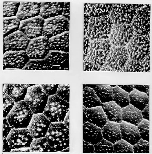
Body surface: melanica specimens presented a large number of perforations on each polygonal cell surface (Fig. 1B), with an average of 57.2 per-forations per cell. This average is 40.6% greater than the observed for the brasiliensis population which presented an average of 34 perforations per cell (Fig. 1A). The specimens of the melanica popu-lation has about 69.6% more perforations than the specimens originated from Juazeiro population (Fig. 1D) with an average of 17.4, and about 76.6% more than the specimens of the macromelasoma population (Fig. 1C), with an average of 13.4 per-forations per cell. The perper-forations distribution of melanic specimens (Fig. 2B) was peculiar. They were packed and close to the edges. In the brasiliensis population only a few perforations are close to the edges; also as perforations approach the center of the cell, their frequency increase. According to the statistical analysis, it was evi-denced a significant difference on the number of perforations to each group (Table I).
495 495495 495495 Mem Inst Oswaldo Cruz, Rio de Janeiro, Vol. 92(4), Jul./Aug. 1997
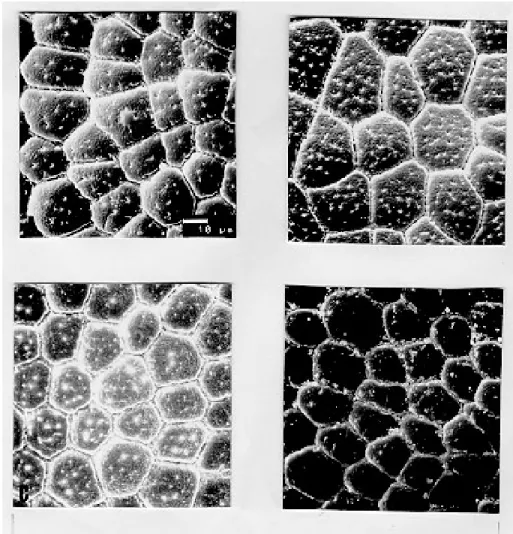
only slightly perceived perforations (Figs 2C and 2D). Specimens of Juazeiro populations have cells with rounded corners, different from the polygo-nal form of the other populations.
Morphometrics - The largest length and width of the egg were observed in the macromelasoma population, with an average of 2.43 mm and 1.41 mm, respectively. The smallest length was shown in the brasiliensis population with an average of 2.29
Fig. 1: exochorion of the egg bodies of four distinct populations of Triatoma brasiliensis. A- brasiliensis; B- melanica; C-macromelasoma; D- Juazeiro.
mm, and the shortest width in the Juazeiro popula-tion with 1.30 mm average value. The greatest mean value length÷width was seen in the Juazeiro speci-mens with an 1.82 average versus the shortest rela-tion in the macromelasoma specimens. Length and width presented significant statistical differences up to the 5% level among all the combinations studied, except for the melanic population versus the Juazeiro population (Table II).
TABLE I
Number of perfurations per cell of eggs of four distinct populations of Triatoma brasiliensis
brasiliensis melanica macromelasoma Juazeiro
min. 28 56 15 11
max 40 59 20 17
X 34 57.2 17.4 13.4
S 3.6765 1.0635 1.7733 1.9226 Kruskal- Wallis X2
3 = 108.67 p= 0.0000
X = average; S = standard deviation
496 496 496 496
496 Genital Structure and Eggs of T. brasiliensis - Different Chromatic Forms • J Costa et al.
Fig. 2: exochorion of the egg lids of four distinct populations of Triatoma brasiliensis. A- brasiliensis; B- melanica; C-macromelasoma; D- Juazeiro.
TABLE II
Egg morphometry of four distinct populations of Triatoma brasiliensis
Populations
Measures brasiliensis melanica macromelasoma Juazeiro Kruskal- Wallis
min. 2.15 2.20 2.24 2.16
max 2.49 2.49 2.58 2.51 X23 = 20.6006
X 2.30 2.36 2.43 2.36 p=0.0001
S 0.0913 0.0675 0.1179 0.0951
min. 1.25 1.27 1.29 1.20
max 1.40 1.44 1.53 1.36 X2
3 =45.4966
X 1.32 1.35 1.41 1.30 p=0.0000
S 0.0366 0.0418 0.0629 0.0469
min. 1.59 1.67 1.56 1.71
max 1.83 1.85 1.95 1.98 X23 =24.2445
X 1.73 1.75 1.72 1.82 p=0.0000
S 0.0517 0.0445 0.0832 0.0828
Length
W
idth
Relation Length/W
497 497497 497497 Mem Inst Oswaldo Cruz, Rio de Janeiro, Vol. 92(4), Jul./Aug. 1997
DISCUSSION
Studies on the male external genital structures, associated with other external morphological char-acters, have been an important tool for Triatominae taxonomy. Similar morphological species have been separated using these structures: Rhodnius stali and R. pictipes (Lent et al. 1993); T. maculata
and T. pseudomaculata (Galvão 1973). Recently, Jurberg and Galvão (1997) used genital structures to describe a new genus in triatomines. At intraspe-cific level, Lent and Jurberg (1985) reported indi-vidual variations for T. infestans and T. dimidiata.
The later species showed chromatic variations and their genital structures did not correlate with the distinct chromatic patterns.
In T. brasiliensis the minor individual varia-tions of the male genitalia clearly did not correlate with the distinct chromatic population or the geo-graphical origins. For example, the most readily quantifiable variable, number of denticles on the lateral endosomal process, varied from 7 to 91 in the sample as a whole and from 15 to 89 in two specimens from the same locality and of the same color pattern. In conclusion, morphological stud-ies of the genitalia are unlikely to be useful for defining these four populations of T. brasiliensis. Along with reports of structural variations in T. infestans (Pires et al. 1995) these results also em-phasize the importance of examining sufficiently large and representative samples before using mi-nor genital variations for taxonomic diagnosis.
In triatomines, several reports on ornamenta-tion of egg exochorion have demonstrated their importance as a tool for taxonomy at specific level (Galliard 1935, Gonçalves et al. 1985, Costa et al. 1991, Barata 1995). Barata (1981), described the exochorion morphology of eggs, using light mi-croscopy and SEM, from 10 species of the genus
Rhodnius, being this the most important contribu-tion.
The previous results on the egg morphology of
T. brasiliensis presented by Barth and Muth (1958) and by Jurberg et al. (1986) are in accordance to our results for the brasiliensis population. Differ-ent environmDiffer-ental conditions (available food sources, temperature and humidity) could affect direct or indirectly the egg morphology. But, in this study, the four populations have been maintained under the same environmental and feeding condi-tions. The number of eggs analyzed and its ran-domly selection from a large stock, originated from more than 30 females for each colony, also cor-roborate the morpho-structural differentiation ob-served at population level.
In the present study, morphometry provided significant differences among the populations of
T. brasiliensis, although overlapping of the mini-mum and maximini-mum values for the different mea-sures were observed. Hence, the possibility to sepa-rate T. brasiliensis populations based on the egg morphology was evidenced. Differences in the structural level, on the exochorion ornamentation, with different number of perforations and also on the shape of cells of the lid were shown.
The population differentiation detected by egg morphology is in agreement with several other data: the four populations showed homogeneity and sta-bility of their chromatic pattern for several years in the colonies reared in the laboratory (Costa et al. 1996b); during the field collections, only one colour pattern was found in each locality. We did not record intermediate forms, reported by Lent and Wygodzinsky (1979). Maybe these intergrading forms could be present in the border lines where crossings among different colour pat-terns could be occuring; isoenzymatical data (Costa et al. 1997) suggested a clear genetic differentia-tion among these populadifferentia-tions of T. brasiliensis, reaching Nei’s genetic distance values correspon-dent to those found at interspecific level, for other phylogenetic related species (Pereira et al. 1996). In order to broaden the knowledge on these different populations of T. brasiliensis, it will be important to define the geographical distribution, emphasizing the overlapping regions, determine the existence or non-existence of reproductive isola-tion mechanisms via directed crosses (Costa et al. 1996a) and to determine the population structures by other genetic markers such as chromosomes and DNA.
ACKNOWLEDGMENTS
To Dr Teresa Cristina M Gonçalves, for permission to analyze the insects of Herman Lent Hemiptera col-lection. To Drs Pedro Cabello and Nelson Chagas for the guidance on statistical procedures. To Dr Herman Lent for his valuable comments on the manuscript.
REFERENCES
Barata JMS 1981. Aspectos morfológicos de ovos de Triatominae. II Características macroscópicas e exocoriais de dez espécies do gênero Rhodnius Stal, 1859 (Hemiptera, Reduviidae). Rev Saúde Públ 15: 490-542.
Barata JMS 1995. Aspectos morfológicos de ovos de Triatominae, p. 55-58. In Proc Inter Workshop on Population Genetics and Control of Triatominae, Ec-uador.
Barth R, Muth H 1958. Estudos anatômicos e his-tológicos sobre a família Triatominae (Heteroptera, Reduviidae). VII Parte: Observações sobre a superfície dos ovos das espécies mais importantes.
Mem Inst Oswaldo Cruz 56: 197-208, figs. 1-23. Costa JM, Jurberg J, Barth O 1991. Estudos morfológicos
(Hemi-498 498 498 498
498 Genital Structure and Eggs of T. brasiliensis - Different Chromatic Forms • J Costa et al.
ptera, Reduviidae, Triatominae). Mem Inst Oswaldo Cruz 86: 247-263.
Costa J, Marchon-Silva V, Almeida JR 1996a. Does hybridization clarify the taxonomic status of differ-ent melanic forms of Triatoma brasiliensis
Neiva,1911 (Hemíptera- Reduviidae- Triatominae)?
Mem Inst Oswaldo Cruz 91 (Suppl. I): 128. Costa J, Marchon-Silva V, Freitas-Sibajev MGR, Pires
MQ, Pacheco RS 1996b. Estudos isoenzimáticos detectam variação intra-especifica em Triatoma brasiliensis Neiva, 1911 (Reduviidae, Triatominae).
Rev Soc Bras de Med Trop 29 (Suppl. I): 129. Costa J, Freitas-Sibajev MGR, Marchon-Silva V, Pires
MQ, Pacheco RS 1997. Isoenzymes detect variation in populations of Triatoma brasiliensis. Mem Inst Oswaldo Cruz 92 (in press).
Galliard H 1935. Recherches sur les Reduviidés Hématophages Rhodnius et Triatoma. V. Mor-phologie de l’oeuf des Triatomes. Ann Parasit hum comp XIII: 511-527.
Galvão AB 1956. Triatoma brasiliensis macromelasoma
n. subsp. (Hemiptera, Reduviidae). Rev brasil de Malariol D trop 7: 455-457.
Galvão AB 1973. Contribuição ao conhecimento do Tri-atoma maculata (Erichson, 1848) e do Triatoma pseudomaculata Corrêa & Espínola, 1964 (Hemi-ptera, Reduviidae). Rev Soc Bras Med Trop 7: 367-380.
Gonçalves TCM, Jurberg J, Costa JM, Souza W 1985. Estudo morfológico comparativo de ovos e ninfas de Triatoma maculata (Erichson, 1848) e Triatoma pseudomaculata Corrêa e Espínola, 1964 (Hemi-ptera, Reduviidae, Triatominae). Mem Inst Oswaldo Cruz 80: 263-276.
Jurberg J, Galvão C 1997. Hermanlentia n. gen. da tribo Triatomini, com um rol de espécies de Triatominae
(Hemiptera, Reduviidae). Mem Inst Oswaldo Cruz 92: 181-185.
Jurberg J, Gonçalves TCM, Costa JM, Souza W 1986. Contribuição ao estudo morfológico de ovos e ninfas de Triatoma brasiliensis Neiva, 1911 (Hemiptera, Reduviidae, Triatominae). Mem Inst Oswaldo Cruz 81: 111-120.
Lent H, Jurberg J 1978. Estudo comparativo da genitália externa masculina de seis espécies de Triatoma
Laporte, 1832 que mais freqüentemente habitam o domicílio humano no Brasil (Hemiptera, Reduvi-idae). Rev Brasil Biol 38: 931-944.
Lent H, Jurberg J 1985. Sobre a variação intra-específica em Triatoma dimiata (Latreille) e Triatioma infestans
(Klug) (Hemiptera, Reduviidae). Mem Inst Oswaldo Cruz 80: 285-299.
Lent H, Jurberg J, Galvão C 1993. Rhodniusstali n.sp., afim de Rhodniuspictipes Stal, 1872 (Hemiptera, Reduviidae, Triatominae). Mem Inst Oswaldo Cruz 88: 605-614.
Lent H, Wygodzinsky P 1979. Revision of Triatominae (Hemiptera, Reduviidae), and their significance as vectors of Chagas disease. Bull Amer Mus Nat His-tory, New York, 163: 520.
Pereira J, Dujardin JP, Salvatella R, Tibayrenc M 1996. Enzymatic variability and phylogenetic relatedness among Triatoma infestans, T. platensis, T. delpontei
and T. rubrovaria. Heredity 77: 47-54.
Pires HHR, Barbosa SH, Jurberg J, Diotaiuti L 1995. Comparative studies of distinct populations of Tri-atoma infentans. V. Morphological patterns of the male genitalia. Mem Inst Oswaldo Cruz 90 (Suppl. I): 227.