From the Rheumatology Unit of the Children’s Institute and the Laboratory of Immunology of the Heart Institute, Hospital das Clínicas, Faculty of Medicine, University of São Paulo.
ANALYSIS OF HUMAN LEUKOCYTE ANTIGENS OF
CLASS II-DR IN BRAZILIAN CHILDREN AND
ADOLESCENTS WITH SYSTEMIC LUPUS
ERYTHEMATOSUS
Bernadete de L. Liphaus, Anna Carla Goldberg, Maria Helena B. Kiss and Clovis A. A. Silva
RHCFAP/3106
LIPHAUS B de L et al. - Analysis of human leukocyte antigens of class II-DR in Brazilian children and adolescents with systemic lupus erythematosus. Rev. Hosp. Clín. Fac. Med. S. Paulo 57(6):277-282, 2002.
OBJECTIVE: To analyze the frequency of human leukocyte antigens class II-DR in children and adolescents with systemic lupus erythematosus.
PATIENTS AND METHODS: Fifty-fiveBrazilian systemic lupus erythematosus children and adolescents and 308 healthy individuals were studied. Gender, race, and age of onset of systemic lupus erythematosus were recorded. The human leukocyte antigens typing of class II-DR was carried out by polymerase chain reaction amplification with sequence-specific primers (PCR-SSP). Data were analyzed statistically using the chi square test with Yates’ correction, Fisher’s exact test, and Bonferroni’s correction.
RESULTS: Human leukocyte antigen-DR 15 was the most frequently detected antigen in this group of children and adolescents, and it also occurred more frequently in the female group, in children with onset of systemic lupus erythematosus between 0 and 9 years and between 10 to 14 years, and in the Black race group, but these associations were not statistically significants.
CONCLUSION: In this group of children and adolescents with a high degree of racial admixture, we could not verify a significant association between human leukocyte antigens class II-DR and systemic lupus erythematosus.
DESCRIPTORS: Systemic lupus erythematosus. Human leukocyte antigens. Children and adolescents.
INTRODUCTION
A complex genetic and immuno-logic network participates in the pathogenesis of systemic lupus ery-thematosus (SLE)1,2. Among the
ge-netic factors, the human leukocyte an-tigens (HLA) have earned special at-tention in the literature, and a series of works have reported an increased fre-quency of the HLA-DR2 (15/16), DR3 (17/18), DR7, and DR9 in patients with SLE3-7.
The relationships established to date between the HLA and SLE were in adult populations or in mixed populations of adults and children,
with few works in literature involving only children.
The objective of this study was to analyze the frequency of the HLA class II-DR in Brazilian children and adoles-cents with SLE.
PATIENTS AND METHODS
A total of 55 children and
adoles-cents with a diagnosis of SLE8 were
studied and followed up. Patients were included in the study after informed and written consent and following ap-proval of the study by the ethics com-mission. The patients’ demographic characteristics are presented in table 1. The control group comprised 308 healthy and unrelated individuals; it was established using the DNA bank from the Laboratory of Immunology of the Heart Institute, Hospital das Clínicas, Faculty of Medicine, Univer-sity of São Paulo.
sequence-spe-cific primers (PCR-SSP). The method was based on the procedure devel-oped by Olerup and Zetterquist9.
Statistical analysis included the chi square test with Yate’s correction of continuity and Fisher’s exact test. Con-fidence limits of 95% (P = 0.05) were used. The significant results were mul-tiplied by the total number of tested an-tigens (14 anan-tigens), thereby obtaining a corrected value of P, termed Pc (Bonferroni’s correction)10-12.
RESULTS
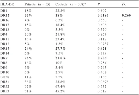
In this group of 55 Brazilian chil-dren and adolescents with SLE, the HLA-DR15 was detected most fre-quently; it was present in 18 patients (33%). The second most frequently de-tected antigen was HLA-DR7, present in 14 patients (26%), and the third was HLA-DR13, present in 13 patients (24%).
The comparisons between the fre-quencies of HLA-DR in the group of patients with SLE and in the control group are presented in table 2.
It was not possible to establish an association between HLA and the group of children and adolescents with SLE under study, in spite of the sug-gestion of an association with HLA-DR15 (P = 0.0186), which was not con-firmed after Bonferroni’s correction (Pc = 0.260).
In the 15 patients with age at dis-ease onset between 0 and 9 years, HLA-DR13 and HLA-DR15 were detected most frequently (P = 0.309 and P = 1.0, respectively) and were present in 33% of the patients. In the 35 patients aged between 10 and 14 years, HLA-DR15 was the most frequently detected (P = 0.978) and was present in 34% of the patients. In the 5 patients with ages between 15 and 19 years, HLA-DR13 (P = 0.581), HLA-DR4 (P = 0.258), and HLA-DR7 (P = 0.592) were the most frequently detected antigens.
The relationships between HLA-DR, gender, and ethnic group are pre-sented in table 3. HLA-DR 15 was the most frequently detected antigen in the female group and in the Black race
group, but again, after Bonferroni’s correction, there was no association be-tween HLA-DR and gender or ethnic group.
Table 1 - Demographic characteristics of the 55 children and adolescents with SLE.
Demographic characteristics
Sex (n) Female 4 6
Male 0 9
Age at disease onset (years) Mean / range 10.4 / 1 to 17 Age at disease onset Stratified (n) 0 to 9 years (n=15) Female 1 4
Male 0 1
10 to 14 years (n=35) Female 2 9
Male 0 6
15 to 19 years (n=05) Female 0 3
Male 0 2
Age at inclusion to study (years) Mean / range 14 / 8 to 20
Follow up (years) Mean / range 3.5 / 1 to 13
Race – Caucasian (n = 25) Female 2 2
Male 0 3
Black (n = 07) Female 0 5
Male 0 2
Asian (n = 01) Female 0 1
Male 0 0
Mixed (n = 22) Female 1 8
Male 0 4
Table 2 - Frequency of HLA-DR in the 55 children and adolescents with SLE and in the 308 individuals comprising the control group.
HLA-DR Patients (n = 55) Controls (n = 308)* P Pc
DR1 18% 22.2% 0.602
-DR15 33% 18% 0.0186 0.260
DR16 4% 6.3% 0.550
-DR17 15% 18.4% 0.606
-DR18 0% 3.3% 0.370
-DR4 20% 21.8% 0.867
-DR11 13% 23.4% 0.112
-DR12 5% 1.3% 0.0737
-DR13 24% 27.7% 0.621
-DR14 5% 7.5% 0.779
-DR7 26% 21.8% 0.706
-DR8 16% 10% 0.254
-DR9 5% 5.4% 0.763
-DR10 5% 2.9% 0.402
-Blank 11% 5.2% 0.136
-DR51 36% 23.8% 0.0696
-DR52 62% 67.4% 0.532
-DR53 51% 45.2% 0.518
-Blank = Non-identified antigens
DISCUSSION
One of the main problems in analyzing the studies regarding HLA is related to the methodology adopted. The characterization of the HLA can be ac-complished using the serologic method (for detecting DR2) or PCR amplifica-tion with sequence-specific primers (for detecting DR15 and DR16) or using the specific sequences of oligopeptides (for detecting DRB1*1501 to 1506 and DRB1*1601 to 1608). These techniques have different sensitivities, which hin-ders an effective comparison between the studies.
The Brazilian population, and es-pecially those who reside in the city of São Paulo, is characterized by a high degree of racial admixture. The ethnic origin of these individuals is very difficult to establish. The major-ity possesses different percentages of Caucasian, Black, and Amerindian ancestry. Therefore, it is necessary to determine which HLA are associated with SLE in a population characterized by this degree of miscegenation.
Despite exhaustive studies in pa-tients with SLE, the sensitivity and specificity of the associations between HLA and SLE vary in the diverse stud-ies and different ethnic groups, render-ing the results inconclusive14,15.
Analy-sis of various studies suggests a strong association between the HLA-B8/ DR3/DQ2/C4A0 and B7/DR2/DQ1 haplotypes and SLE3,15-24. HLA-DR2
(15/16) and HLA-DR3 (17/18), taken individually, are also associated with SLE, but the relative risk conferred by these is low (from 2 to 3)16.
SLE is rare in childhood, yet the risk of SLE in the children of patients with SLE is about 10%, demonstrating a genetic component of the disease.
In infants, SLE develops with clini-cal manifestations that maintain some differences when compared to those observed in adults1,2. These differences
in manifestation raise some questions regarding the possibility that SLE in childhood is a disease with its indi-vidual genetic characteristics and may be different from SLE in adults.
Lehman et al.25 studied 71 children
with SLE and observed that the onset of the disease before 10 years of age was strongly related to the presence of antibodies to Ro/SSA in the maternal blood, suggesting that the antibody to Ro/SSA can influence the immunologi-cal development of the fetus.
In this group of children with SLE, we observed that the distribution of the various HLA-DR was similar to the control group, except for HLA-DR15 which was present in 33% of the chil-dren with SLE and in 18% of the con-trol group; however, this relationship was not statistically significant after Bonferroni’s correction (P = 0.0186 and Pc = 0.260). HLA-DR7 and DR13 were the second and the third most fre-quently detected antigens, respec-tively, but again, these correlations were not statistically significant.
In Brazil, Silva et al.26 studying 93
patients with SLE with ages between 13 and 66 years, observed a higher fre-quency of HLA-DR3 (17/18) (P = 0.003), a result that differs from that observed in our study and from that re-ported by Fernandes et al.27 in a study
Table 3 - Frequency of HLA-DR in the 55 children and adolescents with SLE, according to gender and ethnic group.
HLA- DR Number of patients Female Male Caucasian Black Asian Mixed
DR1 1 0 7 3 (P=0.339) 8 (P=0.0315 - - 2
Pc=0.412)
DR 15 1 8 17 (P=0.240) 1 7 5 (P=0.0316 - 6
Pc=0.412)
DR 16 2 2 - 2 - -
-DR 17 8 5 3 (P=0.113) 4 1 - 3
DR 18 0 - - -
-DR 4 1 1 9 2 8 (P=0.0905) - - 3
DR 11 7 7 - 2 2 - 3
DR 12 3 3 - 1 - - 2
DR 13 1 3 1 0 3 (P=0.427) 4 3 - 6
DR 14 3 3 - 2 - - 1
DR 7 1 4 1 1 3 (P=0.678) 4 2 - 8 (P=0.229)
DR 8 9 7 2 3 1 1 4
DR 9 3 3 - 1 - - 2
DR 10 3 3 - 2 - - 1
Blank 6 5 1 2 - 1 3
DR 51 2 0 1 9 1 9 5 - 6
DR 52 3 4 2 8 6 1 3 6 - 1 5
DR 53 2 8 2 3 5 1 3 2 - 1 3
Total 5 5 4 6 9 2 5 7 1 2 2
with 56 adult SLE patients, in which HLA-DR2 (15/16) was the most fre-quently detected antigen (P <0.005).
The association of HLA-DR7 and HLA-DR13 with SLE was observed in a few studies, such as that by Wilson et al.7 in African Americans and that by
Cowland et al.28 in Denmark.
Regarding the age of SLE onset, some authors describe an increased fre-quency of the HLA-B8/DR3/DQ2/ C4A0 haplotype in patients among which the onset of the disease occurred after 35 years of age. Other authors have described an increased frequency of the HLA-B7/DR2/DQ1 haplotype in patients whose SLE appeared before 25 years of age and that presented se-rious nephritis3. However, Barron et
al.29 in a study of children with SLE
did not corroborate these findings. Likewise, we did not observe any relationship between the age at onset of SLE and HLA. However, all of our patients presented onset of the SLE before 25 years of age, and HLA-DR2 (15/16) was present in 37%, therefore demonstrating a behavior similar to that described above.
It is interesting to underscore that in this study, HLA-DR15 was the most frequently detected antigen in the 0 to 9-year-old and 10 to 14-year-old age groups, but this was not the case among the adolescents from 15 to 18 years of age.
The associations between the HLA and SLE vary between the different eth-nic groups. HLA-DR3 is associated
with SLE in Caucasians, and HLA-DR2 is associated with Black, Chinese, and Japanese populations6,15. In Caucasian
children with SLE, an association was observed with HLA-DR3(DRB1*0301), and in Blacks, there was an association with HLA-DR2(DRB1*1503/ DRB1*1501) / DRB5*0101 / DQA1*0102 / DQB1*060229.
In our study, DR1 and HLA-DR4 were the most frequently detected antigens in the Caucasian patients. There is no report of an association be-tween HLA-DR1 and SLE in the litera-ture, either among Caucasians, Black, or Asians6, while HLA-DR4 is
de-scribed as being associated with SLE only in Indians30. According to the
lit-erature, 75% of Caucasian patients with SLE present the HLA-DR2 and/ or HLA-DR3 antigens. We observed that 52% of our Caucasian patients presented these antigens.
HLA-DR15 was the most frequently antigen in the Black race patients of this study. The HLA-DR2 (15/16) has been associated with Black race SLE patients both in Black race people from America as from South Africa24, 31, 32.
Among the Asian patients, an asso-ciation has been described between SLE and HLA-DR9 in Chinese pa-tients from Singapore 5 and in
Kore-ans20 and with HLA-DR15 in
Japa-nese19, Chinese23, and Korean
pa-tients20. Our only Asian patient
pre-sented HLA-DR8 in homozygosis. There is no association described be-tween SLE and HLA-DR8.
In the patients of the mixed-race group, HLA-DR7 was the most fre-quently occurring antigen. There are descriptions of an association between HLA-DR7 and SLE only in Black race people from America6,7.
It is interesting to observe that in our study, there was a prevalence of HLA-DR7 in the mixed-race group and of HLA-DR15 in the Black race group, suggesting that perhaps the group con-sidered to be mixed race here may in fact not be characterized by a predomi-nant miscegenation of Black race with other races.
In the present study, we observed associations between HLA and SLE ethnic groups that were different from those observed in the literature. This difference is probably due to the fact, as already stated, that racial admixture in Brazil is very prominent, which hin-ders a comparison of our ethnic groups with those of other countries. (Table 4) SLE occurs more frequently among women, mainly after puberty, with a female:male ratio of 8:1. The higher in-cidence of SLE in women has been as-sociated with hormonal factors, such as estrogen, which could influence the on-set of this pathology. In children, female prevalence also occurs; perhaps genetic factors, especially concerning HLA, could have some influence on the higher incidence of SLE in the female sex. The ratio between the female and male sex among the children and adolescents with SLE in this study was 5.1:1, a find-ing similar to the ratio reported in the
Table 4 - Relationship between the HLA and SLE racial groups.
Author Ethnic Groups
Caucasian Black Asian Mixed
Arnett3 HLA-DR2/DR3 - -
-Fernades et al.27 HLA-DR2 - -
-Reivelle et al.32 - HLA-DR2 -
-Rudwaleit et al.31 - HLA-DR2 -
-Barron et al. 29 HLA-DR3 HLA-DR2 -
-Rudwalet et al. 5 and Hong et al.20 - - HLA-DR9
-Hashimoto et al.19 and Doherty et al.23 - - HLA-DR15 (2)
-Wilson et al.7 - HLA-DR7 -
literature, and there was a uniform dis-tribution of HLA-DR in both the female and male groups. (Table 3)
The majority of studies do not re-port a relationship between the HLA and gender; however, Doherty et al.23
observed that the HLA-DR15 antigen
was detected in all the men in their study population.
In view of the discrepancies ob-served in the various studies referred to in the literature, it is possible that HLA have little or no influence on the mecha-nisms of etiopathology of SLE. While
possible, this fact seems improbable be-cause of the close relationship between HLA and the immunological system. Perhaps in the near future, methods with greater sensitivity and more specific probes will establish clearer correla-tions than those seen to date.
RESUMO RHCFAP/3106
LIPHAUS B de L e col. - Análise dos antígenos de histocompatibilidade leucocitária de classe II-DR em cri-anças e adolescentes brasileiros com lúpus eritematoso sistêmico. Rev. Hosp.Clín. Fac. Med. S. Paulo 57(6):277-282 2002.
OBJETIVO: Analisar a freqüência
dos antígenos de histocompatibi-lidade leucocitária de classe II-DR em crianças e adolescentes com o lúpus eritematoso sistêmico.
PACIENTES E MÉTODOS:
Cin-qüenta e cinco crianças e adolescen-tes lúpicos brasileiros e 308 indivídu-os sadiindivídu-os foram estudadindivídu-os. Os sexindivídu-os,
os grupos étnicos e as idades de iní-cio da doença foram anotados. A tipagem de histocompatibilidade leucocitária de classe II-DR foi reali-zada pela reação de polimerase em ca-deia com amplificação de sondas de seqüência específica (PCR-SSP). Na análise estatística foram utilizados o teste de qui-quadrado com correção de Yates, o teste exato de Fisher e a cor-reção de Bonferroni.
RESULTADOS: A
histocompati-bilidade leucocitária-DR15 foi a mais freqüente neste grupo de crianças e adolescentes, sendo também mais fre-qüente nas mulheres, nas crianças com idade de início da doença entre zero e
nove anos e entre 10 e 14 anos e nas crianças de raça negra, mas estas cor-relações não foram estatisticamente significativas.
CONCLUSÃO: Neste grupo de
cri-anças e adolescentes com alto grau de miscigenação não pudemos observar associação significativa entre os antígenos de histocompatibilidade leucocitária de classe II-DR e o lúpus eritematoso sistêmico.
DESCRITORES: Lúpus eritema-toso sistêmico. Antígenos de histo-compatibilidade leucocitária. Crian-ças e adolescentes.
REFERENCES
1 . CASSIDY, J.T. - Systemic lupus erythematosus, juvenile dermatomyositis, scleroderma and vasculitis. In: KELLEY WN; HARRIS ED; RUDY S et al. - (Ed) - Textbook of Rheumatology. 5th ed. Philadelphia, Pennsylvania, Saunders, 1997. p1241-5. 2 . CASSIDY JT & PETTY RE - Systemic lupus erythematosus. In: Textbook of Pediatric Rheumatology. 3rd ed. Philadelphia, Pennsylvania, Saunders, 1995. p 260-322.
3 . ARNETT FC - The genetics of human lupus. In: WALLACE DJ & HAM BH ed. - Dubois Lupus Erythematosus. 5th ed. Philadelphia, Lea, 1997. p 77-117.
4 . HAHN BH - An overview of the pathogenesis of systemic lupus erythematosus. In: WALLACE DJ & HAM BH ed. - Dubois Lupus Erythematosus. 5th ed. Philadelphia, Lea, 1997. p 69-75.
5 . RUDWALEIT M; GIBSON K; WORDSWORTH P et al. - HLA associations of systemic lupus erythematosus in Chinese from Singapore. Ann Rheum Dis 1995; 54(8): 686-7.
6 . SCHUR PH - Genetics of systemic lupus erythematosus. Lupus 1995; 4: 425-37.
7 . WILSON WA; SCOPELITIES E; MICHALKI JP - Association of HLA-DR7 with antibody to SSA(Ro) and disease susceptibility in blacks with systemic lupus erythematosus. J Rheumatol 1984; 11: 653-7.
9 . OLERUP O & ZETTERQUIST H - HLA-DR typing by PCR amplification with sequence-specific primers (PCR-SSP) in 2 hours: An alternative to serological DR typing in clinical practice including donor-recipient matching in cadaveric transplantation. Tissue Antigens 1992; 39: 225-35.
10.BENGTSSON BO & THOMSON G - Measuring the strength of associations between HLA antigens and diseases. Tissue Antigens 1981; 18: 356-63.
11.FUKUDA K; SUGAWA K; WAKISAKA A et al. - Statistical detection of HLA and disease association. Tissue Antigens 1985; 26: 81-6.
12.SVEJGAARD A; JERSILD C; STAUB NIELSEN L et al. - HLA antigens and disease statistical and genetic considerations. Tissue Antigens 1974; 4: 95-105.
13.GOLDBERG AC; CHIARELLA JM; MARIN MLC et al. -Molecular typing of HLA class II antigens in a São Paulo population. Genet Mol Biol 1998; 21(3): 301-5.
14.ADAMS D - How the immune system works and why it causes autoimmune diseases. Immunology Today 1996; 17(7): 300-2 .
15.ARNETT FC - Genetic aspects of human lupus. Clin Immunol Immunopathol 1992; 63(1): 4-6.
16.HAHN BH - Pathogenesis of systemic lupus erythematosus. In: KELLEY WN; HARRIS ED; RUDY S et al. – (Ed.) - Textbook of Rheumatology.5th ed. Philadelphia, Saunders, 1997. p. 1015-27.
17.DAVIES EJ; HUTCHINGS CJ; HILLARBY MC et al. - HLA-DP does not contribute towards susceptibility to systemic lupus erythematosus. Ann Rheum Dis 1994; 53(3): 188-90. 18.DAVIES EJ; STEERS G; OLLIER WER et al. - Relative
contributions of HLA-DQA and complement C4a loci in determining susceptibility to systemic lupus erythematosus. Br J Rheumatol 1995; 34: 221-5.
19.HASHIMOTO H; NISHIMURA Y; DONG RP et al. - HLA antigens in Japanese patients with systemic lupus erythematosus. Scand J Rheumatol 1994; 23(4): 191-6.
20.HONG GH; KIM HY; TAKEUCHI F et al. - Association of complement C4 and HLA-DR alleles with systemic lupus erythematosus in Koreans. J Rheumatol 1994; 21(3): 442-7. 21.BATCHELOR JR - Systemic lupus erythematosus and genes within
the HLA region. Br J Rheumatol 1993; 32(1):13-5.
22.GOLDSTEIN R; SENGAR DPS - Comparative studies of the major histocompatibility complex in French Canadian and non-French Canadian Caucasians with systemic lupus erythematosus. Arthritis Rheum 1993; 36(8): 1121-7.
23.DOHERTY DG; IRELAND R; DEMAINE AG et al. - Major histocompatibility complex genes and susceptibility to systemic lupus erythematosus in southern Chinese. Arthritis Rheum 1992; 35(6): 641-6.
24.HARTUNG K; BAUR MP; COLDEWEY R et al. - Major histocompatibility complex haplotypes and complement C4 alleles in systemic lupus erythematosus. Results of a multicenter study. JClin Invest 1992; 90(4): 1346-51.
25.LEHMAN TJA; REICHLIN M; SANTNER TJ et al. - Maternal antibodies to Ro(SSA) are associated with both early onset of disease and male sex among children with systemic lupus erythematosus. Arthrtis Rheum 1989; 32(11): 1414-20. 26.SILVA LM & DONADI EA - Is immunogenetic susceptibility to
neuropsychiatric systemic lupus erythematosus (SLE) different from non-neuropsychiatric SLE ? Ann Rheum Dis 1996; 55: 544-7.
27.FERNANDES SRM; PERSOLI LB; MARQUES SBD et al. - HLA antigens and susceptibility to systemic lupus erythematosus in Brazilian patients. Lupus 1995; 4:43s.
28.COWLAND JB et al. - DNA polymorphism of HLA class II genes in systemic lupus erythematosus. Tissue Antigens 1994; 43: 34-7.
29.BARRON KS; SILVEMAN ED; GONZALES J et al. - Clinical, serologic, and immunogenetic studies in childhood-onset systemic lupus erythematosus. Arthritis Rheum 1993; 36(3): 348-54.
30.MEHRA NK; PANDE I; TANEJA V et al. - Major histocompatibility complex genes and susceptibility to systemic lupus erythematosus in northern India. Lupus 1993; 2(5): 313-4.
31.RUDWALEIT M; TIKLY M; GIBSON K et al. - HLA class II antigens associated with systemic lupus erythematosus in black South Africans. Ann Rheum Dis 1995; 54: 678-80.
32.REVEILLE JD; BARGER BO; HODGE TW - HLA-DR2-DRB1 allele frequencies in DR2-positive black Americans with and without systemic lupus erythematosus. Tissue Antigens 1991; 38(4): 178-80.