Odontogenic tumors: clinical
and pathology study of 238
cases
Summary
Rafael Linard Avelar1, Antonio Azoubel Antunes2,
Thiago de Santana Santos3, Emanuel Sávio de
Souza Andrade4, Edwaldo Dourado5
1 Dental surgeon, resident in buccomaxillofacial surgery and traumatology, Hospital Universitário Oswaldo Cruz.
2 Dental surgeon, student in the specialization course on buccomaxillofacial surgery and traumatology, Universidade de Pernambuco - FOP/UPE. 3 Dental surgeon, resident in buccomaxillofacial surgery and traumatology, Hospital Universitário Oswaldo Cruz - HUOC/UPE. 4 Doctor, adjunct professor of buccal pathology, Faculdade de Odontologia de Pernambuco - Universidade de Pernambuco - FOP/UPE.
5 Doctor, adjunct professor of buccomaxillofacial surgery and traumatology, Faculdade de Odontologia de Pernambuco - Universidade de Pernambuco - FOP/UPE. Discipline of buccal pathology, Faculdade de Odontologia da Universidade de Pernambuco - FOP/UPE.
Address for correspondence: Emanuel Sávio de Souza Andrade - Faculdade de Odontologia de Pernambuco, Disciplina de Patologia Bucal. Av. Gal. Newton Cavalcanti 1650 Tabatinga Camaragibe PE 54753-220.
This paper was submitted to the RBORL-SGP (Publishing Manager System) on 10 May 2007. Code 4513. The article was accepted on 14 July 2007.
O
dontogenic tumors are neoplasms that develops exclusively in the gnathic bones; they originate from odontogenic tissues, by epithelial or mesenchymal proliferation, or both. Aim: To evaluate the incidence of odontogenic tumors in a specific institution, and to compare these findings with other studies in the literature.Study format: A cross-sectional cohort retrospective study.
Material and method: The sample was obtained from the files of patients with odontogenic tumors diagnosed between January 1992 and March 2007 (15 years). Cases in which the diagnosis could be adapted to the new World Health Organization (WHO) of 2005 were included. Data such as gender, age, anatomical site, histological type and symptomatology were analyzed. Results: Odontogenic tumors were 4.76% of all biopsied lesions within the studied period. The mean age was 30.7 years; 57% of the patients were male. The keratocystic odontogenic tumor was the most prevalent histological type (30%), followed by the ameloblastoma (23,7%). The rate of asymptomatic cases was 75.7%. Conclusion: Odontogenic tumors occurred more frequently in females, in the second and third decades of life, and more commonly in the mandible; most cases were asymptomatic.
Keywords: classification, epidemiology, odontogenic tumors.
original article Rev Bras Otorrinolaringol
INTRODUCTION
Odontogenic tumors are a heterogeneous group of lesions with variable clinical and pathohistological features. The biological behavior of these tumors inclu-des hamartomatous proliferation, non-aggressive benign
tumors, and aggressive and malignant tumors.1 There has
been considerable interest in odontogenic tumors by oral pathologists, who have studied and catalogued these tu-mors for decades. These tutu-mors are 2.5% of all biopsied
lesions in dental offices.2,3
Although many retrospective studies have been
conducted in Africa,4,5,6 Asia,7 Europe,8,9 and North
America,10 unanswered question still remain about the
relative frequency and the incidence of certain
odonto-genic tumors.11
The geographical distribution of these lesions is
variable.2 Many studies in different part of the world have
shown differences in the relative prevalence of these
tu-mors.12 Few reports have been published in English about
the frequency of odontogenic tumors in Latin America,
particularly in Brazil.13 The frequency was 1.29%14 in a
Chilean study of 362 cases.
Various attempts at classification of these tumors have been published to define diagnostic criteria, given the diversity of lesions that may arise from odontogenic tissues.1
The first classification of these tumors was published in 1971, based on a 5-year joint effort coordinated by the World Health Organization (WHO).12 An updated edition
of this classification was published in 1992.15 A new
classi-fication was proposed in 2005, which included the
odon-togenic keratocyst as a benign odonodon-togenic tumor.16
The purpose of this study was to investigate the epidemiological behavior of this heterogeneous group of tumors across a 15-year period (1992-2007) and to compare these data with those in the literature.
MATERIAL AND METHODS
A retrospective study was made of cases of odonto-genic tumors recorded at our institution between January 1992 and March 2007. The variables gender, age, anato-mical site, histological type and symptoms were analyzed in 238 histopathology reports.
In reports with recurring tumors, the histological appearance of the first and recurring tumors was compared and considered as a single case.
The diagnoses were reassessed and adapted to the
2005 WHO classification.16
After the sample was obtained, a database was generated using the SPSS (v. 13.0) statistics software; the chi-square test was applied to check the statistical signifi-cance of the findings. A p value below 0.05 was considered statistically significant.
This study was duly recorded by the Research Ethics Committee of our institution (protocol number 135717/07).
RESULTS
Patients were distributed according to the data as follows:
Table 1. Distribution of patients according to gender.
Histological types
Gender
Male Female
N % n %
Ameloblastoma 30 52,6 27 47,4
Cementoblastoma 1 25 3 75
Keratocystic Odontogenic Tumor 30 43,4 39 56,6
Calcifying Epithelial Odontogenic Cyst 7 46,6 8 53,4
Ameloblastic Fibroma 3 75 1 25
Ameloblastic Fibrodontoma - - 1 100
Mixoma 8 53,4 7 46,6
Odontoma 17 31,4 37 68,6
Adenomatoid Odontogenic Tumor 3 23 10 77
Calcifying Epithelial Odontogenic Tumor 3 60 2 40
Squamous Odontogenic Tumor - - 1 100
TOTAL 102 136
Table 2. Age of patients with odontogenic tumors. Histological types
Age Groups Total
Age ranges
Mean
ages 00-10 11-20 21-30 31-40 41-50 51-60 61-70 70 n %
Ameloblastoma 10-99 34 1 14 16 8 5 9 3 1 57 23,7
Cementoblastoma 40-57 44 - - - 2 1 1 - - 4 1,7
Keratocystic
Odon-togenic Tumor 10-84 31 2 14 23 15 9 3 2 1 69 30,0
Calcifying Epithelial
Odontogenic Cyst 10-70 32 1 4 5 - 1 3 1 - 15 6,3
Ameloblastic
Fi-broma 38-45 42 - - - 1 3 - - - 4 1,7
Ameloblastic
Fibro-dontoma 11 11 - 1 - - - 1 0,4
Mixoma 7-45 29 1 4 3 4 3 - - - 15 6,3
Odontoma 4-84 27 8 17 10 9 4 2 2 2 54 22,1
Adenomatoid
Odontogenic Tumor 10-45 20 1 7 2 2 1 - - - 13 5,4
Calcifying Epithelial
Odontogenic Tumor 11-46 26 - 3 - 1 1 - - - 5 2,0
Squamous
Odonto-genic Tumor 16 16 - 1 - - - 1 0,4
p>0,05 - chi-square test
Table 3. Anatomical sites of odontogenic tumors.
Histological types
Anatomical site
Mandible Maxilla
N % n %
Ameloblastoma 48 84,2 09 15,8
Cementoblastoma 04 100 -
-Keratocystic Odontogenic
Tumor 52 75,3 17 14,7
Calcifying Epithelial
Odontogenic Cyst 10 66,6 05 33,3
Ameloblastic Fibroma 04 100 -
-Ameloblastic
Fibrodontoma 01 100 -
-Mixoma 07 46,6 08 53,4
Odontoma 23 42,5 31 57,5
Adenomatoid
Odontogenic Tumor 06 46,1 07 53,9
Calcifying Epithelial
Odontogenic Tumor 04 80 01 20
Squamous Odontogenic
Tumor 01 100 -
-TOTAL 160 78
DISCUSSION
Odontogenic tumors are infrequent in gnathic bo-nes, and should be included in the differential diagnosis
of gnathic bone lesions.2 Few published studies have been
done on large series in any country or region to assess age, gender and site of odontogenic tumors, based on the 1992
classification of the World Health Organization.3,6,7,14,17
As of 2005, a new classification included the odon-togenic keratocyst as one of the odonodon-togenic tumors, renaming it as a keratocystic odontogenic tumor. This was the most prevalent lesion in this study (30%) (Table 2); given its recent reclassification, it was not possible to compare its prevalence with that of other odontogenic tu-mors or with findings published elsewhere. The incidence was slightly higher in females (Table 1); this finding has not been demonstrated in other studies, such as that by
Mosqueda-Taylor et al.18 There were two age-related peaks
in both genders; the highest peak was in the second and third decades of life, and a lower peak occurred in the seventh decade of life (Table 2). These findings are similar
to those of Ahlfors et al.19
The ameloblastoma was the second most common tumor in this study (23.7%) (Table 2); its frequency was
lower compared to published reports by Lu et al.7 (59%),
Oduyoka6 (58%) and Adebayo et al.5 (48%). The
amelo-blastoma is considered as the most common histological
type in Africa, followed by the odontogenic mixoma.6,15 In
countries like Chile18 and Canadá,10 they are respectively
20 and 18% of such tumors; the most frequent tumor in these countries is the odontoma (Chile - 45 and Canada
- 46%).10,19 In our study, the frequency of ameloblastoma
was higher in males (52.6%) (Table 1), as also seen in
studies conducted in Nigeria5 and Turkey;8 most of these
patients had no tumor-related symptoms (79%; p=0.023) (Table 4). Findings in African studies have shown that the prevalence of this tumor is higher in the second to fourth
decades of life,5,17 and that they are located preferably in
the mandible,7,17 both of which were confirmed in our
study (Tables 2 and 3).
Odontomas are 4% to 67% of odontogenic
tumors.5,6,10 This was the most common of these tumors in
the Americas, as reported by Ochsenius et al.,14
Mosqueda-Taylor et al.,l3 and Daley et al.;10 odontomas are least
frequent in Africans6 and Chinese.7 There were 54 cases
of odontomas in our 15-year study (22.1%) (Table 2). The
lower incidence of odontomas in Africans4 is due probably
to the lack of symptoms of many of these lesions6 or by
genetic factors. This neoplasm was diagnosed mostly in patients aged below 30 years; published papers have re-ported that new odontomas are discovered up to the third
decade of life.5,6 Odontomas were more prevalent in the
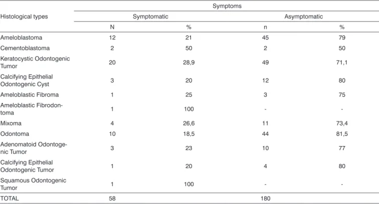
Table 4. Distribution of patients according to symptoms.
Histological types
Symptoms
Symptomatic Asymptomatic
N % n %
Ameloblastoma 12 21 45 79
Cementoblastoma 2 50 2 50
Keratocystic Odontogenic
Tumor 20 28,9 49 71,1
Calcifying Epithelial
Odontogenic Cyst 3 20 12 80
Ameloblastic Fibroma 1 25 3 75
Ameloblastic
Fibrodon-toma 1 100 -
-Mixoma 4 26,6 11 73,4
Odontoma 10 18,5 44 81,5
Adenomatoid
Odontoge-nic Tumor 3 23 10 77
Calcifying Epithelial
Odontogenic Tumor 1 20 4 80
Squamous Odontogenic
Tumor 1 100 -
-TOTAL 58 180
maxilla (57.5%) (Table 3) and in females (68.6%) (Table
1); this is similar to the findings of Santos et al.13
The incidence of the odontogenic mixoma was 6.3% of all odontogenic tumors in our study (Table 2), which is about two times less than that reported by
Mosqueda-Taylor et al.,3 but comparable to North-American studies.10
Our study revealed a slightly higher prevalence of this
tumor in males (Table 1); Ladeinde et al.12 and Odukoia
et al.6 have reported a higher frequency in females. This
study showed that the mean age of these patients was 29 years, higher than the age reported in two previous studies
in Nigeria5 and Mexico,11 in which the mean age was 19
years. A slight preference for the maxilla (53.4%) (Table 3) has been reported in a number of studies, such as those
by Mosqueda-Taylor et al.;3 Lu et al.,7 on the other hand,
have reported differently.
The incidence of the adenomatoid odontogenic tumor was 5.4% of all odontogenic tumors in our study (Table 2). Some papers have reported that this tumor is
more frequently found in the maxilla4,13,17 and in female
patients.17 (Table 1); these findings were confirmed in our
study, in which 53.9% of these lesions were found in the maxilla (Table 3) and 77% of these patients were female (Table 1), mostly in the second decade of life (7 of 13 cases) (Table 2).
The calcifying epithelial odontogenic cyst was seen in 15 cases (6.3%) (Table 2); there was a slightly higher incidence in females (53.4%) (Table 1), and 66% of these cases were in the mandible (Table 3). These findings are
similar to those in most studies13 except for Hiroyuki et
al.’s17 report. This neoplasm is usually found accidentally
in routine exams.5 In our study, 80% of these cases were asymptomatic (p=0.023) (Table 4) and showed no age preference (Table 2).
The incidence of the ameloblastic fibroma and the cementoblastoma was 1.7% each, and the incidence of the calcifying epithelial odontogenic tumor was 2% of the odontogenic tumors in our study (Table 2). The cemento-blastoma was more frequent in females (75%) (Table 1); the ameloblastic fibroma was more frequent in males (75%) (Table 1), as was the calcifying epithelial odontogenic tumor (60%) (Table 1). These lesions were more frequent in the mandible in our study (Table 3), contrary to some studies that have reported a higher frequency these tumors
in the maxilla.13
Lesions such as the squamous odontogenic tumor and the ameloblastic fibrodontoma were found in a single case each; both were females and presented mandibular pain. Our study confirmed the low frequency of these
neoplasms as shown in other studies,3,7,12,14 which
under-lines their rarity.
It is essential to define the epidemiology of these tumors to improve our knowledge about their behavior, which allows us to optimize the diagnosis and therapy.
CONCLUSION
There is a slight predominance of odontogenic tu-mors in women and during the first decades of life; most of them occur in the mandible, and most are asymptomatic. There were statistically significant differences among the variables histological type and symptoms.
In Brazil, specifically in this study, which was done in a region where population miscegenation is significant, there were some differences compared to published stu-dies undertaken in other parts of the world.
REFERENCES
1. Buchner A, Merrell PW, Carpenter WM. Relative frequency of central odontogenic tumors: a study of 1,088 cases from Northern Califor-nia and comparison to studies from other parts of the world. J Oral Maxillofac Surg 2006;64(9):1343-52.
2. Antunes AA, Silva JL, Silva PV, Antunes AP. Tumores odonto-gênicos: Análise de 128 casos. Rev Bras Cir Cabeça Pescoço 2006;35(3):160-3.
3. Mosqueda-Taylor A, Ledesma-Montes C, Caballero-Sandoval S, Portilla-Robertson J, Ruíz-Godoy Rivera LM, Meneses-García A. Odontogenic tumors in Mexico: A collaborative retrospective study of 349 cases. Oral Surg Oral Med Oral Pathol Oral Radiol Endod 1997;84(6):672-5.
4. Arotiba JT,Ogunbiyi JO,Obiechina AE. Odontogenic tumours: A 15-year review from Ibadan, Nigeria.Br J Oral Maxillofac Surg 1997;35(5):363-7.
5. Adebayo ET, Ajike SO, Adekeye EO. A review of 318 odontogenic tumors in Kaduna, Nigeria.J Oral Maxillofac Surg 2005;63(6):811-9. 6. Odukoya O. Odontogenic tumors: Analysis of 289 Nigerian cases.J
Oral Pathol Med 1995;24(10):454-7.
7. Lu Y,Xuan M,Takata T,Wang C,He Z,Zhou Z et al. Odontogenic tu-mors: A demographic study of 759 cases in a Chinese populationOral Surg Oral Med Oral Pathol Oral Radiol Endod 1998;86(6):707-14. 8. Olgac V, Koseoglu BG, Aksakalli N. Odontogenic tumours in Istanbul:
527 cases.Br J Oral Maxillofac Surg 2006;44(5):386-8.
9. Tamme T, Soots M, Kulla A, Karu K, Hanstein S-M, Sokk A et al. Odontogenic tumours, a collaborative retrospective study of 75 cases covering more than 25 years from Estonia. J CranioMaxillofac Surg 2004;32(3):161-5.
10. Daley TD,Wysocki GP,Pringle GA. Relative incidence of odontogenic tumors and oral and jaw cysts in a Canadian population.Oral Surg Oral Med Oral Pathol 1994;77(3):276-80.
11. Simon ENM, Merkx MAW, Vuhahula E, Ngassapa D, Stoelinga PJW. A 4-year prospective study on epidemiology and clinicopathological presentation of odontogenic tumors in Tanzania.Oral Surg Oral Med Oral Pathol Oral Radiol Endod 2005;99(5):598-602.
12. Ladeinde AL, Ajayi OF, Ogunlewe MO, Adeyemo WL, Arotiba GT, Bamgbose BO et al.. Odontogenic tumors: A review of 319 cases in a Nigerian teaching hospital. Oral Surg Oral Med Oral Pathol Oral Radiol Endod 2005;99(2):191-5.
13. Santos JN, Pinto LP, Figueiredo CRLV, Souza LB. Odontogenic tumors - Analysis of 127 cases. Pesqui Odontol Bras 2001;15(4):308-13. 14. Ochsenius G,Ortega A,Godoy L,Pẽafiel C,Escobar E.
Odontoge-nic tumors in Chile: A study of 362 cases.J Oral Pathol Med 2002; 31(7):415-20.
15. Kramer IRH, Pindborg JJ, Shear M. WHO histological typing of odon-togenic tumours. 2nd ed. Geneva: Springer-Verlag; 1992.
17. Hiroyuki O, Hirotsugu Y, Tilakaratne WM, Odontogenic Tumors in Sri Lanka: Analysis of 226 Cases. J Oral Maxillofac Surg 2007;65:875-882.
18. Mosqueda Taylor A, Irigoyen Camacho ME, Díaz Franco MA, To¬rres Tejero MA. Odontogenic cysts. Analysis of 856 cases. Med Oral 2002;7:89-96.