Key words:
Urolithiasis; Calcium Oxalate; Madin Darby Canine Kidney Cells; Electrophoresis, Gel, Two-Dimensional
Int Braz J Urol. 2013; 39: 128-36
__________________ Submitted for publication: August 14, 2012
__________________ Accepted after revision: November 30, 2012 Purpose: Proteins constitute a major portion of the organic matrix of human calcium
oxalate (CaOx) renal stones and the matrix is considered to be important in stone for-mation and growth. The present study evaluates the effect of these proteins on oxalate injured renal epithelial cells accompanied by a 2D map of these proteins.
Materials and Methods: Proteins were isolated from the matrix of kidney stones contai-ning CaOx as the major constituent using EGTA as a demineralizing agent. The effect of more than 3kDa proteins from matrix of human renal (calcium oxalate) CaOx stones was investigated on oxalate induced cell injury of MDCK renal tubular epithelial cells. A 2D map of >3kDa proteins was also generated followed by protein identification using MALDI-TOF MS.
Results: The >3kDa proteins enhanced the injury caused by oxalate on MDCK cells. Also, the 2D map of proteins having MW more than 3kDa suggested the abundance of proteins in the matrix of renal stone.
Conclusion: Studies indicate that the mixture of >3kDa proteins in the matrix of human renal stones acts as promoter of calcium oxalate crystal nucleation and growth as it aug-ments the renal epithelial cell injury induced by oxalate. The effect of promoters masks the inhibitors in the protein mixture thereby leading to enhanced renal cell injury. 2D map throws light on the nature of proteins present in the kidney stones.
INTRODUCTION
With its multifactor aetiology and high rate of recurrences, urinary tract stone disease provides a medical challenge. Depending on the socio-economic conditions and subsequent chan-ges in the dietary habits, the overall probability of stone formers differs in various parts of the world: 1-5% Asia, 5-9% Europe, 13-15% USA and 20% Saudi Arabia (1). Calcium containing stones are
the most common, consisting of about 75% of all urinary calculi, which may be in the form of pure calcium oxalate (50%) or calcium phosphate (5%) and a mixture of both (45%). The controlling in-fluence of macromolecules in the construction of healthy biomineralised tissues is undisputed (2-4). It is now well recognised that the organic com-ponent of such tissues, which in animals include bone, shell, dentin and enamel, is crucial to the biomineralization process. Some macromolecules
2D map of proteins from human renal stone matrix
and evaluation of their effect on oxalate induced renal
tubular epithelial cell injury
_______________________________________________
K.P. Aggarwal, S. Tandon, S.K. Singh, C. Tandon
Department of Biotechnology and Bioinformatics, Jaypee University of Information Technology (KPA, CT, ST), Waknaghat, Solan, HP and Department of Urology, Post Graduate Institute of Medical Education & Research (PGIMER) (SKS), Chandigarh-160012 India
ABSTRACT
ARTICLE
INFO
in these systems are responsible for initiating mi-neralisation, defining its physical limits and dicta-ting its cessation, but others provide an architec-tural framework upon which the inorganic salts are laid down. Less clearly defined, however, are the roles played by macromolecules in the forma-tion of human uroliths, a process possessing all the hallmarks of uncontrolled mineralisation (5,6).
Many proteins occur in stone, but their role in urolithiasis remains unknown. Calculi con-tains some proteins normally present in urine, in addition to others arising from injury inflicted by the stones themselves, making it impossible to discriminate between those that bind to the sto-ne as it grows, but play no role in its develop-ment (7); the inhibition is generally understood to arise mainly from the non-dialyzable molecu-les of urine, particularly acid glycoproteins, and acidic glycoproteins and glycosaminoglycans (8,9). Some inhibitor molecules have been iden-tified, including Tamm-Horsefall Protein, uro-pontin (10,11), calgranulin (12), bikunin (13), and prothrombin F1 fragment (14). Thus, in order to understand the mechanism of stone genesis, it is essential to determine the characteristics of mo-lecules constituting the urinary stone matrix. In the present study, we analysed a 2D map (2- di-mensional polyacrylamide gel electrophoresis) of human renal stone matrix proteins by MALDI-TOF (Matrix-assisted laser desorption/ionization-Time of Flight) to throw light on the matrix proteins and also study their effect on oxalate injured renal epithelial cells.
MATERIALS AND METHODS
Human Renal stones collection
Stones surgically removed by Percuta-neous nephrolithotomy (PNL) from the kidney stone patients were obtained from the Department of Urology, Postgraduate Institute of Medical Edu-cation and Research (PGIMER), Chandigarh, India. Stones were preserved at 4º C before study. Stones were of non-infectious nature and were collected from those patients who were more than 25 ye-ars of age and were suffering from no other ab-normality. After FTIR (Fourier transform infrared spectroscopy) analysis, the stones with calcium
and oxalate as their major components were se-lected for present study. Thirty stones with cal-cium and oxalate, as the major components were used for further studies. Thirty stone samples were randomly pooled into 5 groups, each group con-taining 6 stone samples.
Protein extraction from Human Renal stones Proteins were isolated from the matrix of kidney stones containing calcium oxalate (CaOx) as the major constituent using EGTA as a demine-ralising agent. Stones were washed in 0.15 M so-dium chloride (NaCl) and were then dried and pul-verized with a mortar and pestle. For extraction of the organic matrix of powdered stone, each gram of stone was suspended in 10 mL of 0.05 M EGTA (ethylene glycol tetraacetic acid), 1 mM PMSF (phenylmethanesulfonylfluoride or phenylmethyl-sulfonyl fluoride) and 1% β-mercaptoethanol. The extraction was carried out for 4 days at 4º C with constant stirring. The suspension was centrifuged for 30 minutes at 10,000 g and at 4º C. The super-natant of EGTA extract was filtered through Ami-con ultra centrifugal filter device (Catalog UFC 800324) with a molecular weight cut off 3kDa at 4º C and concentrated to a known volume. The excess of the 3kDa fractions were stored at -20º C for further studies (15).
Protein determination
Total protein concentration was determined by Lowry’s method using BSA as a standard (16).
Assay to measure inhibitory activity of protein w.r.t Calcium Oxalate (CaOx) crystal nucleation
absorbance of the solution was monitored at 620 nm after every 60 sec. The percentage inhibition produced by the protein extract was calculated as [1-(Tsi/Tsc)] X 100, where Tsc was the turbidity slope of the control and Tsi the turbidity slope in the presence of the inhibitor.
Assay to measure activity of protein w.r.t. CaOx crystal growth
Activity against CaOx crystal growth was measured using the seeded, solution-depletion as-say, according to the method described by Naka-gawa et al. (18).
Cell Culture
Madin-Darby Canine kidney (MDCK) cells were obtained from National Centre of Cell Scien-ces (NCCS, Pune). The cells were maintained as monolayers in Dulbecco’s Modified Eagle’s Me-dium (DMEM) with 2.0 mM L-glutamine adjusted to contain 3.7 g/L sodium bicarbonate, 4.5 g/L glucose. Media was supplemented with 1% Peni-cillin (100 units/mL)-Streptomycin (10,000 µg/mL) and 10% fetal bovine serum. Cells were cultured in 25 cm2 tissue-culture treated flasks at 37º C and
5% CO2 in humidified chambers (19).
Oxalate-induced Cell Injury
MDCK cells were incubated in DMEM con-taining 1 mM sodium oxalate in the presence of different concentrations of protein samples for 48 hours (20). Cell injury was assessed by measuring the cell viability through MTT and LDH (Lactate dehydrogenase) Assay.
Preparation of the protein samples
For cell culture studies, the proteins was dialysed through Millipore Amicon Ultra Centri-fugal Filters, 3kDa and desalted by ReadyPrep 2-D Cleanup Kit (catalog 163-2130) and it was recons-tituted in 0.22 µm filtered distilled water using Millipore Millex GV Filter Unit 0.22 µm (Catalog SLGU033RS).
MTT Assay
Cell viability studies via MTT test were conducted by the method described by Fulya Ka-ramustafa et al. with slight modifications (21).
MDCK cells were suspended in DMEM with serum and plated into the microwells of 96-well tissue culture plates. Plates were incubated for 24 h at 37º C in a humidified incubator containing 5% CO2. Then the medium was removed from wells. 200 µL DMEM (without serum) containing diffe-rent concentrations of proteins with and without sodium oxalate were added into the wells. After 48 hours, the medium was removed. Each well was treated with 100 µL medium and 13 µL MTT so-lution, and incubated for a further 3 hours. Then, plates were emptied and 100 µL isopropanol was added to dissolve the formazan precipitate. The developed colour was read at a wavelength of 570 nm with spectrophotometer.
LDH Leakage Assay
MDCK cells were suspended in DMEM with serum and plated into the microwells of 96-well tissue culture plates. Plates were incubated for 24 h at 37º C in a humidified incubator containing 5% CO2. Then the medium was removed from wells. 200 µL DMEM (without serum) containing diffe-rent concentrations of proteins with and without sodium oxalate were added into the wells for 48 hours. LDH leakage assay was performed by the LDH Cytotoxicity Assay Kit (Cayman 10008882) according to the manufacturer’s instructions (22).
Statistical analysis
Data were expressed as mean values of three independent experiments (each in triplica-te) and analyzed by the analysis of variance (p < 0.05) to estimate the differences between values.
2-D Gel Electrophoresis
equilibra-tion buffer II (6 M Urea, 2% SDS, 0.375 M Tris HCl (pH 8.8), 20% Glycerol, 135mM Iodoaceta-mide). Equilibrated IPG strips were loaded onto a 10% polyacrylamide gel sealed with overlay aga-rose, and electrophoresed at a constant voltage of 100 V. The gel was stained by silver staining and analysed using Biorad PD Quest Advanced 2D Analysis Software. The spots of interest were manually excised from the gel and were destai-ned using destainer provided in the ProteoSilver™ Plus Silver Stain Kit (PROTSIL2, Sigma-Aldrich Co.) followed by in-gel digestion using Trypsin profile IGD kit (PPO100, Sigma-Aldrich Co.). The proteins were identified by matrix assisted laser desorption/ionization-time of flight (MALDI-TOF) MS followed by MASCOT database search.
Tryptic in-gel digestion of purified protein Single band detected after molecular-sieve chromatography was excised from the gel and was destained with destainer provided in the Prote-oSilver™ Plus Silver Stain Kit (PROTSIL2, Sigma--Aldrich Co.). Trypsin profile IGD kit (PPO100, Sigma-Aldrich Co.) was used for in-gel digestion of purified protein. Destained gel piece was dried for approximately 15 to 30 min. Trypsin solubilised in 1 mmoL/L HCl and mixed with 40 mmoL/L ammo-nium bicarbonate and 9% acetonitrile was added to the destained gel piece. Gel piece was fully covered by the addition of 40 mmoL/L ammonium bicarbo-nate and 9% acetonitrile (pH 8.2) solution and was incubated for 5 hours at 37º C. After the incubation, liquid was removed from the gel piece and trans-ferred to a new labeled Eppendorf tubes and was preserved for mass spectroscopic analysis (23).
Peptide mass fingerprinting by MALDI-TOF-MS Peptides were extracted into the extrac-tion soluextrac-tion and dried by speedvac. Dried spots samples were spotted onto the MALDI plate with thorough mixing with matrix at 1:1 concentration and analyzed by MALDI TOF/TOF ULTRALFLEX III (Bruker Daltonics).
Mascot Protein Identification
The mass/charge spectra obtained were se-arched in MASCOT search engine (http://www.ma-trixscience.com) using all the 3 databases (MSDB,
SwissProt, NCBInr). For search, peptides were as-sumed monoisotopic, oxidized at methionine re-sidues and carbamidomethylated at cysteine resi-dues. An Homo sapiens taxonomy restriction was used, only one missed cleavage was allowed, and peptide mass tolerance of 1.2 kDa was used for peptide mass fingerprinting.
RESULTS
Activity study of proteins from human renal sto-ne on CaOx assay system
Whole EGTA extract and >3kDa were as-sayed to measure the activity against CaOx crystal nucleation and growth. Different concentrations of whole extract showed both promoter and inhibitory activity against nucleation of CaOx crystal as well as CaOx growth.
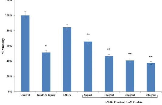
Bioactivity of >3kDa fraction on Oxalate Injured MDCK cells
Figure 1 - Effect of >3kDa fraction on MTT assay. Data are mean ± SEM of three independent observations.
*P < 0.01 versus untreated control. **P < 0.05 versus oxalate control (the experiment was done thrice in triplicates each time).
* p < 0.05 versus untreated control, ** p < 0.05 versus oxalate control.
oxalate alone (from 100% of control to 179.92% of oxalate injured).
MTT assay also reflected the same pattern. % viability of cells decreased in cells from 100% to 51.23% injured with oxalate. By these tests it can be suggested that the proteins that lead to cell injury by oxalate mask the activity of the proteins that inhibit the same.
2D-PAGE
2D PAGE of >3kDa proteins revealed the abundance of proteins in the matrix of human CaOx containing kidney stones (Figure-3). A total of 66 spots were detected using Biorad PD Quest Advanced 2D Analysis Software. 2D-PAGE revealed that the proteins present in the stone matrix were of both high and low molecular weight. The pro-teins were distributed throughtout the gel indicating the presence of both cationic and anionic proteins. Out of the 66 spots, 7 most prominent spots spread across the isoelectric points (pI) were further analy-sed using MALDI-TOF MS and MASCOT server. The identified proteins are given in Table-1.
DISCUSSION
The role played by the proteins present in the human CaOx renal stones in the course of
crys-tallization is yet not clear. Some of these proteins promote crystal formation, growth, aggregation and retention, while others inhibit these processes. Their activity is often complex and depends on the urine conditions prevailing at the time of crystallization or retention. The same protein can both promote as well as inhibit a process. Under normal conditions, the crystals of calcium oxalate that form are small and well protected from crystal growth and crystal aggregation by a cover of inhibitory macromolecu-les. If inhibitors of crystal formation were not able to act and control their size, the final result will be nephrolithiasis (24,25). Proteins which cover crystal surface and may lead to inhibition of its growth or ability to aggregate while the same proteins bound to a surface may act to accumulate salt ions and for-ms a template for the first nucleus. The latter will play a role when stone formation involves processes at cell surfaces and in the sub-epithelial space (26). The purpose of the present study was to explore the 2D map of human renal stone matrix proteins by MALDI-TOF-MS to throw light on the matrix pro-teins and also to study their effect on oxalate injured renal epithelial cells.
Proteins have a strong affinity for CaOx crystals (27). The role of matrix compounds is diffe-rent in the formation of the stone center and in the subsequent build-up of the stone (28). The same pro-tein which inhibits crystal formation might promote
Figure 3 - 2D page of >3kDa proteins from human renal CaOx stones.
M O L E C U L A R
IBJU
|
EXPLORING THE MA
TRIX PRO
TEINS IN HUMAN RENAL ST
ONE MA
TRIX
134
coverage
1 Disheveled-associated activator
of morphogenesis
1,23,976 6.81 9% Binds to disheveled (Dvl) and Rho, and mediates Wnt-induced Dvl-Rho complex formation. May play a role as a caffolding protein to recruit Rho-GDP and Rho-GEF, thereby enhancing Rho-GTP formation. Can direct nucleation and elongation of new
actin filaments.
2 Glutamate receptor delta-1 subunit 113046 6.23 17% Receptor for glutamate. L-glutamate acts as an excitatory neurotransmitter at many synapses in the central nervous system. The postsynaptic actions of Glu are mediated
by a variety of receptors that are named according to their selective agonists.
3 Caspase recruitment domain-containing protein 114880 5.65 13% Activates NF-kappa-B via BCL10 and IKK. Stimulates the phosphorylation of BCL10. Contains 1 CARD domain.
Contains 1 guanylate kinase-like domain. Contains 1 PDZ (DHR) domain.
4 ALBU_BOVIN 71279 5.82 3% Serum albumin, the main protein of plasma, has a good binding capacity for water,
Ca(2+), Na(+), K(+), fatty acids, hormones, bilirubin and drugs. Its main function is the regulation of the colloidal osmotic pressure of blood. Major zinc transporter in plasma,
typically binds about 80% of all plasma zinc.
5 VP7 glycoprotein precursor 37523 4.68 34% Outer capsid protein involved in attachment and possibly entry into the host epithelial
cell. It is subsequently lost, together with VP4, following virus entry into the host cell. The outer layer contains 780 copies of VP7, grouped as 260 trimers. Rotavirus attachment and entry into the host cell probably involves multiple sequential contacts
between the outer capsid proteins VP4 and VP7, and the cell receptors
6 Chymotrypsinogen A 26230 8.52 14% Chymotrypsinogen is a precursor (zymogen) of the digestive enzyme chymotrypsin.
7 Plasminogen 15782 8.73 54% Plasminogen is th inactive precursor of the trypsin-like serine protease lpasmin. It is
normally found circulating through the blood stream. When plasminogen becomes ac-tivated and is converted to plamin, it unfolds a potent enzymatic domain that dissolves
the growth of the crystal. Thus, the same protein acts differently at different stages of stone formation.
In the present study, when the >3kda frac-tion was examined for its effect on oxalate injured MDCK cells, it majorly reflected promoter activi-ties thereby leading to an increased cell death in a dose dependent manner. These results suggest that the proteins which are promoters of crystalli-zation in nature mask the activity of the proteins which are inhibitory in nature, thereby leading to an enhanced cell injury, and consequently cell de-ath. Our observations are in conformity with the observations that that renal epithelial damage can lead to increased crystal attachment (29).
The 2D map suggests that abundant pro-teins are present in the matrix propro-teins which are of both high and low molecular weight. Also both anionic and cationic proteins are present. Solution depletion (30) and examination of crystals incu-bated in protein solutions by transmission elec-tron microscopy (31) tested the theory of physical adsorption of urine proteins on surfaces of CaOx crystals. Results showed proteins have a strong affinity for CaOx crystals. Adsorption of anionic proteins was sensitive to calcium ion concentra-tion, whereas cationic protein adsorption depen-ded upon the oxalate ion concentration with tem-perature and pH playing only a minor role.
A pathological crystallization leading to stone formation might be the net result of one or se-veral abnormalities or defects in the control of this process. Low concentrations or structural abnorma-lities of crystallization modifying macromolecules or small molecules will cause increased growth and aggregation of crystals (32). Therefore, any crys-tallization that occurs most certainly is facilitated by promoters and it has been suggested that lipo-protein membranes from the brush border of proxi-mal tubular cells might serve this purpose (33).
CONCLUSIONS
Studies indicate that the mixture of >3kDa proteins in the matrix of human renal stones aug-ment the renal epithelial cell injury induced by oxalate and thereby may act as promoters of cal-cium oxalate crystal nucleation and growth. The effect of promoters masks the inhibitors in the
protein mixture thereby leading to enhanced renal cell injury. 2D map throws light on the nature of proteins present in the kidney stone matrix, howe-ver, their exact role in the mechanism of kidney stone formation warrant further investigations.
ABBREVIATIONS
CaOx: calcium oxalate
MDCK: madin darby canine kidney DMEM: dulbecco’s modified eagle’s media
MTT: 3-(4, 5-dimethylthiazol-2-yl)-2, 5-diphenyl-tetrazolium bromide
LDH: lactate dehydrogenase
2D PAGE: 2 dimensional polyacrylamide gel elec-trophoresis
MALDI-TOF: Matrix-assisted laser desorption/io-nization- Time of Flight
FTIR: Fourier transform infrared spectroscopy
ACKNOWLEDGEMENTS
The authors would like to thank Indian Council of Medical Research (ICMR), India and Jaypee University of Information Technology (JUIT), Solan, HP, India for providing funds to carry out this research work. We also express our gratitude to the Department of Urology, Post Graduate Institute of Medical Education and Research (PGIMER) Chan-digarh, India for providing kidney stones.
CONFLICT OF INTEREST
None declared.
REFERENCES
1. López M, Hoppe B: History, epidemiology and regional di-versities of urolithiasis. Pediatr Nephrol. 2010; 25: 49-59. 2. Margolis HC, Beniash E, Fowler CE: Role of
macromolecu-lar assembly of enamel matrix proteins in enamel forma-tion. J Dent Res. 2006; 85: 775-93.
3. Wesson JA, Ward MD: Medical Mineralogy and Geochem-istry: Pathological Biomineralization of Kidney Stones. Ele-ments. 2007; 3: 415-421.
5. Tiselius HG: A hypothesis of calcium stone formation: an inter-pretation of stone research during the past decades. Urol Res. 2011; 39: 231-43.
6. Coe FL, Evan AP, Worcester EM, Lingeman JE: Three pathways for human kidney stone formation. Urol Res. 2010; 38: 147-60. 7. Ryall RL, Chauvet MC, Grover PK: Intracrystalline proteins and
urolithiasis: a comparison of the protein content and ultrastruc-ture of urinary calcium oxalate monohydrate and dihydrate crystals. BJU Int. 2005; 96: 654-63.
8. Fleisch H: Inhibitors and promoters of stone formation. Kidney Int. 1978; 13: 361-71.
9. Dussol B, Berland Y: Urinary kidney stone inhibitors. What is the news? Urol Int. 1998; 60: 69-73.
10. Hess B, Nakagawa Y, Parks JH, Coe FL: Molecular abnormality of Tamm-Horsfall glycoprotein in calcium oxalate nephrolithia-sis. Am J Physiol. 1991; 260: F569-78.
11. Asplin JR, Arsenault D, Parks JH, Coe FL, Hoyer JR: Contribu-tion of human uropontin to inhibiContribu-tion of calcium oxalate crystal-lization. Kidney Int. 1998; 53: 194-9.
12. Pillay SN, Asplin JR, Coe FL: Evidence that calgranulin is pro-duced by kidney cells and is an inhibitor of calcium oxalate crystallization. Am J Physiol. 1998; 275: F255-61.
13. Atmani F, Khan SR: Characterization of uronic-acid-rich in-hibitor of calcium oxalate crystallization isolated from rat urine. Urol Res. 1995; 23: 95-101.
14. Stapleton AM, Ryall RL: Blood coagulation proteins and uro-lithiasis are linked: crystal matrix protein is the F1 activation peptide of human prothrombin. Br J Urol. 1995; 75: 712-9. 15. Aggarwal S, Tandon CD, Forouzandeh M, Singla SK, Kiran R,
Jethi RK: Role of biomolecules from human renal stone matrix on COM crystal growth. Mol Cell Biochem. 2000; 210: 109-19. 16. Lowry OH, Rosebrough NJ, Farr AL, Randall RJ: Protein mea-surement with the Folin phenol reagent. J Biol Chem. 1951; 193: 265-75.
17. Hennequin C, Lalanne V, Daudon M, Lacour B, Drueke T: A new approach to studying inhibitors of calcium oxalate crystal growth. Urol Res. 1993; 21: 101-8.
18. Nakagawa Y, Abram V, Parks JH, Lau HS, Kawooya JK, Coe FL: Urine glycoprotein crystal growth inhibitors. Evidence for a molecular abnormality in calcium oxalate nephrolithiasis. J Clin Invest. 1985; 76: 1455-62.
19. Aggarwal A, Tandon S, Singla SK, Tandon C: Diminution of oxa-late induced renal tubular epithelial cell injury and inhibition of cal-cium oxalate crystallization in vitro by aqueous extract of Tribulus terrestris. Int Braz J Urol. 2010; 36: 480-8; discussion 488, 489. 20. Moriyama MT, Miyazawa K, Noda K, Oka M, Tanaka M, Suzuki
K: Reduction in oxalate-induced renal tubular epithelial cell in-jury by an extract from Quercus salicina Blume/Quercus steno-phylla Makino. Urol Res. 2007; 35: 295-300.
21. Karamustafa F, Çelebi N, Değim Z, Şyilmaz Ş: Evaluation Of The Viability Of L-929 Cells In The Presence Of Alendronate And Absorption Enhancers. Fabad J Pharm Sci. 2006; 31: 1-5.
22. Lee HG, Li MH, Joung EJ, Na HK, Cha YN, Surh YJ: Nrf2-Mediated heme oxygenase-1 upregulation as adaptive survival response to glucose deprivation-induced apoptosis in HepG2 cells. Antioxid Redox Signal. 2010; 13: 1639-48.
23. Priyadarshini, Singh SK, Tandon C: Mass spectrometric iden-tification of human phosphate cytidylyltransferase 1 as a novel calcium oxalate crystal growth inhibitor purified from human renal stone matrix. Clin Chim Acta. 2009; 408: 34-8.
24. Carvalho M, Mulinari RA, Nakagawa Y: Role of Tamm-Horsfall protein and uromodulin in calcium oxalate crystallization. Braz J Med Biol Res. 2002; 35: 1165-72.
25. Kok DJ, Schell-Feith EA: Risk factors for crystallization in the nephron: the role of renal development. J Am Soc Nephrol. 1999; 10(Suppl 14): S364-70.
26. Leal JJ, Finlayson B: Adsorption of naturally occurring poly-mers onto calcium oxalate crystal surfaces. Invest Urol. 1977; 14: 278-83.
27. Coe F, Parks JH: Defenses of an unstable compromise: crystal-lization inhibitors and the kidney’s role in mineral regulation. Kidney Int. 1990; 38: 625-31.
28. Khan SR, Hackett RL: Identification of urinary stone and sedi-ment crystals by scanning electron microscopy and x-ray mi-croanalysis. J Urol. 1986; 135: 818-25.
29. Verkoelen CF, van der Boom BG, Houtsmuller AB, Schröder FH, Romijn JC: Increased calcium oxalate monohydrate crystal binding to injured renal tubular epithelial cells in culture. Am J Physiol. 1998; 274: F958-65.
30. Khan SR, Maslamani SA, Atmani F, Glenton PA, Opalko FJ, Thamilselvan S, et al.: Membranes and their constituents as promoters of calcium oxalate crystal formation in human urine. Calcif Tissue Int. 2000; 66: 90-6.
31. Thamilselvan S, Khan SR: Oxalate and calcium oxalate crystals are injurious to renal epithelial cells: results of in vivo and in vitro studies. J Nephrol. 1998; 11(Suppl 1): 66-9.
32. Lieske JC, Coe FL: Urinary inhibitors and renal stone formation. In: Kidney Stones-Medical and Surgical Management (ed.), F. L. Coe, M. J. Favus, C. Y .C. Pak, J. H. Parks, and G. M. Preminger. Lippincott-Raven, Philadelphia. 1996; pp. 65-113.
33. Bigelow MW, Wiessner JH, Kleinman JG, Mandel NS: Surface exposure of phosphatidylserine increases calcium oxalate crystal attachment to IMCD cells. Am J Physiol. 1997; 272: F55-62.
_____________________
Correspondence address: Dr. C. Tandon Professor of Biotechnology and Bioinformatics Jaypee University of Information Technology,