ISSN 1414-431X
www.bjournal.com.br
www.bjournal.com.br
Volume 45 (11) 995-1101 November 2012
Braz J Med Biol Res, November 2012, Volume 45(11) 1017-1024
doi: 10.1590/S0100-879X2012007500123
Antimicrobial peptides and nitric oxide production by neutrophils
from periodontitis subjects
F.S. Mariano, A.P. Campanelli, F.H. Nociti Jr., R.O. Mattos-Granerand R.B. Gonçalves
Institutional Sponsors
The Brazilian Journal of Medical and Biological Research is partially financed by
Faculdade de Medicina de Ribeirão Preto Campus
Ribeirão Preto
Explore High - Performance MS Orbitrap Technology In Proteomics & Metabolomics
analiticaweb.com.br S C I E N T I F I C
BIOMEDICAL SCIENCES
AND
Antimicrobial peptides and nitric oxide
production by neutrophils from
periodontitis subjects
F.S. Mariano
1, A.P. Campanelli
2, F.H. Nociti Jr.
3, R.O. Mattos-Graner
1and R.B. Gonçalves
1,41Departamento de Diagnóstico Oral, Área Microbiologia e Imunologia,
Faculdade de Odontologia de Piracicaba, Universidade de Campinas, Piracicaba, SP, Brasil
2Departamento de Ciências Biológicas, Faculdade de Odontologia de Bauru,
Universidade de São Paulo, Bauru, SP, Brasil
3Departamento de Prótese e Periodontia, Área Periodontia,
Faculdade de Odontologia de Piracicaba, Universidade de Campinas, Piracicaba, SP, Brasil
4Department of Periodontics, Faculty of Dentistry, Laval University,
Quebec City, Quebec, Canada
Abstract
Neutrophils play an important role in periodontitis by producing nitric oxide (NO) and antimicrobial peptides, molecules with microbicidal activity via oxygen-dependent and -independent mechanisms, respectively. It is unknown whether variation in the
production of antimicrobial peptides such as LL-37, human neutrophil peptides (HNP) 1-3, and NO by neutrophils influences
the pathogenesis of periodontal diseases. We compared the production of these peptides and NO by lipopolysaccharide (LPS)-stimulated neutrophils isolated from healthy subjects and from patients with periodontitis. Peripheral blood neutrophils were cultured with or without Aggregatibacter actinomycetemcomitans-LPS (Aa-LPS), Porphyromonas gingivalis-LPS (Pg-LPS) and Escherichia coli-LPS (Ec-LPS). qRT-PCR was used to determine quantities of HNP 1-3 and LL-37 mRNA in neutrophils. Amounts of HNP 1-3 and LL-37 proteins in the cell culture supernatants were also determined by ELISA. In addition, NO levels in neutrophil culture supernatants were quantitated by the Griess reaction. Neutrophils from periodontitis patients cultured with
Aa-LPS, Pg-LPS and Ec-LPSexpressed higher HNP 1-3 mRNA than neutrophils from healthy subjects. LL-37 mRNA expression was higher in neutrophils from patients stimulated with Aa-LPS. Neutrophils from periodontitis patients produced significantly
higher LL-37 protein levels than neutrophils from healthy subjects when stimulated with Pg-LPSand Ec-LPS, but no difference was observed in HNP 1-3 production. Neutrophils from periodontitis patients cultured or not with Pg-LPS and Ec-LPS produced
significantly lower NO levels than neutrophils from healthy subjects. The significant differences in the production of LL-37 and NO between neutrophils from healthy and periodontitis subjects indicate that production of these molecules might influence
individual susceptibility to important periodontal pathogens.
Key words: Antimicrobial peptides; Nitric oxide; Neutrophils; Periodontitis; Innate immunity
Introduction
Correspondence: F.S. Mariano, Departamento de Diagnóstico Oral, Área Microbiologia e Imunologia, Faculdade de Odontologia de Piracicaba, UNICAMP, Caixa Postal 52, 13414-903 Piracicaba, SP, Brasil. Fax: +55-19-2106-5322.
E-mail: flaviasmariano@yahoo.com.br
Received March 12, 2012. Accepted July 23, 2012. Available online August 3, 2012. Published October 5, 2012.
Periodontitis is a chronic inflammatory disease char -acterized by connective tissue and alveolar bone destruc-tion, eventually leading to tooth loss. The recruitment of neutrophils and other leukocytes in the periodontal pocket
is an important feature of the inflammatory process in peri -odontal disease (1). Important periodontopathogens, e.g.,
Aggregatibacter actinomycetemcomitans and
Porphyromo-nas gingivalis, seem to induce destruction of periodontal
tissues through lipopolysaccharide (LPS)-dependent mechanisms or by eliciting the production of a variety of biologically active substances by host immune cells (2). In general, LPS activates different cells, including epithelial
1018 F.S. Mariano et al.
promoting phagocytosis, production of reactive oxygen species, cytokines, and release of antimicrobial peptides from azurophil granules (5).
Neutrophils respond to bacterial products or inflamma -tory mediators by chemotaxis, phagocytosis, and microbial killing through oxygen radical- and non-oxygen-dependent mechanisms (6). The non-oxidative antibacterial mecha-nisms involve a diverse group of antimicrobial peptides including alpha-defensins (7) and human cathelicidin, LL-37 (8), which are stored in azurophil granules. The defensins are divided into alpha, beta and theta defensins and are found in vertebrates and invertebrates. Alpha-defensins include a group of six types of small peptides (7), four of which are produced by human neutrophils, thus being designated human neutrophil peptides (HNP) -1, -2, -3, and -4 (7). HNP 1-3 are the most abundant, and individual
analysis of these peptides is difficult because of the high
similarity of their amino acid sequences (1,9).HNP 1-3 are
detected in whole saliva (10)and gingival crevicular fluid of
both healthy and periodontal disease subjects (1). LL-37 is known to play an important role in the lysis of periodontal pathogens, which is consistent with the frequent develop-ment of severe periodontal diseases in subjects with Morbus Kostmann syndrome. In this congenital disease, neutrophils
are deficient in LL-37, and this antimicrobial peptide is also
present in abnormally low levels in saliva (11). Additionally, neutrophils from these subjects produce reduced amounts
of HNP 1-3 (8). LL-37 impairs the in vitro growth of several
bacterial species of the oral cavity, including the periodontal
pathogens A. actinomycetemcomitans (12),Fusobacterium
nucleatum and Prevotella intermedia (13). Additionally,
LL-37 shows high affinity for LPS from different bacteria and
thus can neutralize these endotoxins (14).
Defensins and LL-37 have the ability to kill and/or
inacti-vate several bacterial species (including A.
actinomycetem-comitans and P. gingivalis) (12),fungi, and some enveloped
viruses (15). These microbicidal properties lie in the ability of these peptides to form pores in the membranes, promoting
bacterial lysis or affecting viral envelopes (16).In addition,
HNP 1-3 and LL-37 induce and/or amplify subsequent in-nate and adaptive immune responses against pathogens, such as mast cell degranulation, production of interleukins (IL) such as IL-10 and tumor necrosis factor, and dendritic cell activation (17,18). The production of nitric oxide (NO) represents another mechanism of pathogen destruction in activated neutrophils (19). Production of NO or expression of inducible NO synthase (iNOS) by peripheral neutrophils or in gingival tissues was associated with periodontal disease in human and animal models, respectively (20,21). Other stud-ies have shown that production of NO and reactive oxygen species is enhanced by peripheral neutrophils stimulated
in vitro with A. actinomycetemcomitans and P. gingivalis
(22,23). Despite the massive presence of neutrophils and their enhanced activity at sites of periodontal disease (24), the roles of these leukocytes and their antimicrobial
prod-ucts in the susceptibility and/or pathogenesis of periodontal disease remain to be examined. Although neutrophils have been considered to be responsible for the destruction of periodontal tissues (24), some studies have suggested that neutrophils play protective roles in controlling pathogenic bacteria involved in periodontal disease (25,26).
In the present study, we investigated the expression of genes encoding HNP 1-3, LL-37, NO, and HNP 1-3/LL-37 production in neutrophils isolated from generally healthy subjects with and without periodontitis, in response to bacterial LPS from species considered or not to be peri-odontal pathogens.
Subjects and Methods
Subjects
All subjects enrolled in this study were clinically and radiographically examined at the Graduate Clinic of the Faculdade de Odontologia de Piracicaba, Universidade Estadual de Campinas (UNICAMP), Brazil. Periodontal examination included full-mouth probing depth, plaque index, gingival index, and gingival recession. Inclusion cri-teria were: subjects diagnosed with generalized moderate chronic periodontitis (at least 4 teeth with probing depth
≥5 mm) who had not received periodontal treatment and/
or antibiotics in the last 6 months preceding the study, with at least 14 natural teeth. Exclusion criteria were: systemic modifying factors affecting the immune response, such as diabetes mellitus, immune and hormone disorders, smok-ers and former smoksmok-ers, alcoholics and former alcohol-ics, pregnant and lactating women, and those taking oral contraceptive drugs.
The subjects were separated into two experimental groups. The chronic periodontitis group included 6 patients, 3 males and 3 females (mean age: 47.5 ± 11.8 years) with moderate chronic periodontitis according to the criteria proposed by the 1999 International Workshop for a
Clas-sification of Periodontal Diseases and conditions (27). The
healthy control groupincluded 6 subjects, three males and
three females (mean age: 31.4 ± 3.4 years). All subjects were clinically healthy (probing depth <3 mm, without bleeding on probing, with no detectable radiographic alveolar bone loss on radiography). All procedures performed were approved by the Ethics Committee of Faculdade de Odontologia de Piracicaba, UNICAMP, and all volunteers involved in this study signed a consent form.
Bacterial strains, growth conditions and antigens
Periodontopathogens A. actinomycetemcomitans strain
Y4 and P. gingivalis strain ATCC 33277 were obtained from
the Bacterial Collection of the Research Group in Oral Biol-ogy (GREB), School of Dentistry, Laval University, Quebec, Canada. These microorganisms were cultured at 37°C in brain heart agar (Difco Co., USA) supplemented with
mL menadione (Sigma, USA) under anaerobic conditions
(10% CO2, 10% H2 and 80% N2) in a Mini MACS Anaerobic
Workstation (Don Whitley Scientific, UK) (28).
Purification of LPS from A. actinomycetemcomitans
and P. gingivalis was performed as described (29). LPS
samples were freeze-dried and stored at -20°C. Protein contamination of these samples was lower than 0.001% in all preparations, as evaluated using a Quick Start™ Bradford protein assay with bovine serum albumin standard (Bio-Rad
Laboratories, Canada). Escherichia coli-LPS (Ec-LPS)
obtained from Sigma was used as positive control.
Isolation and in vitro stimulation of human neutrophils
Whole blood (20 mL) from control and periodontitis subjects was collected into lithium heparin tubes (BD
Vacu-tainer™, USA). Neutrophils purified from 5 mL Histopaque
1119 (Sigma-Aldrich Brazil Ltda., Brazil) were poured into a 15-mL round bottom tube and overlaid with 3 mL Histopaque 1083 (Sigma-Aldrich Brazil Ltda.), and 6 mL whole blood was layered over the gradients. Tubes containing gradients
and blood were centrifuged at 460 g for 28 min at 25°C.
The layer containing the neutrophils was aspirated and washed twice with cold RPMI 1640 (Invitrogen™, Brazil), supplemented with 10% fetal bovine serum and antibiotics. The erythrocytes were eliminated by hypotonic lysis for 30 s. The viability of blood neutrophils was determined by Trypan
blue exclusion using a Neubauer chamber. One x 107 cells/
well were incubated at 37°C in the presence of 5% CO2 for
1 h. Neutrophils were then stimulated with 100 ng/mL of A.
actinomycetemcomitans-LPS (Aa-LPS), P. gingivalis-LPS
(Pg-LPS) or Ec-LPS. In order to examine the effects of LPS
on gene expression and production of HNP 1-3 or LL-37 by neutrophils, the cells isolated from blood were stimulated with LPS for 6 and 12 h. Neutrophils from similar cultures not exposed to LPS were used as negative controls. After
incubation, cells were centrifuged (290 g, at 4°C for 10 min),
and the culture supernatants were stored at -70°C until use for the determination of HNP 1-3 and LL-37 production. The neutrophils were used for the total mRNA extraction for the analysis of HNP 1-3 and LL-37 gene expression.
RNA extraction and reverse transcription
Neutrophils from cultures exposed or not to LPS were washed twice with PBS and
the total RNA was immediately extracted using Trizol reagent (Invitrogen, USA), according to manufacturer instructions. After treatment of RNA samples with DNase (Turbo DNA-free, Ambion Inc., USA), a total of 0.5 µg RNA was used for cDNA synthesis. Reverse transcription was car-ried out using the Transcriptor First-Strand cDNA Synthesis kit
(Roche Diagnostic Co., USA) according to manufacturer instructions.
Quantitative real-time polymerase chain reaction (qRT-PCR)
The levels of HPN 1 and HPN 3 transcripts were
as-sessed using a primer set specific for a conserved sequence
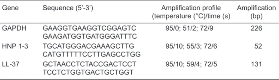
of these peptides (30). Table 1 shows the primer sequences applied in qRT-PCR for HPN 1-3 and LL-37.
The sequences of the primers for qRT-PCR analysis were obtained using the Light-Cycler software (Roche Diagnostics GmbH, Germany). The qRT-PCR assays were performed using the Light Cycler system (Roche Diagnostics GmbH). The reaction mixture (15 µL) included template cDNA (0.5 µg), 0.75 µM of each primer, and 1X SYBR-Green mix (Fast Start DNA Master plus, Roche Di-agnostic Co.). The thermal cycling conditions were: 95°C for 10 min for the initial denaturation, followed by 40 cycles of three steps consisting of denaturation at 95°C for 10 s, primer annealing at 59°C for 4 s, and primer extension at 72°C for 5 s. Controls included reaction mixtures without template cDNA. The housekeeping gene,
glyceraldehyde-3-phosphate dehydrogenase (GAPDH), was amplified in
parallel to the gene of interest. Relative copy numbers
compared with GAPDH were calculated using 2∆Ct. Assays
with RNA samples were performed in triplicate.
Enzyme-linked immunosorbent assay (ELISA)
The amounts of HNP 1-3 and LL-37 produced by neutrophils exposed or not to LPS samples were
quanti-fied by ELISA using kits from Hycult Biotechnology (The
Netherlands), according to manufacturer instructions.
Individual quantification of each HNP subtype (1, 2, and
3) is not possible because of the high structural similarity of these defensins. The concentrations of HNP 1-3 and
LL-37 in culture fluids are reported as pg/mL and ng/mL,
respectively. Assays were performed in triplicate with the supernatants of neutrophil culture.
Flow cytometry analysis
Cell acquisition was performed with a FACSort flow
cytometer using the CellQuest software (BD Biosciences, USA). The absolute leukocyte values/20 mL peripheral blood
Table 1. Primer sequences for the genes, amplification profile, and amplicon length studied
here.
Gene Sequence (5’-3’) Amplification profile
(temperature (°C)/time (s)
Amplification
(bp)
GAPDH GAAGGTGAAGGTCGGAGTC
GAAGATGGTGATGGGATTTC
95/0; 51/2; 72/9 226
HNP 1-3 TGCATGGGACGAAAGCTTG
CATGTTTTTCCTTGAGCCTGG
95/10; 55/3; 72/6 52
LL-37 GCTAACCTCTACCGACTCCT
TCCTCTGGTGACTGCTGGT
1020 F.S. Mariano et al.
were calculated according to characteristics of granularity
and size (side scatter vs forward scatter) based on the
per-centage obtained by FACS (50,000 events), and the amount of cells was determined in a Neubauer chamber.
Nitric oxide production
Nitrate was reduced to nitrite with nitrate reductase and the concentration of nitrite was determined by mixing culture supernatants of neutrophils exposed or not to LPS
with Griess reagent, as described below. Briefly, neutrophils
(1 x 10⁷ cells/mL) from subjects with periodontal disease or
healthy ones were cultured in the presence or absence of
Aa-LPS, Pg-LPS or Ec-LPS (100 ng/mL) for 6 and 12 h, at
37°C and in the presence of 5% CO2. A total of 50 µL culture
supernatant was then incubated at room temperature with an equal volume of the Griess reagent on 96-well plates
(Corning, USA) for 30 min. The absorbance (A540 nm) of the
samples was measured using a plate scanner (VersaMax Tunable Microplate Reader; Molecular Devices, USA). The
amounts of NO2 in the samples were then calculated using
a standard curve of NaNO2 (1-200 µM) within a linear range.
Assays were performed in triplicate with supernatants of neutrophil cultures from each subject.
Statistical analysis and software
The Student t-test was used to determine the
statisti-cal significance of the differences observed between the
volunteers with and without periodontitis and periodontally healthy volunteers. The statistical analyses were performed with the help of the GRAPHPAD PRISM software version 5.00 for Windows (http://www.graphpad.com). Differences
were considered to be statistically significant at P values
of <0.05.
Results
Expression of HNP 1-3 and LL-37 in peripheral blood neutrophils
The purity of cells isolated from peripheral blood of patients with periodontitis and healthy subjects was
as-sessed by flow cytometry (Figure 1A). Approximately 94%
total gated cells were polymorphonuclear neutrophils, thus
ensuring a minimum influence of cell contaminants in the experiments performed. Subsequently, it was verified that
Aa-LPS, Pg-LPS and Ec-LPS could induce different levels
of HNP 1-3 and LL-37 mRNA in neutrophils from patients and healthy subjects. According to our data, neutrophils from patients with periodontitis express higher amounts of mRNA corresponding to HNP 1-3 than control cells after
Pg-LPS stimuli for 6 and 12 h (Figure 1B). Similarly, Aa-LPS
and Ec-LPS induced high levels of HNP 1-3 transcripts in
neutrophils from periodontitis patients compared to neutro-phils from control subjects after 12 h of incubation (Figure
1B). However, no significant difference was observed in the
levels of HNP 1-3 transcripts between neutrophils from the
two groups that were cultured with Aa-LPS for a shorter
period of time (6 h). No significant differences in HNP 1-3
transcripts were detected between neutrophils from the periodontitis and healthy groups when cells were cultured without the LPS stimulus for 12 h (Figure 1B).
The LL-37 transcript levels were closely similar for neutrophils from periodontitis patients and healthy subjects after stimulation or not with LPS for 6 and 12 h (Figure 1C). An exception were the neutrophils of periodontitis patients
exposed to Aa-LPS for 12 h, which showed significantly
higher levels of LL-37 transcripts compared to neutrophils
from healthy controls exposed to Aa-LPS under the same
conditions (Figure 1C).
Production of HNP 1-3 and LL-37 proteins by peripheral blood neutrophils
The neutrophils from healthy subjects tended to produce higher concentrations of alpha-defensin proteins than cells from periodontitis subjects, but the differences were not
statistically significant (Figure 2A). Additionally, similar levels
of LL-37 protein were produced by neutrophils from perio-dontitis and healthy subjects regardless of LPS stimulation for 6 h (Figure 2B). Neutrophils from periodontitis subjects
stimulated with Pg-LPS and Ec-LPS for 12 h produced
significantly higher amounts of LL-37 protein compared to
neutrophils from healthy subjects similarly stimulated (Fig-ure 2B). However, neutrophils from both groups produced similar amounts of total protein after a 12-h stimulation with
Aa-LPS or without LPS exposure (Figure 2B).
Differentiated production of NO by neutrophils
Neutrophils from periodontitis patients cultured with
Pg-LPS or Ec-LPS for 6 h or cultured without LPS
stimula-tion produced significantly lower levels of NO levels when
compared to neutrophils from healthy subjects (Figure 3).
After 12 h of incubation, NO production was still significantly
lower in cultures of neutrophils from periodontitis patients
stimulated with Pg-LPS and Ec-LPS compared to cells
of control subjects. However, equivalent NO levels were produced by neutrophils isolated from periodontitis and
healthy subjects stimulated with Aa-LPS for 6 and 12 h. No
significant differences in the amounts of NO were observed
between neutrophils from the two groups not stimulated with LPS (Figure 3).
Discussion
In periodontal disease, neutrophil deficiency may be
Figure 1. HNP 1-3 and LL-37 mRNA expression in peripheral neutrophils from periodontitis and healthy
subjects. Neutrophils were isolated from peripheral blood and analyzed by flow cytometry, showing more
than 94% purity according to side scatter (SSC) vs forward scatter (FSC) parameters (A). Neutrophils were incubated for 6 and 12 h with culture medium only or with medium containing Aa-LPS (100 ng/ mL), Pg-LPS (100 ng/mL) or Ec-LPS (100 ng/mL). The relative mRNA expression of HNP 1-3 (B) and LL-37 (C) in neutrophils was assessed by qRT-PCR. Data are reported as means ± SD for N = 6 in each group. Aa-LPS = Aggregatibacter actinomycetemcomitans-lipopolysaccharide; Pg-LPS = Porphyromo-nas gingivalis-LPS; Ec-LPS = Escherichia coli-LPS. *P ≤ 0.05, periodontitis compared to healthy subjects
(Student t-test).
Figure 2. HNP 1-3 and LL-37 production by peripheral neutrophils. Neutrophils were incubated for 6 and 12 h with culture medium only or with medium containing Aa-LPS (100 ng/mL), Pg-LPS (100 ng/mL) or
Ec-LPS (100 ng/mL), and production of HNP 1-3 (A) and LL-37 (B) was determined by ELISA. Data are
reported as means ± SD for N = 6 in each group. *P ≤ 0.05, periodontitis compared to healthy subjects
1022 F.S. Mariano et al.
neutrophils to normal blood counts and reduces recurrent infections, but does not prevent severe periodontitis (31).
Peripheral neutrophils from these patients are deficient in
LL-37 production and produce reduced amounts of HNP 1-3 defensins. However, whether neutrophils from periodontitis patients have atypical responses to components of peri-odontal pathogens (e.g., LPS) compared to periperi-odontally healthy subjects remains to be investigated. In the present study, we show that neutrophils from subjects with perio-dontitis respond differently to stimuli with LPS from
differ-ent bacterial sources (periodontal pathogens and E. coli)
compared to neutrophils from healthy subjects, expressing
significantly higher levels of HNP 1-3 and LL-37 genes and
showing reduced amounts of NO.
Although the number of subjects with periodontitis studied was relatively small, given the complex nature of chronic periodontitis, the data presented here open a new
line of investigation about the influence of neutrophil phe -notypes on the course of periodontal diseases. Previous studies using a similar number of patients with aggressive and chronic periodontitis (6 and 12 subjects, respectively)
were able to detect significant differences in neutrophil
phagocytosis and killing of A. actinomycetemcomitans and
P. gingivalis when compared to neutrophils from healthy
subjects (20). In addition, the profiles of transcription and
protein production of LL-37 were previously determined in gingival tissues of the same number of volunteers with chronic periodontitis (32). Further studies will be necessary to compare the phenotypes of peripheral and periodontal
neutrophils between subjects with and without periodontal diseases.
It is known that the amounts of mRNA from peripheral
blood neutrophils can be consistently quantified by mo -lecular biology methods (33). We measured the amounts of HNP 1-3 and LL-37 transcripts in neutrophils from blood samples exposed to different types of LPS. We observed that neutrophils harvested from peripheral blood of periodontitis patients expressed higher HNP 1-3 mRNA levels when
stimulated with Aa-LPS, Pg-LPS and Ec-LPS, specifically
during 12 h of exposure. However, the protein levels of HNP
1-3 produced did not vary significantly between neutrophils
from healthy versus periodontitis subjects. In a previous study, the amounts of HNP 1-3 were determined in the
gingival crevice fluid of sites affected or not by periodontitis
from periodontally healthy subjects or subjects with perio-dontitis using mass-spectrometry analysis (1). The authors
demonstrated no significant differences in the amounts of
these peptides detected between healthy and diseased sites, although these defensins seem to be more abundant
in healthy gingival crevicular fluid (1). In another study, no
association was observed between HNP 1-3 levels in
gin-gival fluid and periodontal status (34). On the other hand,
low levels of HNP 1-3 in neutrophils from Morbus Kostmann syndrome patients were associated with the development of chronic periodontal disease in these patients (11). The discrepancies between these studies might be due to dif-ferent models used to obtain clinical samples and protein
quantification. It has been recognized that periodontal infec
-tions elicit systemic inflammatory responses likely due to
transient bacteremias from periodontitis-affected sites (35).
These events may ultimately influence peripheral neutro -phil concentration and activation (35). The present study shows that circulating neutrophils from periodontitis-affected subjects are more responsive to LPS, expressing higher levels of antimicrobial peptides compared to neutrophils
from periodontally healthy subjects. Although no significant
differences were observed in the amounts of HNP 1-3 in the supernatants of neutrophil cultures between these two groups, it is possible that alpha-defensin produced after LPS stimulation remained stored in neutrophil azurophil granules, which contain up to 50% HNP 1-3 (1,36).
To our knowledge, there are no published studies that correlate the cellular expression and release of antimicrobial peptides in neutrophil culture supernatants. Furthermore, no correlation between levels of transcripts and antimicrobial peptides released by neutrophils may be expected, since
these cells even at rest have significant amounts of pre -formed antimicrobial peptides in cytosolic granules. Thus, newly activated neutrophils can secrete LL-37 and HNP
1-3 without de novo synthesis of these peptides. Further
studies will be necessary to characterize the contents of
azurophilic and specific granules of peripheral neutrophils
from subjects with periodontitis. High LL-37 expression and production was previously reported for local neutrophils
Figure 3. NO production by neutrophils stimulated with LPS. Neutrophils isolated from peripheral blood of periodontitis and healthy subjects were cultured for 6 and 12 h in the presence and absence of Aa-LPS (100 ng/mL), Pg-LPS (100 ng/mL) and
Ec-LPS (100 ng/mL). Nitric oxide (NO) concentrations were mea-sured by the Griess reaction in the cell culture supernatant. Data
are reported as means ± SD for N = 6 in each group. *P ≤ 0.05,
from gingival tissues of periodontitis patients, as evaluated by qRT-PCR and immunohistochemistry (34,37), as also observed in our study. Concentrations of LL-37 in gingival tissue homogenates were also positively correlated with the depth of the gingival pockets (37). LL-37 has variable bactericidal activity against diverse bacterial species such
as A. actinomycetemcomitans, P. gingivalis, F. nucleatum,
Streptococcus sobrinus, and Streptococcus mutans (13).
There is evidence that LPS from different bacterial
spe-cies differs regarding the ability to activate TLR. P. gingivalis
expresses a heterogeneous mixture of lipid A species that can induce cell activation through TLR2 or TLR4, while
Aa-LPS and Ec-LPS interact with TLR4 only (5,38). The
heterogeneity that characterizes P. gingivalis lipid A moieties
is environmentally regulated and the resulting changes in
Pg-LPS structure may determine the type of host response
(8). Differences in biological activity of lipid A from various organisms have been recognized (39). However, the key
structural differences between E. coli lipid A and P.
gingi-valis lipid A that account for the differences in activity have
not been defined (39). In the present study, we observed
some differences in neutrophil stimulation between
differ-ent bacterial sources of LPS, i.e., Aa-LPS vsPg/Ec-LPS.
However, the biological basis of these differences remains to be elucidated, since there is no information regarding
specific structural traits of Aa-LPS compared to Pg-LPS and
Ec-LPS. The similar effects of Pg-LPS and Ec-LPS further
suggest that the phenotypic changes induced in neutrophils by these two types of LPS might involve a common signal pathway of neutrophil activation. Besides differences in LPS structures, neutrophil responses to these components might be a result of a complex variation in the array of TLR/ ligand complexes.
In addition to the antimicrobial peptides, neutrophils also apply oxygen-dependent microbicidal mechanisms, with the NO production during pathogen phagocytosis being one of
the most studied (19).Periodontopathogenic bacteria such
as A. actinomycetemcomitans, P. gingivalis and F.
nuclea-tum are able to induce iNOS enzyme activation, resulting in
high NO production in gingival tissues and blood of healthy
subjects (22,23).Corroborating these data, we showed that
neutrophils from healthy subjects were able to produce
elevated quantities of this toxic radical after Pg-LPS and Ec
-LPS stimulation. In contrast, low NO levels were produced by neutrophils from chronic periodontitis patients cultured in
the presence of Pg-LPS and Ec-LPS or in the absence of
LPS stimuli. These results contrast with previous studies in that NO production was enhanced in peripheral neutrophils from chronic periodontitis subjects after exposure of these cells with opsonized bacteria by the components from the patient’s own serum (20). Also, animals with experimental periodontitis expressed more iNOS in periodontal tissue than controls (21). Although the criteria for selection of subjects with or without periodontitis were similar to those adopted in other studies (20), difference in methods for NO
quantification and in conditions/time used for neutrophil exposure to antigens could influence the results.
The precise role of NO during the development of inflam -matory processes such as periodontitis remains unknown. We worked with the hypothesis that variation in NO
produc-tion by neutrophils might influence individual susceptibility
to periodontal infections, i.e., subjects with neutrophils with low NO synthesis might have increased susceptibility to periodontal disease. This hypothesis is strengthened
by studies showing that iNOS inhibition or deficiency al -lows uncontrolled growth of diverse periodontopathogenic bacteria and periodontal disease development (21-23). However, to determine the clinical/biological relevance of the data presented here, further studies are being conducted by our research group.
This study indicates that peripheral neutrophils from sub-jects with chronic periodontitis differ from neutrophils from periodontally healthy subjects regarding their responses to different LPS types, which affects the expression and production of antimicrobial peptides and NO production. Low NO production by neutrophils of periodontitis patients might ultimately represent an increased susceptibility to periodontal pathogen infections.
Acknowledgments
We thank Dr. Fatiha Chandad from the Research Group in Oral Biology (GREB), School of Dentistry, Laval
Univer-sity, Quebec, Canada, for providing LPS purified from A.
actinomycetemcomitans and P. gingivalis. We also thank Dr.
Thaís Helena Gasparoto from the Faculdade de Odontolo-gia de Bauru, USP, Bauru, SP, Brazil, and Dr. Thaisângela Lopes Rodrigues from the Faculdade de Odontologia de Piracicaba, UNICAMP, Piracicaba, SP, Brazil, for their help in setting up the assays applied in this study. Research supported by FAPESP (#2007/00219-1).
References
1. Lundy FT, Orr DF, Shaw C, Lamey PJ, Linden GJ. Detection of individual human neutrophil alpha-defensins (human neutrophil
peptides 1, 2 and 3) in unfractionated gingival crevicular fluid - a
MALDI-MS approach. Mol Immunol 2005; 42: 575-579. 2. Trevani AS, Chorny A, Salamone G, Vermeulen M, Gamberale
R, Schettini J, et al. Bacterial DNA activates human neutrophils by a CpG-independent pathway. Eur J Immunol 2003; 33: 3164-3174.
actino-1024 F.S. Mariano et al.
mycetemcomitans. J Med Microbiol 2002; 51: 581-588. 4. Remer KA, Brcic M, Jungi TW. Toll-like receptor-4 is involved in
eliciting an LPS-induced oxidative burst in neutrophils. Immunol Lett 2003; 85: 75-80.
5. Kinane DF, Demuth DR, Gorr SU, Hajishengallis GN, Martin MH. Human variability in innate immunity. Periodontol 2000
2007; 45: 14-34.
6. Kobayashi SD, Voyich JM, DeLeo FR. Regulation of the
neu-trophil-mediated inflammatory response to infection. Microbes Infect 2003; 5: 1337-1344.
7. Miles K, Clarke DJ, Lu W, Sibinska Z, Beaumont PE, Davidson
DJ, et al. Dying and necrotic neutrophils are anti-inflammatory
secondary to the release of alpha-defensins. J Immunol 2009; 183: 2122-2132.
8. Puklo M, Guentsch A, Hiemstra PS, Eick S, Potempa J. Analysis of neutrophil-derived antimicrobial peptides in gingival crevicular
fluid suggests importance of cathelicidin LL-37 in the innate
immune response against periodontogenic bacteria. Oral Mi-crobiol Immunol 2008; 23: 328-335.
9. Lehrer RI. Primate defensins. Nat Rev Microbiol 2004; 2: 727-738.
10. Goebel C, Mackay LG, Vickers ER, Mather LE. Determination of defensin HNP-1, HNP-2, and HNP-3 in human saliva by using LC/MS. Peptides 2000; 21: 757-765.
11. Putsep K, Carlsson G, Boman HG, Andersson M. Deficiency
of antibacterial peptides in patients with Morbus Kostmann: an observation study. Lancet 2002; 360: 1144-1149.
12. Ji S, Hyun J, Park E, Lee BL, Kim KK, Choi Y. Susceptibility of various oral bacteria to antimicrobial peptides and to phagocy-tosis by neutrophils. J Periodontal Res 2007; 42: 410-419. 13. Ouhara K, Komatsuzawa H, Yamada S, Shiba H, Fujiwara T,
Ohara M, et al. Susceptibilities of periodontopathogenic and cariogenic bacteria to antibacterial peptides, {beta}-defensins and LL37, produced by human epithelial cells. J Antimicrob Chemother 2005; 55: 888-896.
14. Ciornei CD, Sigurdardottir T, Schmidtchen A, Bodelsson M. Antimicrobial and chemoattractant activity, lipopolysaccharide neutralization, cytotoxicity, and inhibition by serum of analogs of human cathelicidin LL-37. Antimicrob Agents Chemother 2005; 49: 2845-2850.
15. Lucchese A, Guida A, Petruzzi M, Capone G, Laino L, Serpico R. Peptides in oral diseases. Curr Pharm Des 2012; 18: 782-788.
16. Brogden KA. Antimicrobial peptides: pore formers or metabolic inhibitors in bacteria? Nat Rev Microbiol 2005; 3: 238-250. 17. Fábián TK, Hermann P, Beck A, Fejérdy P, Fábián G. Salivary
defense proteins: their network and role in innate and acquired oral immunity. Int J Mol Sci 2012; 13: 4295-4320.
18. Choi KY, Chow LN, Mookherjee N. Cationic host defence
pep-tides: multifaceted role in immune modulation and inflammation. J Innate Immun 2012; 4: 361-370.
19. Bogdan C. Nitric oxide and the immune response. Nat Immunol
2001; 2: 907-916.
20. Guentsch A, Puklo M, Preshaw PM, Glockmann E, Pfister
W, Potempa J, et al. Neutrophils in chronic and aggressive periodontitis in interaction with Porphyromonas gingivalis and
Aggregatibacter actinomycetemcomitans. J Periodontal Res
2009; 44: 368-377.
21. Garlet GP, Cardoso CR, Campanelli AP, Garlet TP, Avila-Campos MJ, Cunha FQ, et al. The essential role of IFN-gamma in the control of lethal Aggregatibacter actinomycetemcomitans
infection in mice. Microbes Infect 2008; 10: 489-496.
22. Miyasaki KT, Wilson ME, Brunetti AJ, Genco RJ. Oxidative and nonoxidative killing of Actinobacillus actinomycetemcomitans
by human neutrophils. Infect Immun 1986; 53: 154-160. 23. Alayan J, Ivanovski S, Gemmell E, Ford P, Hamlet S, Farah CS.
Deficiency of iNOS contributes to Porphyromonas gingivalis -induced tissue damage. Oral Microbiol Immunol 2006; 21: 360-365.
24. Kantarci A, Van Dyke TE. Neutrophil-mediated host response to Porphyromonas gingivalis. J Int Acad Periodontol 2002; 4: 119-125.
25. Miller DR, Lamster IB, Chasens AI. Role of the polymorpho-nuclear leukocyte in periodontal health and disease. J Clin Periodontol 1984; 11: 1-15.
26. Scott DA, Krauss J. Neutrophils in periodontal inflammation. Front Oral Biol 2012; 15: 56-83.
27. Armitage GC. Development of a classification system for
periodontal diseases and conditions. Ann Periodontol 1999; 4: 1-6.
28. Cogo K, Calvi BM, Mariano FS, Franco GC, Goncalves RB, Groppo FC. The effects of nicotine and cotinine on Porphy-romonas gingivalis colonisation of epithelial cells. Arch Oral Biol
2009; 54: 1061-1067.
29. Darveau RP, Hancock RE. Procedure for isolation of bacterial lipopolysaccharides from both smooth and rough Pseudomo-nas aeruginosa and Salmonella typhimurium strains. J Bacteriol
1983; 155: 831-838.
30. Sthoeger ZM, Bezalel S, Chapnik N, Asher I, Froy O. High alpha-defensin levels in patients with systemic lupus erythema-tosus. Immunology 2009; 127: 116-122.
31. Carlsson G, Wahlin YB, Johansson A, Olsson A, Eriksson T, Claesson R, et al. Periodontal disease in patients from the original Kostmann family with severe congenital neutropenia. J Periodontol 2006; 77: 744-751.
32. Turkoglu O, Kandiloglu G, Berdeli A, Emingil G, Atilla G. Antimi-crobial peptide hCAP-18/LL-37 protein and mRNA expressions in different periodontal diseases. Oral Dis 2011; 17: 60-67. 33. Jonsson S, Lundberg A, Kalvegren H, Bergstrom I,
Szyman-owski A, Jonasson L. Increased levels of leukocyte-derived MMP-9 in patients with stable angina pectoris. PLoS One 2011; 6: e19340.
34. Turkoglu O, Emingil G, Kutukculer N, Atilla G. Evaluation of
gingival crevicular fluid adrenomedullin and human neutrophil
peptide 1-3 levels of patients with different periodontal diseases.
J Periodontol 2010; 81: 284-291.
35. Loos BG. Systemic markers of inflammation in periodontitis. J Periodontol 2005; 76: 2106-2115.
36. Ganz T. Defensins: antimicrobial peptides of innate immunity.
Nat Rev Immunol 2003; 3: 710-720.
37. Hosokawa I, Hosokawa Y, Komatsuzawa H, Goncalves RB, Karimbux N, Napimoga MH, et al. Innate immune peptide LL-37 displays distinct expression pattern from beta-defensins in
in-flamed gingival tissue. Clin Exp Immunol 2006; 146: 218-225. 38. Li X, Zhou L, Takai H, Sasaki Y, Mezawa M, Li Z, et al.
Aggregati-bacter actinomycetemcomitans lipopolysaccharide regulates bone sialoprotein gene transcription. J Cell Biochem 2012; 113: 2822-2834.
39. Bainbridge BW, Coats SR, Pham TT, Reife RA, Darveau RP. Expression of a Porphyromonas gingivalis lipid A palmityla-cyltransferase in Escherichia coli yields a chimeric lipid A with altered ability to stimulate interleukin-8 secretion. Cell Microbiol